
Stimuli-responsive carriers for mpi-guided drug delivery

a carrier and stimuli technology, applied in the field of stimuli-responsive carriers for mpi-guided drug delivery, can solve the problems of bringing about, significant side effects for patients, and therapeutic efficiency, and achieve the effect of high sensitivity and resolution

Inactive Publication Date: 2012-04-26
KONINKLIJKE PHILIPS ELECTRONICS NV
View PDF6 Cites 8 Cited by
  • Summary
  • Abstract
  • Description
  • Claims
  • Application Information

AI Technical Summary

Benefits of technology

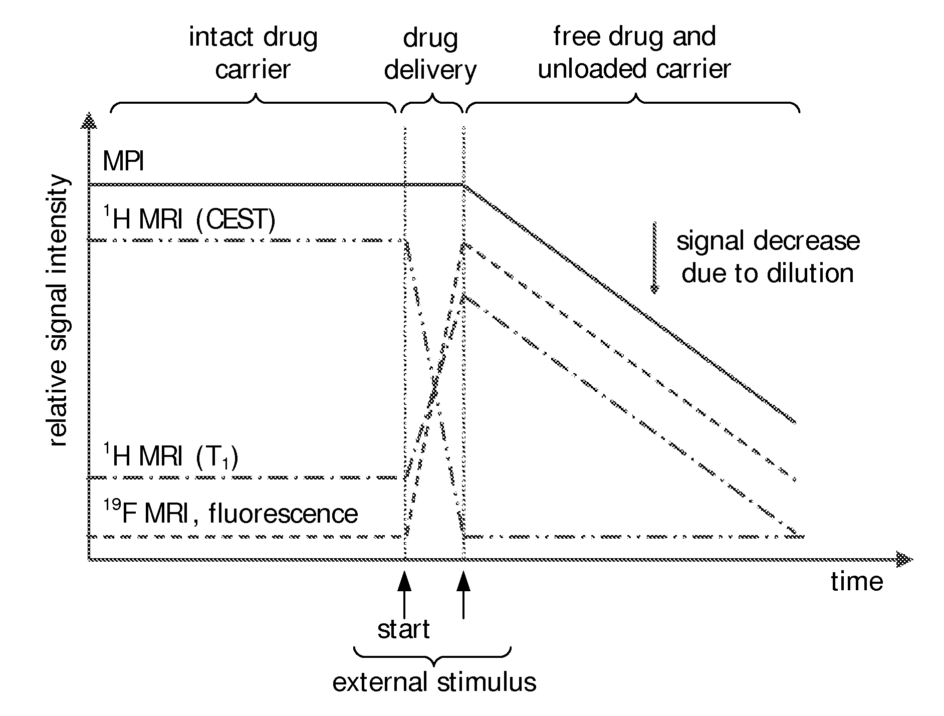
[0009]Such compositions combine the advantageous properties of stimuli-responsive carriers, i.e. the possibility of releasing substances, in particular drugs, at predetermined locations after the application a suitable signal, stimulus or action, and the advantageous properties of the Magnetic Particle Imaging (MPI) technology which allows the direct detection of the spatial distribution of magnetic nanoparticles from a non-linear remagnetization analysis resulting in a high sensitivity and resolution. It could, in particular, be shown that

Problems solved by technology

Typical drug delivery strategies are based on a systemic application of the drug often bringing about significant side effects for the patient due to undesired biodistribution and toxicity.
An important drawback of such systemic approaches is the fact that the therapeutic efficiency is dependent on the minimal required drug concentration in the diseased or target tissue or organ on the one hand and the toxic effects of the drug in non-targeted parts of the body on the other.
However, in such an approach the initial contrast agent concentration inside the carrier is so high that the concentration of these carriers cannot be determined easily in the beginning of the intervention due to a significant T2 shortening and diffusion effects.
These alternative approaches provide either problems with contrast prior to drug release or subsequent to drug release and generally offer no quantifiable signal values.
Thus none of the hitherto clinically used imaging methods is suitable to provide quantitative information over the entire treatment process when using stimuli-responsive carriers for drug delivery.

Method used


Image

  • Stimuli-responsive carriers for mpi-guided drug delivery
  • Stimuli-responsive carriers for mpi-guided drug delivery
  • Stimuli-responsive carriers for mpi-guided drug delivery

Examples


example 1

Preparation of DNA-Loaded Thermosensitive Liposomes

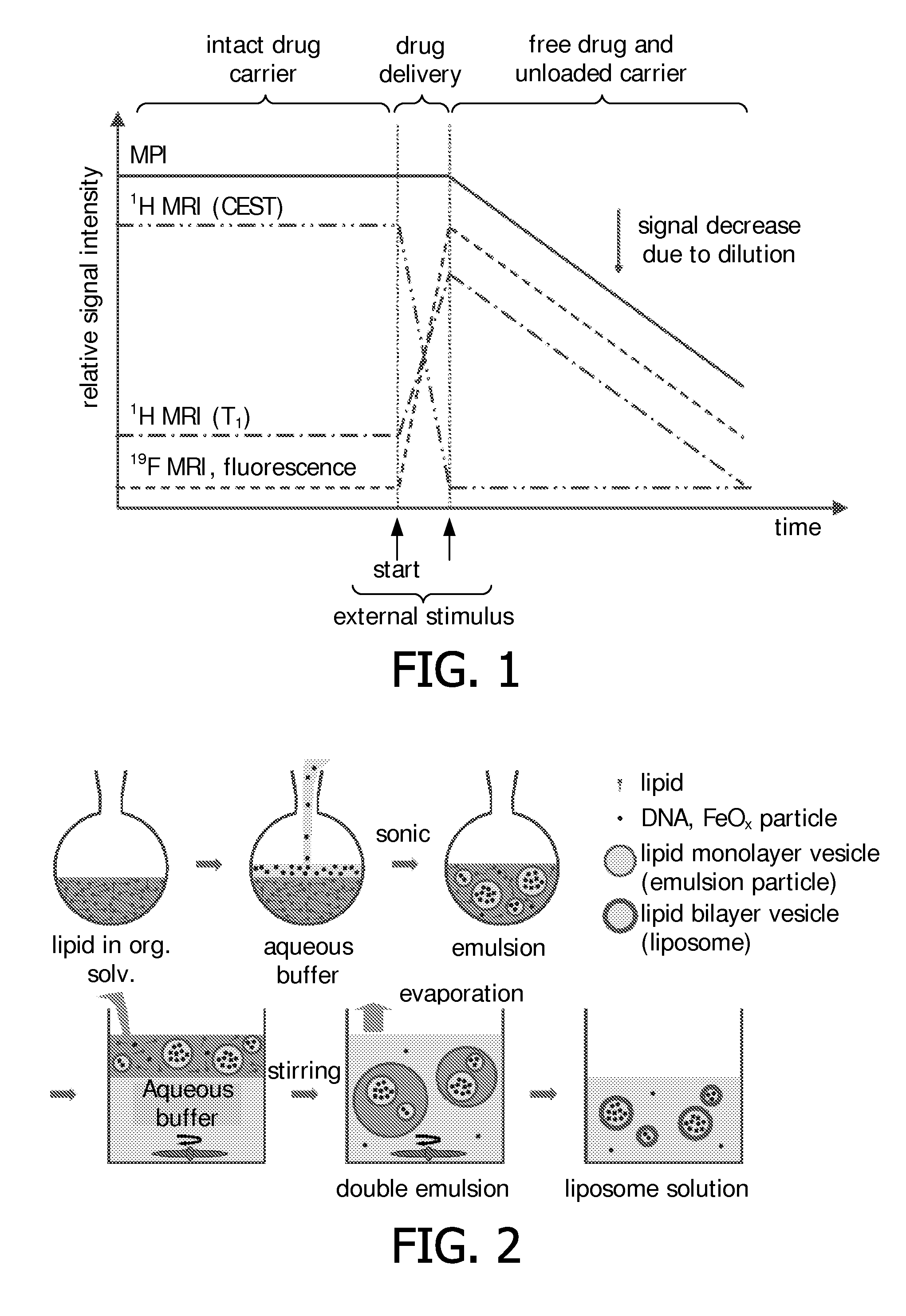
[0152]In a typical preparation of DNA-loaded thermosensitive liposomes 6.3 mg (8.5 μmol) of DPPC, 0.5 mg (1.0 μmol) of MPPC, 1.4 mg (0.5 μmol) of DPPE-PEG2000, and 25 μL of a 1 mg / mL solution of Liss Rhod PE in CHCl3 were dissolved in CHCl3 to obtain 1.0 mL of a CHCl3 solution comprising a 10 mM concentration of lipids. DNA (herring sperm, Sigma-Aldrich) was dissolved in HEPES buffer (135 mM NaCl, 20 mM HEPES, pH 7.40), Resovist stock solution, or a mixture of both. 0.3 mL of the so obtained aqueous solution were mixed with the CHCl3 solution to obtain a 0.3:1 water / oil (W / O) ratio. The constitution of the aqueous phase varied as described in the following Table 1:

TABLE 1Composition of Examples 1-3.c(DNA) / V(DNA) / c(Resovist) / V(Resovist) / nmg / mLmLmMmLlabel*1300.30−−−2300.30−−+3300.150.500.15−*Sample was fluorescently labeled with Liss Rhod PE 0.1% in the lipid composition

[0153]The obtained mixture was sonicated using a QEX 600 sonicati...

example 2

Alternative Preparation of DNA-Loaded Thermosensitive Liposomes

[0157]Liposomes were prepared as in Example 1, with the difference of adding Liss Rhod PE fluorescent lipid (0.1% replacing 0.1% of DPPC) to the lipid composition in order to visualize the presence of liposomes on the agarose gel as outlined above. The chosen initial lipid concentration was 10 mM (CHCl3), and a DNA solution containing 30 mg / mL DNA was used to form the inner water compartment. The obtained purified liposome solution was up-concentrated by a factor of 10 using 100 kDa Amicon centrifugal units.

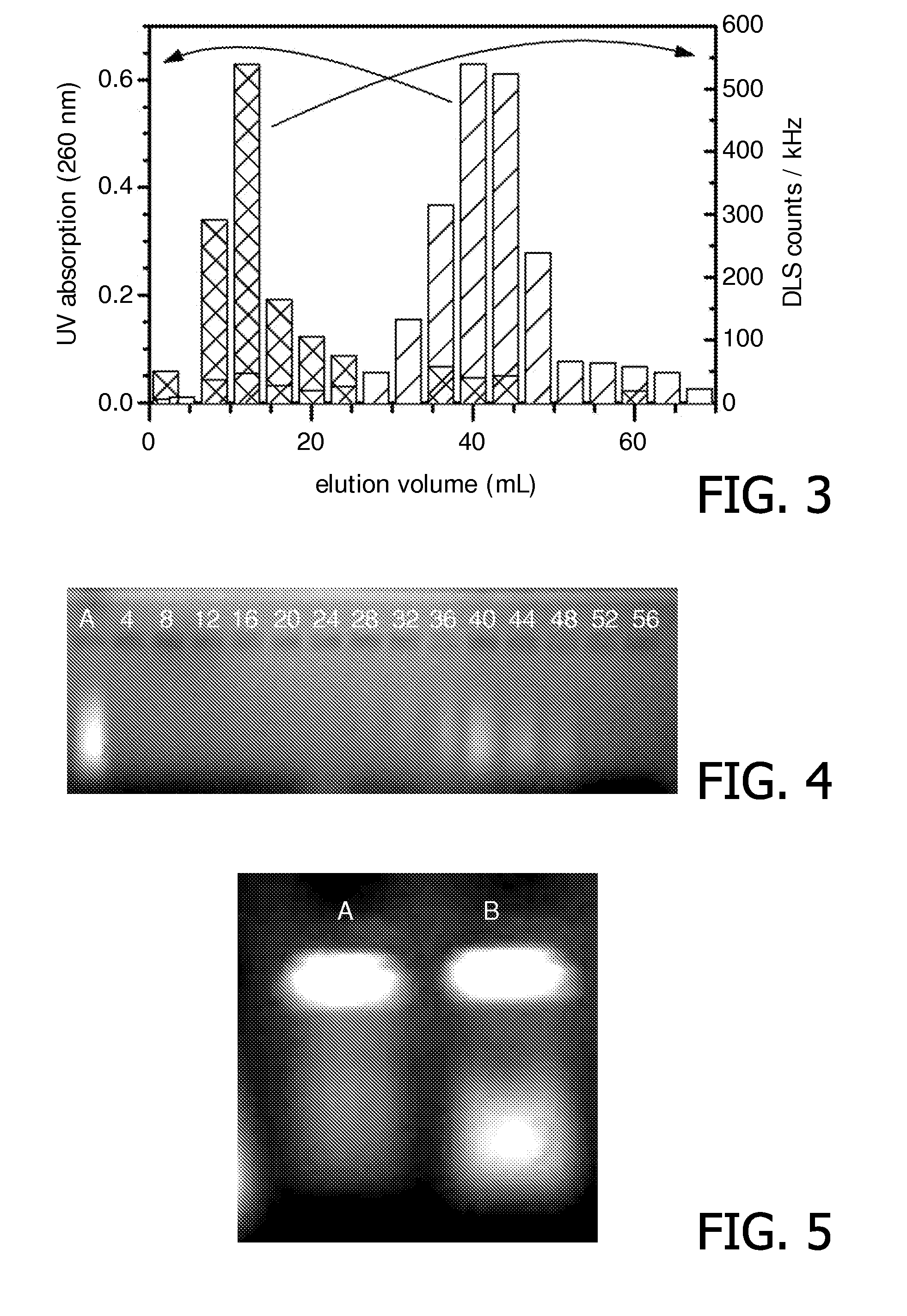
[0158]Temperature-induced DNA delivery was tested by heating the above solution to 50° C. for 30 minutes. Gel electrophoresis was performed of solution samples before and after heating in order to investigate the efficiency of the release of entrapped DNA (see FIG. 5). Before heating (Line A) only one main spot remained at the origin of the gel and a weak background signal could be detected across the respective line....

example 3

Verification of Drug Release

[0159]DNA / Resovist-loaded liposomes were prepared as described in Example 1 using a mixture of DNA and Resovist in the loading aqueous phase. The chosen initial lipid concentration was 10 mM (CHCl3), and the used inner water compartment contained 15 mg / mL DNA and Resovist (0.25 mM Fe) (see Table 1). After purification, the sample was up-concentrated by a factor of 10 using 100 kDa Amicon centrifugal units.

[0160]The melting phase transition temperature of the liposomal lipid bilayer was determined by means of differential scanning calorimetry (DSC). A sample was subjected to a heating / cooling cycle between 20° C. and 60° C. at a heating and cooling rate of 15° C. / min and the associated heat flow was monitored. From the obtained thermogram (see FIG. 6), the melting phase transition temperature was determined to be 40.8° C. in two subsequent heating cycles, which is in good agreement with the for this lipid composition expected melting phase transition of 41...


PUM

PropertyMeasurementUnit
Fractionaaaaaaaaaa
Magnetic momentaaaaaaaaaa
Temperatureaaaaaaaaaa

Abstract

The present invention relates to a composition comprising a shell structure forming a cavity, wherein said shell structure comprises a drug and wherein said composition is associated with at least one contrast agent; wherein said shell structure is capable of releasing its contents into the exterior upon the application of an external stimulus and wherein said contrast agent comprises magnetic particles which are capable of being detected by Magnetic Particle Imaging (MPI), wherein at least more than 5% (w / w) of the magnetic particles comprised in said contrast agent have a magnetic moment of at least −18 m 2 A, 10 wherein said magnetic particles are preferably composed of Fe, Co, Ni, Zn or Mn or alloys thereof or oxides of any of these. The present invention further relates to the use of such a composition or a composition comprising a shell structure forming a cavity, wherein said shell structure comprises a drug and wherein said composition is associated with at least one contrast agent, wherein said contrast agent is capable of being detected by MPI and wherein said shell structure is capable of releasing its contents into the exterior upon the application of an external stimulus as a carrier for a controlled delivery of a drug, as well as to a method of data acquisition for the control of a drug delivery process comprising the detection or localization via MPI of such compositions. In a further aspect the present invention relates to such compositions for treating a pathological condition, wherein the treatment comprises the release of the drug by the application of a stimulus.

Description

FIELD OF THE INVENTION[0001]The present invention relates to a composition comprising a shell structure forming a cavity, wherein said shell structure comprises a drug and wherein said composition is associated with at least one contrast agent; wherein said shell structure is capable of releasing its contents into the exterior upon the application of an external stimulus and wherein said contrast agent comprises magnetic particles which are capable of being detected by Magnetic Particle Imaging (MPI), wherein at least more than 5% (w / w) of the magnetic particles comprised in said contrast agent have a magnetic moment of at least 10−18 m2A, wherein said magnetic particles are preferably composed of Fe, Co, Ni, Zn or Mn or alloys thereof or oxides of any of these. The present invention further relates to the use of such a composition or a composition comprising a shell structure forming a cavity, wherein said shell structure comprises a drug and wherein said composition is associated ...

Claims


Application Information

Patent Timeline
IPC IPC(8): A61K49/18A61P41/00
CPCA61K9/0009A61P41/00
Inventor BURDINSKI, DIRKPIKKEMAAT, JEROEN A.SCHMITT, BERTRANDGRUELL, HOLGERLANGEREIS, SANDER
Owner KONINKLIJKE PHILIPS ELECTRONICS NV
Learn More