

Animal Model for Toxicology and Dose Prediction
a toxicology and dose prediction technology, applied in the field of cell biology, can solve the problems of less expensive rodent models, insufficient reflection of human physiology, and even more acute problems, and achieve the effect of reducing the incidence of cancer development and reducing morbidity
- Summary
- Abstract
- Description
- Claims
- Application Information
AI Technical Summary
Benefits of technology
Problems solved by technology
Method used
Image

Examples
example 1
Generation of Non-Human Animal Models
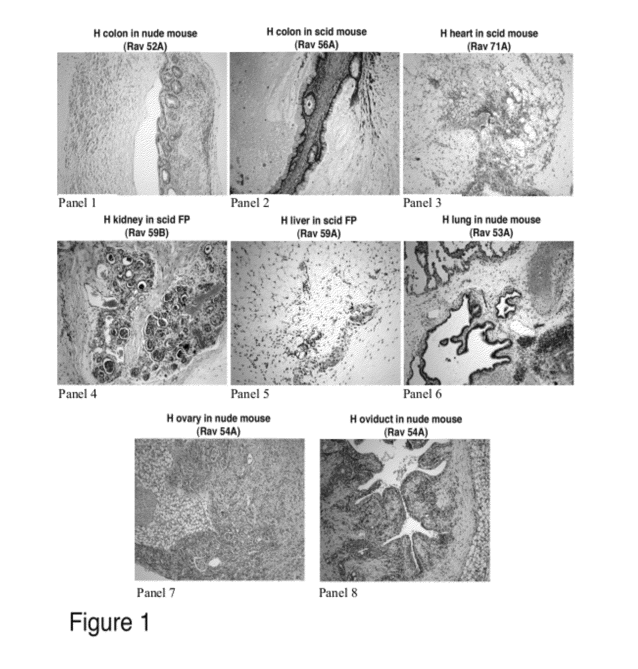
[0076]Tissues from normal fetal organs (colon, heart, kidney, liver, lung, ovary, and oviduct) were trimmed to 1 mm cubed pieces and placed in the kidney capsule or fat pad of nude (nu / nu) or SCID immunocompromised mice. The tissues were left in the animals for 6-40 weeks to allow time for the development into mature tissues. The animal was euthanized and the tissues were removed and sectioned for H&E staining and immunohistochemical evaluation.
[0077]FIG. 1 shows the results of one series of implantations where the tissues were allowed to mature for 4 months. In this example, all references to “Panels” refer to FIG. 1. Panels 1, 2, 3, 6, 7, and 8 show implantation under the kidney capsule while panels 4 and 5 show implantation under the fat pad. Panels 1, 6, 7, and 8 show implantation of normal fetal organs in a nude (nu / nu) mouse while Panels 2, 3, 4, and 5 show implantation of normal fetal organs in a SCID mouse. Kidney, heart and liver tissue ...
example 2
Use of Matured Tissues for Safety / Efficacy Models
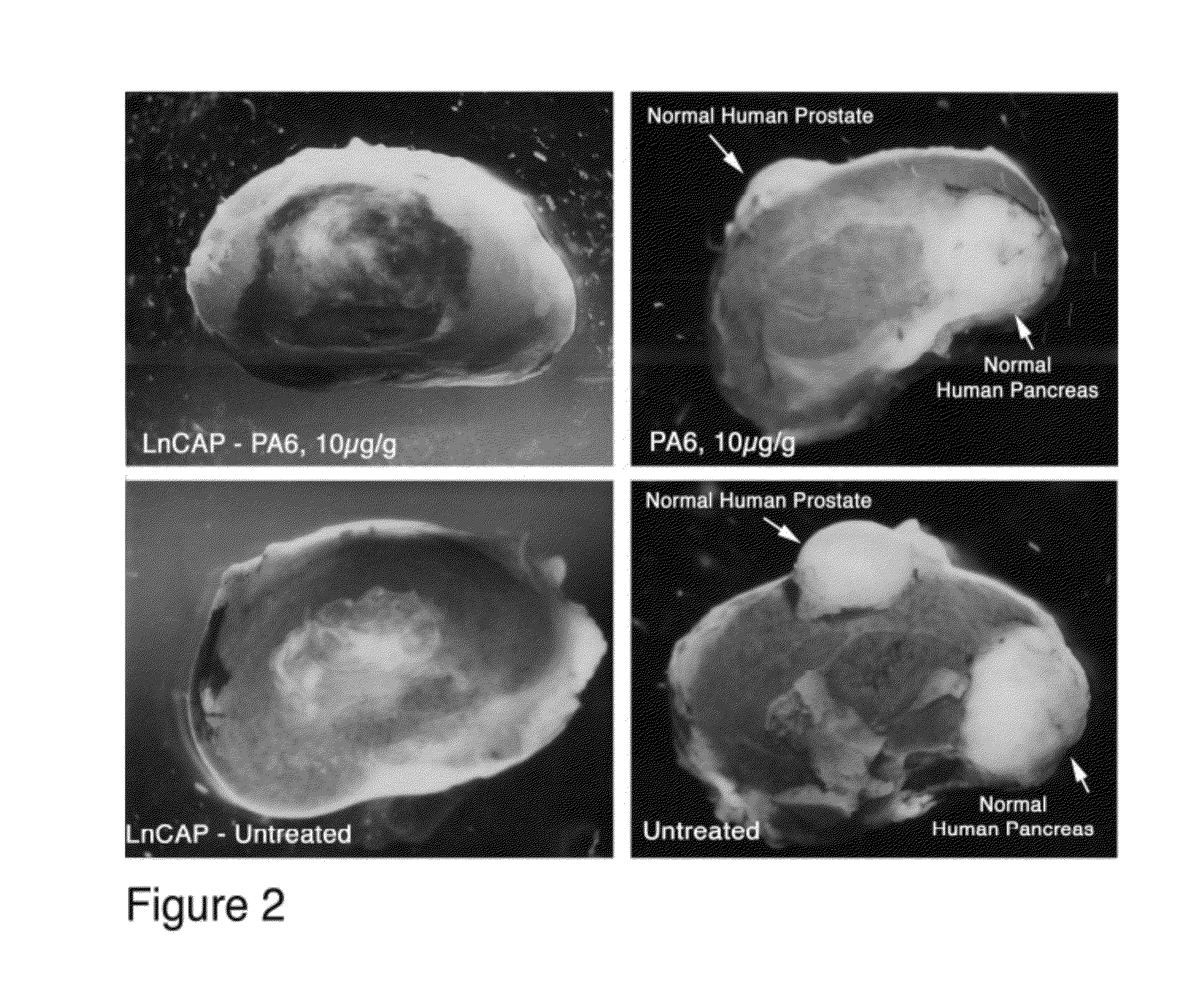
[0078]Normal human prostate and pancreas pieces were placed under the kidney capsule and allowed to mature for 6 weeks. At this time, human prostate cancer cells (LnCAP) were placed under the contralateral kidney capsules of the same animals and allowed to grow for one additional week. At day 7 after implanting the LnCAP tumors, one animal was treated with 10 ugm / gm PA6 antibody (anti-human EpCAM) by i.p. injection. The control animal was treated with saline injections. 4 injections were given over a two week period. At the end of this time, the animals were euthanized and the tumor and normal tissue xenografts examined. The kidneys of the animals are shown in FIG. 2. The left side of FIG. 2 shows LnCAP tumors while the right side shows normal tissues (9 weeks total in the animal). The upper panels are from treated animal while the lower panels are from control animals. Additional treated animals contained normal colon tissue.
example 3
Immunohistochemistry of Human Prostate and Human Colon Matured Tissues
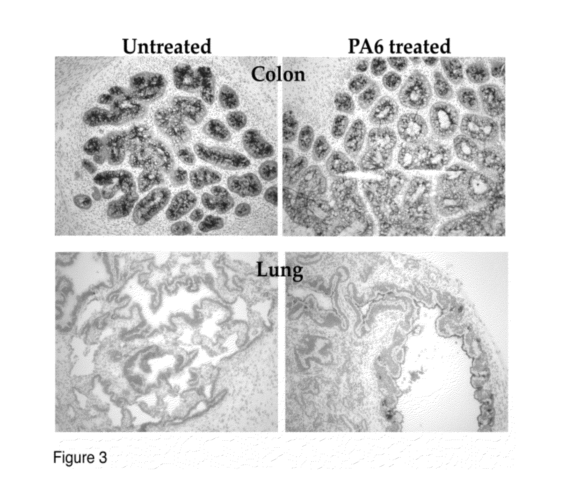
[0079]Immunohistochemistry of human prostate and human colon matured tissues from the experiment described in FIG. 2. Although the tumor was impacted by the antibody treatment with cell death and hemorrhaging, the normal tissues were unaffected by the antibody (A-D). In order to determine whether the tissues contained the antibody target (EpCAM), tissues were stained with directly labeled PA6 (anti-human EPCAM) antibody. The tissues, both treated and untreated show binding of the antibody. The matured human prostate tissue also stained strongly for prostate specific antigen (PSA), a marker for prostate cells.
PUM
| Property | Measurement | Unit |
|---|---|---|
| molecular weight | aaaaa | aaaaa |
| molecular weight | aaaaa | aaaaa |
| time | aaaaa | aaaaa |
Abstract
Description
Claims
Application Information