

Methods for complex tissue engineering
a technology of complex tissues and engineering methods, applied in the field of bioengineered tissue, can solve the problems of complex sourcing problems, only useful attempts, and extremely difficult bioengineering complex tissues
- Summary
- Abstract
- Description
- Claims
- Application Information
AI Technical Summary
Benefits of technology
Problems solved by technology
Method used
Image

Examples
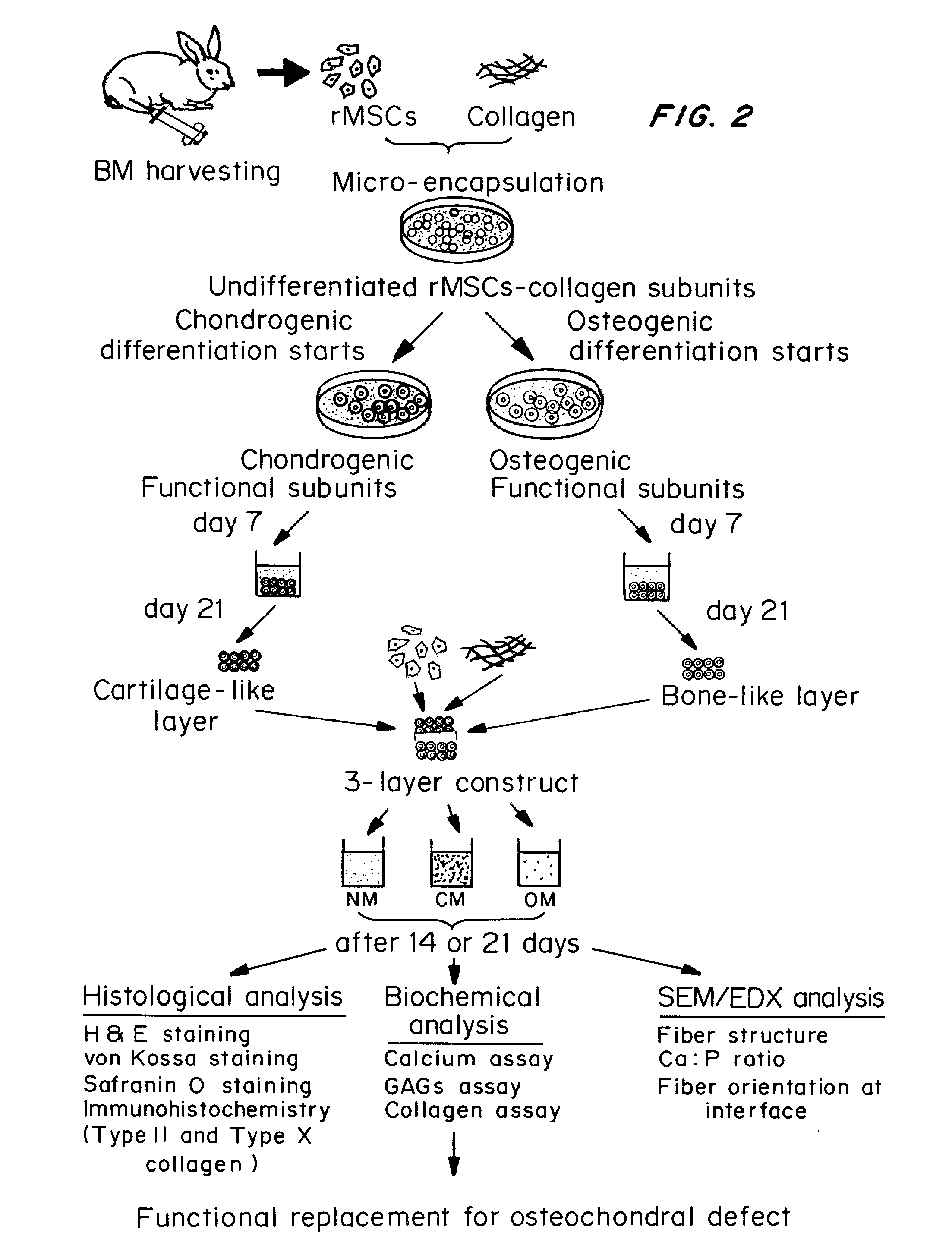
example 1
Bone Marrow Aspiration and Rabbit Mesenchymal Stem Cell (rMSCs) Isolation
[0064]Three month old New Zealand white rabbits weighing an average of 3.5 kg were anesthetized by an intramuscular injection of a mixture of 10% ketamine hydrochloride (0.35 ml / kg) and 2% xylazine (0.25 ml / kg). Approximately 5 ml of bone marrow was aspirated from the tibia. After Ficoll-Hypague gradient separation, mononuclear cells at the interface were collected and cultured in Dulbecco's modified Eagle's medium (DMEM) containing 10% fetal bovine serum (FBS) and antibiotics. The medium was refreshed 10 days after seeding, and replenished every 2 days thereafter. Visible colonies of adhered cells were found about 5 to 7 days after the initial plating. After reaching confluence (about 12 to 14 days after initial plating), the cells were detached by 0.25% trypsin / EDTA for subculture.
example 2
Culture of rMSCs
[0065]rMSCs were cultured in full medium consisting of Dulbecco's modified Eagle's medium high glucose (DMEM-HG), 10% fetal bovine serum (FBS), 100 U / m1 penicillin, 100 mg / ml streptomycin, 1.875 mg / ml sodium hydrogen carbonate (NaHCO3), 0.02M HEPES, and 0.29 mg / ml L-glutamine. The final pH of the medium was adjusted to 7.4 with 1 N sodium hydroxide (NaOH). Live cells were separated from dead cells following 24 hours in cultures, by adherence selection i.e., the cells were cultured for 24 hours and then the adhered cells separated from the dead cells which would be in the culture medium. Cells were maintained in full medium, which was replenished every 3 days. rMSCs at subconfluence were detached by 0.25% trypsin / EDTA. Cells from passage 2-3 were used for the subsequent microencapsulation step.
example 3
Fabrication of Naïve Subunits—Collagen-rMSCs Microspheres
[0066]Ice-cold rat tail collagen type I (Becton Dickenson) was neutralized with 1N NaOH and was further diluted with full medium to give a final concentration of 2 mg / ml. Aliquots of rMSCs at P2-P3 in full medium were rapidly mixed with the neutralized collagen solution in an ice bath, resulting in a cell-matrix mixture with a final cell density of 1250 cells / 2.5 μl droplet. Liquid droplets were dispensed into a 35 mm-diameter Petri dish (Sterlin) with UV irradiated parafilm covering the substratum. After incubation at 37° C. in a humidified atmosphere with 5% CO2 for 1 hour, the liquid droplets gelated to form solid rMSCs-collagen microspheres, which were then gently flushed into Petri dish using full medium and cultured for 3 days before the differentiation step.
PUM
| Property | Measurement | Unit |
|---|---|---|
| volume | aaaaa | aaaaa |
| concentration | aaaaa | aaaaa |
| pH | aaaaa | aaaaa |
Abstract
Description
Claims
Application Information