Method and apparatus for forming the doped cryo-biology specimen of electron microscope

a cryo-biology and electron microscope technology, applied in the field of cryo-biology specimen formation methods and apparatuses, can solve the problems of insufficient contrast of produced image, inability to solve all problems in the biotechnology field, and unclear image displayed, so as to reduce the radiation damage of biomolecules, improve the resolution of electron microscopy, and repair the damage caused by electron radiation quickly

Inactive Publication Date: 2012-08-30
CHAO CHIH YU
View PDF0 Cites 16 Cited by
  • Summary
  • Abstract
  • Description
  • Claims
  • Application Information

AI Technical Summary

Benefits of technology

[0009]The invention relates to a method for doping the proton and the electron into the cryo-biology specimen at the amorphous-ice state. Firstly, freezing the biomolecular aqueous solution rapidly, and making the biomolecular aqueous solution become the cryo-biology specimen at the amorphous-ice state. Then, doping the proton and the electron into the cryo-biology specimen is achieved at the amorphous-ice state under the cryogenic temperature. After doping certain concentration of protons and electrons, the cryo-biology specimen at the amorphous-ice state can be changed into the conductor. The free electrons in the doped cryo-biology specimen can be promptly returned to the protein molecule fragments or water molecular free radicals damaged by the electron beam irradiation. Thus, the radiation damage caused by electron radiation can be repaired quickly by mobile electrons in the doped cryo-biology specimen at any time. The invention can reduce the radiation damage of biomolecules and the surrounding amorphous-ice environment under electron beam exposure. Therefore, the prototype of proteins

Problems solved by technology

Because the protein molecule or the biological cell specimen is made up of lighter elements' mainly, such as carbon, hydrogen, and oxygen, the produced image contrast is not strong enough, therefore the displayed image is not clear.
Even though the present electron microscope and relevant technology can bring the development of biotechnology field into the resolution level of several nanometers, it still cannot solve all problems in the biotechnology field.
However, the cryo

Method used


Image

  • Method and apparatus for forming the doped cryo-biology specimen of electron microscope
  • Method and apparatus for forming the doped cryo-biology specimen of electron microscope
  • Method and apparatus for forming the doped cryo-biology specimen of electron microscope

Examples


Embodiment Construction

[0026]The invention relates to a method for forming the doped cryo-biology specimen of electron microscope, which mainly comprises the following detailed procedures:

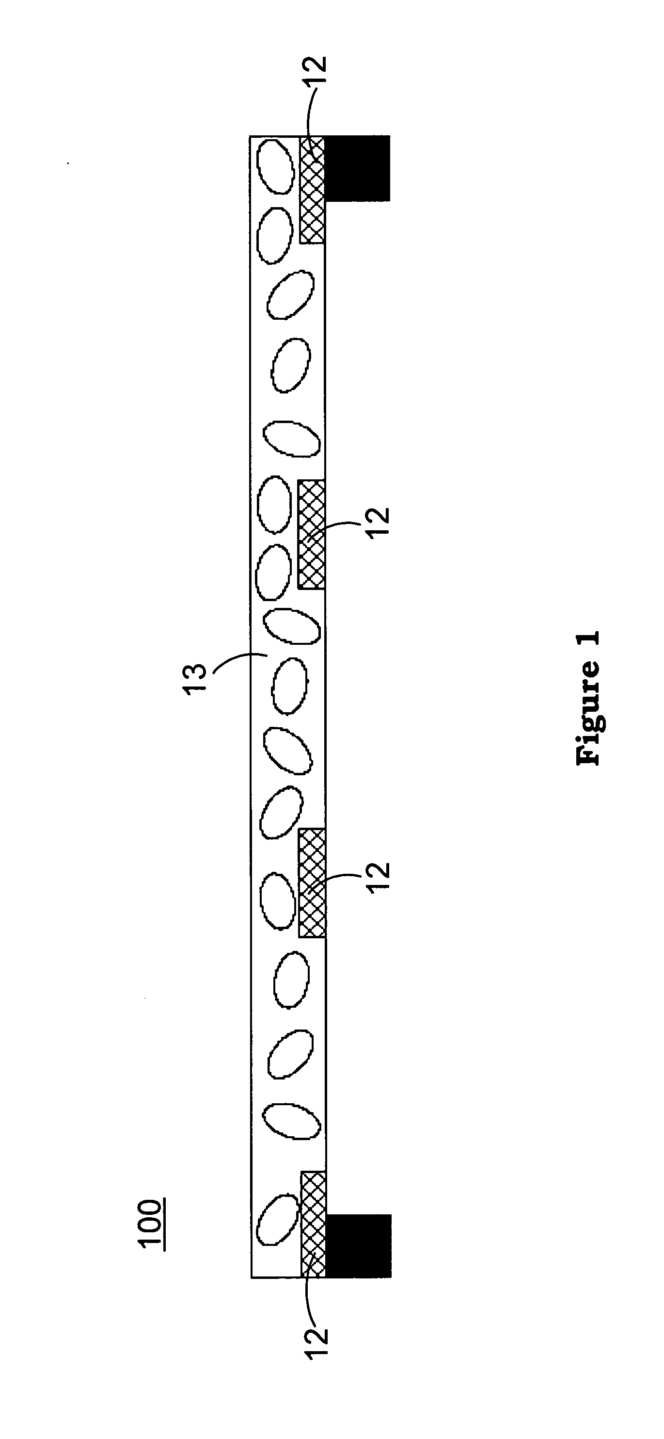
[0027]As shown in FIG. 1 firstly, adding a biomolecule into an aqueous solution to become a biomolecular aqueous solution, and then dropping the biomolecular aqueous solution into the specimen grid 100 is carried out. Then, remove the surplus solution with the filter paper. It is rapidly frozen to about 77K by low-temperature liquid ethane, so that the biomolecular aqueous solution becomes the cryo-biology specimen 13 at the amorphous-ice state. Still as shown in FIG. 1, the cryo-biology specimen 13 is frozen and stored in the specimen grid 100. The specimen grid 100 is composed of amorphous carbon film 12 in the shape as holey carbon film. In addition, in order to increase the electric conductivity of electrons in the amorphous ice during the doping process, the biomolecule can be added into the aqueous solution contain...


PUM

PropertyMeasurementUnit
Temperatureaaaaaaaaaa
Concentrationaaaaaaaaaa
Electric potential / voltageaaaaaaaaaa

Abstract

The invention discloses a method and apparatus for forming the doped cryo-biology specimen of electron microscope. The invention applies rapid cryogenic freezing to the biology specimen, and dopes certain concentration of protons and electrons into the cryo-biology specimen for conducting the observation using electron microscope. The invention reduces the radiation damage of cryogenic biology specimen and amorphous ice caused by the electron radiation, and observes the prototype of biomolecules and biomaterials clearly.

Description

BACKGROUND OF THE INVENTION[0001]1. Field of the Invention[0002]The invention relates to a method and apparatus for forming the doped cryo-biology specimen, particularly to a method and apparatus for forming the doped cryo-biology specimen of electron microscope.[0003]2. Description of the Prior Art[0004]The electron microscope (EM) was invented in the early years of 20th century. After the continuous development for several decades, it has already become an important and often times indispensable tool in the modern science and technology, and has already been applied in the biotechnology field massively. Compared to 0.1 mm resolution of human bare eyes, the resolution of common optical microscope is 500×, and the resolution of electron microscope is almost 500,000×. Besides the electromagnetic lens system in the main column, the electron microscope has the auxiliary vacuum pumping apparatus and many other electric systems. There is similar principle between the electron microscope ...

Claims


Application Information

Patent Timeline
IPC IPC(8): G01N1/00C25B9/00B01J19/08
CPCH01J37/317Y10T436/2525G01N1/42H01J2237/31701
Inventor CHAO, CHIH-YU
Owner CHAO CHIH YU
Why Eureka
Try Eureka