System and method for inserting intracranial catheters

a system and catheter technology, applied in the field of system and method for inserting intracranial catheters, can solve the problems of occupying a lot of floor space, affecting the safety of patients, so as to reduce the complexity of the system and avoid damage to blood vessels

Inactive Publication Date: 2012-11-29
KOHRING GREGORY ALLEN
View PDF3 Cites 38 Cited by
  • Summary
  • Abstract
  • Description
  • Claims
  • Application Information

AI Technical Summary

Benefits of technology

[0021]Accordingly there are several advantages of one or more aspects: a) the in vivo video camera decreases system complexity; b) the IMU allows the software to record the complete trajectory of the catheter for post operative analysis and should a revision be necessary, the new trajectory, together with the old trajectory are available for post-operative analysis; c) the software intelligently combines the data from the ultrasound sensor, video camera and IMU to detect blood vessels, monitor the catheter's trajectory with respect to the location of the ventricles, and suggests course corrections to avoid damaging blood vessels while remaining on target; and d) the entire system is portable to conform to ICU environments. Other advantages of one or more aspects will be apparent from a consideration of the drawings and ensuing description.

Problems solved by technology

Inserting a catheter into a ventricle of the brain is a delicate procedure due to the risks of collateral brain damage.
A drawback of robotic systems is that they tend to be very bulky, taking up quite a bit of floor space.
For the case of trauma patients, pressure within the ventricles can rise to life threatening levels within a short time.
The freehand insertion procedure is not very accurate with studies (Huyette, et al., 2008) showing that freehand placement typically required two attempts before the EVD was satisfactorily placed and CSF was flowing.
(Deaths directly attributable to the procedure are difficult to ascertain as patients are usually in a very critical condition to begin with and CT scans are rarely performed after a patient has died.)
This has several disadvantages: First, extended durations of EVD placement have been implicated as a risk factor in EVD related infections (Kim, et al., 2012).
Second, if several attempts are made to sit the EVD or the EVD must be revised because the CSF flow is week, then only the final catheter placement will be visible on the CT scan; information regarding the position of the previous placements is lost.
However, the exact location of the ventricles within the brain depends upon patient specific factors that cannot be estimated using external information alone.
Simultaneously to far field sensing precise near field sensing is required as arteries having a diameter of 0.5 mm can lead to significant hemorrhaging if damaged.
However, displaying the image from the fiber optic lens on a computer screen requires an external device to convert the optical signal into an electronic signal thereby adding complexity and costs to the overall system.
Using a solid state imaging device embedded in the catheter's distal end decreases the complexity of the system (U.S. Pat. No. 5,989,185; U.S. Pat. No. 5,325,847; U.S. Pat. No. 5,305,736), although, simply displaying both images on the screen does not take advantage the synergies possible with the use of multiple sensors of different modality.
Such approaches are clumsy in practice since they require a second device to be used simultaneously while the physician is inserting the catheter.
None of the previous inventions have used computer aided sensor processing to automatically warn the physician if the catheter is off course, has passed through the ventricle or is in danger of intersecting an artery.
In summary, the heretofore disclosed solutions to the long standing problem of accurately and safely performing a ventriculostomy on trauma patients in an ICU environment suffer from a number of disadvantages: a) they lack an automated system capable of taking advantage of the synergisms arising through the combination of different sensor input in order to provide an intelligent navigation aid for the physician, b) they lack a means to minimize concomitant hemorrhaging, c) they lack a means to record the trajectories of the catheter, thereby hampering post-operative diagnostics, and d) to the extent that robotic systems are available, they do not integrate well into an ICU environment, requiring instead the use of an OP.

Method used


Image

  • System and method for inserting intracranial catheters
  • System and method for inserting intracranial catheters
  • System and method for inserting intracranial catheters

Examples


first embodiment

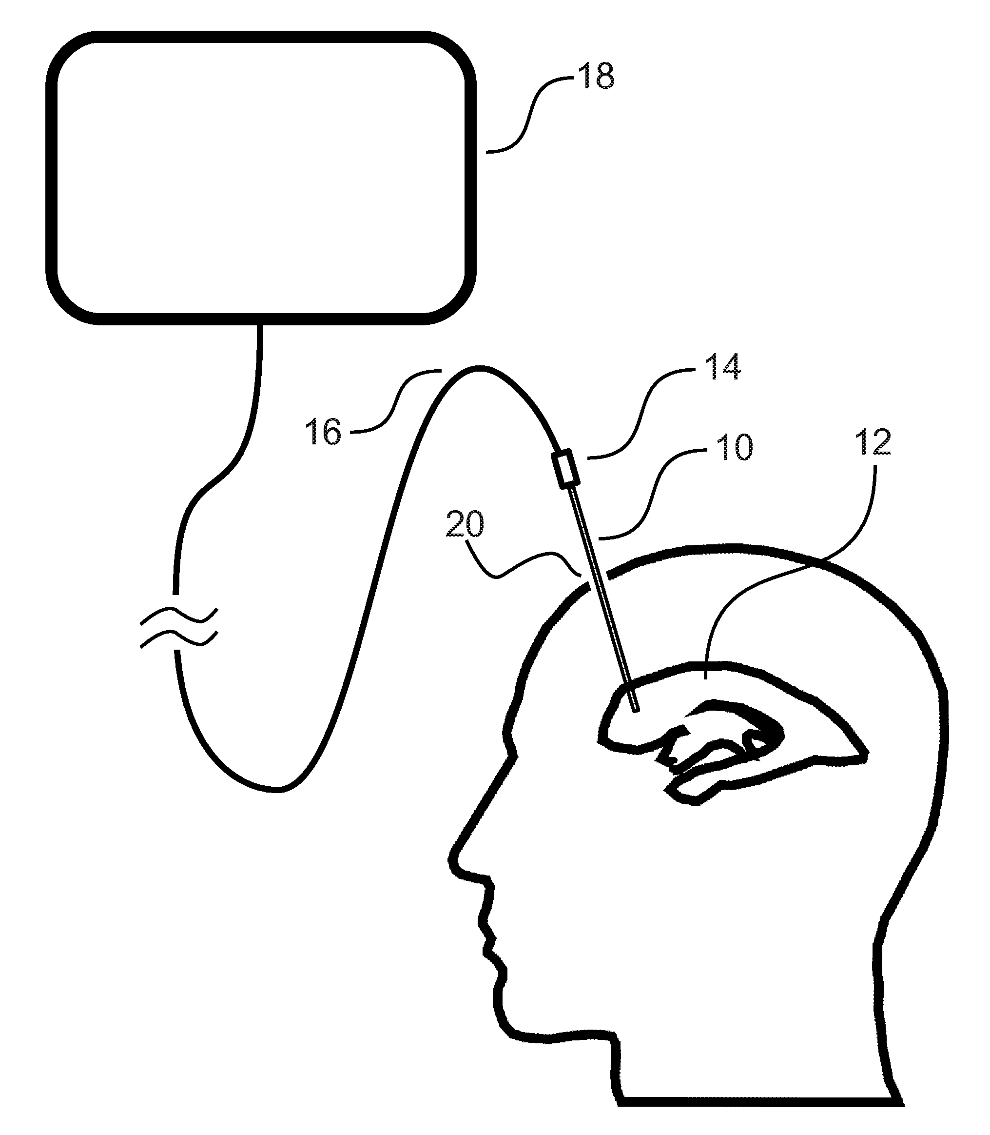
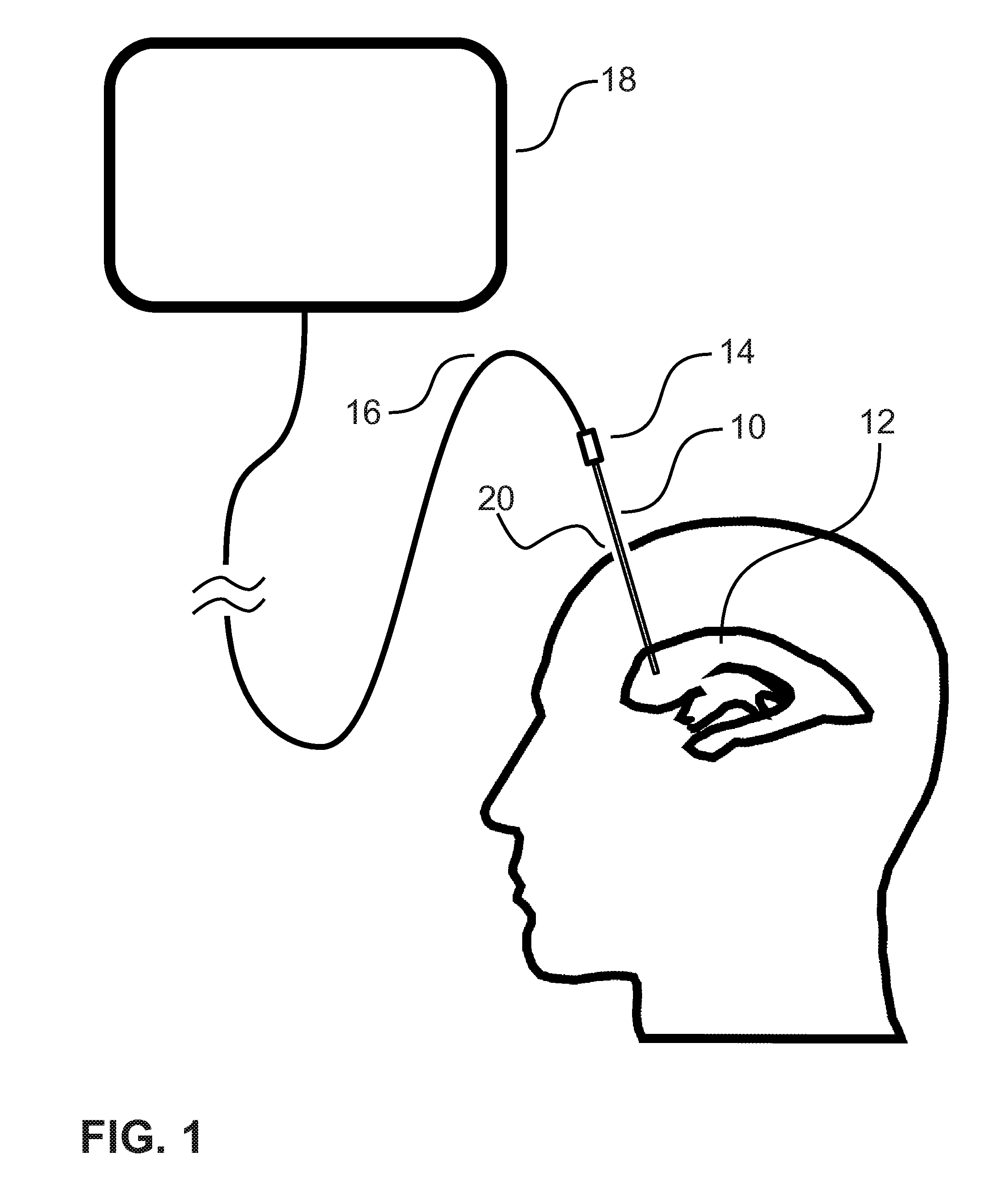
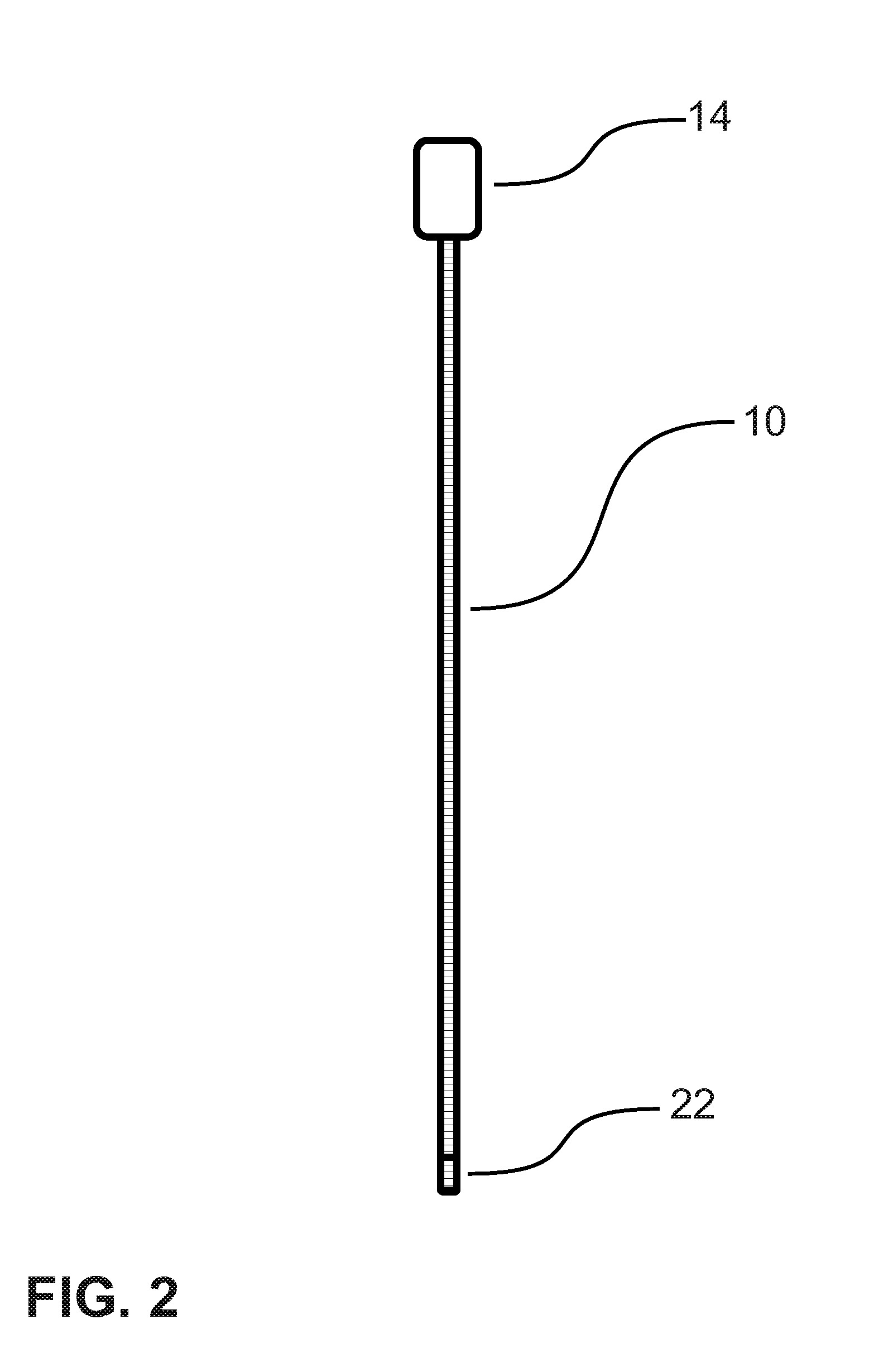
[0034]FIG. 2 shows the catheter 10 drawn to scale. In the first embodiment, the catheter 10 has an inner diameter of 2.5 mm and is manufactured out of a, surgically safe material, e.g., polyurethane. If the material out of which the catheter 10 is manufactured is not sufficiently stiff to allow it to pushed into the brain's soft matter, then a metal stylus can be inserted in the drain 28 (see FIG. 3) to provide the required stiffness. The stylus can be removed once the insertion procedure is over to allow the CSF to flow.

[0035]The catheter 10 has equally spaced markings running from the distal end to the proximal end. As will be explained later, these provide a means for determining the depth of insertion of the catheter 10, i.e., the distance of the catheter's distal end 22 from the burr hole 20. In the first embodiment it is contemplated to put depth markings every millimeter, but other intervals as well as other means of determining the depth of insertion are possible.

[0036]Built...

second embodiment

[0053]Accordingly the reader will see that the embodiments described above provides a number of evident advantages:[0054](a) The synergistic effect of combining the signals from the video camera, with those from the ultrasonic transducer and the IMU enables the creation of a novel, intelligent guidance system that reduces the likelihood of excess hemorrhaging, while increasing the accuracy of final placement.[0055](b) The use of an in vivo video camera in the catheter's distal end simplifies the overall system design compared to fiber optic cameras.[0056](c) The use of an IMU together with the algorithm described in FIG. 9 comprises a means for the system to record the path taken by the catheter en route to the desired location. Information which is invaluable for post-operative diagnostics and treatment.[0057](d) The use of a portable PC allows the entire system to be carried into the ICU when needed and carried out again when no longer required. Furthermore, the second embodiment ...


PUM


Abstract

An improved system for safely and accurately placing intracranial catheters by using techniques from the field of artificial intelligence (AI) which combine the output from in vivo ultrasonic sensors with in vivo video cameras and an embedded inertial measurement unit. The AI subsystem synthesis the output from the three sensors to determine an optimal route to the desired intracranial site while avoiding larger blood vessels en route. Additionally, by using the output from the inertial measurement unit, the catheter's complete trajectory can be recorded and made available for post-operative analysis. The entire system is portable so that it can be used outside of the hospital operating room, for example, in an intensive care unit. Compared to standard freehand methods of placing intracranial catheters, the system embodied here will reduce concomitant hemorrhaging while increasing the accuracy of catheter placement.

Description

CROSS-REFERENCE TO RELATED APPLICATIONS[0001]Not ApplicableSTATEMENT REGARDING FEDERALLY SPONSORED RESEARCH OR DEVELOPMENT[0002]Not ApplicableTHE NAMES OF THE PARTIES TO A JOINT RESEARCH AGREEMENT[0003]Not ApplicableINCORPORATION-BY-REFERENCE OF MATERIAL SUBMITTED ON A COMPACT DISC[0004]Not ApplicableBACKGROUND OF THE INVENTION[0005]1. Field of the Invention[0006]The field of the present invention relates to those systems, methods and devices for accurately and safely inserting a catheter into a body. In particular the system, method and apparatus described herein supports the intracranial insertion of catheters without the attendant complications and disadvantages of known technologies.[0007]2. Description of Related Art[0008]In the field of neurosurgery it is often required to insert devices of various type into the brain. When the device is tubular in shape it is generally referred to as a catheter. Catheters allow the drainage or administration of fluids as well as the placement...

Claims


Application Information

Patent Timeline
Patent Type & Authority Applications(United States)
IPC IPC(8): A61B1/04A61B8/12
CPCA61B8/085A61B8/12A61B8/4254A61B8/4416A61B8/4427A61B2019/5246A61B8/4472A61B2019/5291A61B1/05A61B2019/5206A61B8/445A61B2034/2046A61B2090/306A61B2090/365
Inventor KOHRING, GREGORY ALLEN
Owner KOHRING GREGORY ALLEN
Why Eureka
Try Eureka