Composite support containing silk and collagen, and preparation method thereof

a technology of collagen and silk, applied in the field of biodegradable scaffolds, can solve the problems of affecting the healing effect of ligaments, and inhibiting the regeneration of ligament tissue, and achieves excellent tissue regeneration and mechanical properties, and causes little or no immune response.

Inactive Publication Date: 2013-03-21
DONGGUK UNIV IND ACADEMIC COOPERATION FOUND
View PDF7 Cites 13 Cited by
  • Summary
  • Abstract
  • Description
  • Claims
  • Application Information

AI Technical Summary

Benefits of technology

[0018]A composite scaffold according to embodiments of the present invention is biodegradable, and thus does not require additional removal surgery. In addition, it is excellent in terms of tissue regeneration and mechanical properties and causes little or no immune response after implantation. Thus, it can be effectively used as a matrix for the regeneration of ligaments and tendons and the repair of injured muscles.

Problems solved by technology

This method was not approved by the US Food and Drug Administration (FDA), because it causes exudates, graft failure and synovitis.
This method suffers from various problems, including immune rejection, inhibition of ligament tissue regeneration, infection with disease, and lack of donors.
This therapeutic method also suffers from various problems, including the pain of donor sites, muscle atrophy, and the need for long-term rehabilitation.

Method used


Image

  • Composite support containing silk and collagen, and preparation method thereof
  • Composite support containing silk and collagen, and preparation method thereof
  • Composite support containing silk and collagen, and preparation method thereof

Examples


preparation example 1

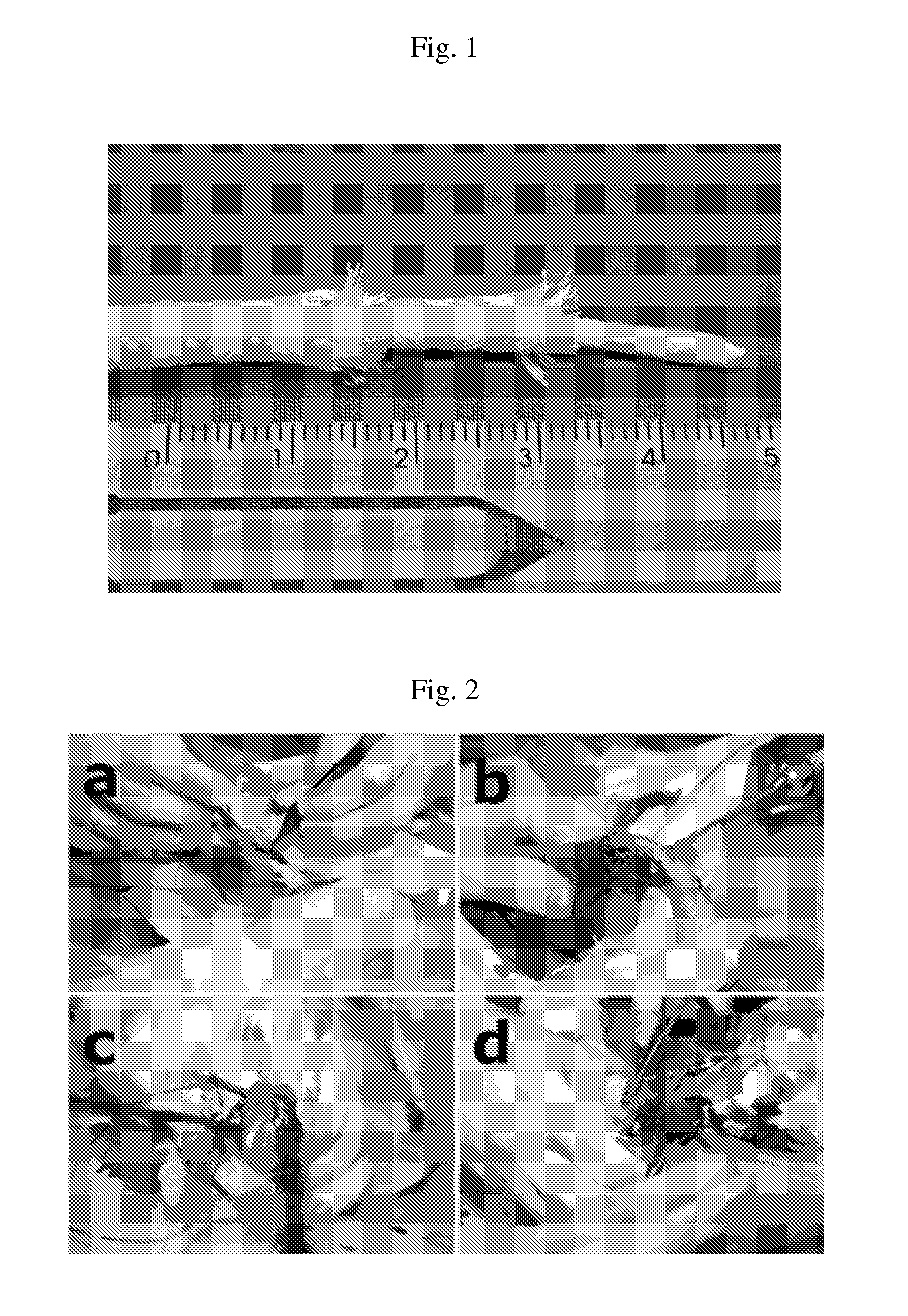
Preparation of Silk Tube Layer

[0078]Silk (Won Corporation, Korea) was woven into tubes having diameters of 2 mm and 4 mm, respectively, using a weaving machine. Then, the smaller-diameter silk tube was inserted onto the larger-diameter silk tube to form a double-layer tube. Then, to remove sericin, the tube was treated with a 0.02M Na2CO3 solution at 100° C. for 8 hours. Then, the tube was clean with 0.3% ivory cleaner, washed with triple-distilled water for 3 days, and dried at 4° C., thereby preparing a silk tube layer from which sericin has been removed.

example 1

Preparation of Composite Scaffold

[0079]Atelocollagen (Bioland, Korea) was dissolved in a 0.05 M aqueous solution of hydrochloric acid to prepare 20 mg / ml of a gel-like solution. To the gel-like solution, 20 mg / ml of chondroitin sulfate (Sigma, USA) was added in an amount of 10 parts by weight based on 100 parts by weight of the gel-like solution to prepare a mixed solution of collagen and chondroitin sulfate.

[0080]The mixed solution was injected into the double-layer silk tube prepared in Preparation Example 1. Specifically, the double-layer silk tube was placed in a silicon tube (mold), and the mixed solution was injected therein, followed by freeze-drying at −80° C.

[0081]The injection and freeze-drying step was repeated four times, and then 20 mg / ml hyaluronic acid (NovaMatrix, Norway) was injected into the tube, followed by freeze-drying at −80° C. Then, the tube was subjected to cross-linking using a carbodiimide crosslinking agent for 20 minutes, after it was washed and freeze-...

example 2

Evaluation of Effect of Composite Scaffold in Animals

[0084]To evaluate the effect of the composite scaffold for ligaments in animals, rabbits with collateral ligament injury were used.

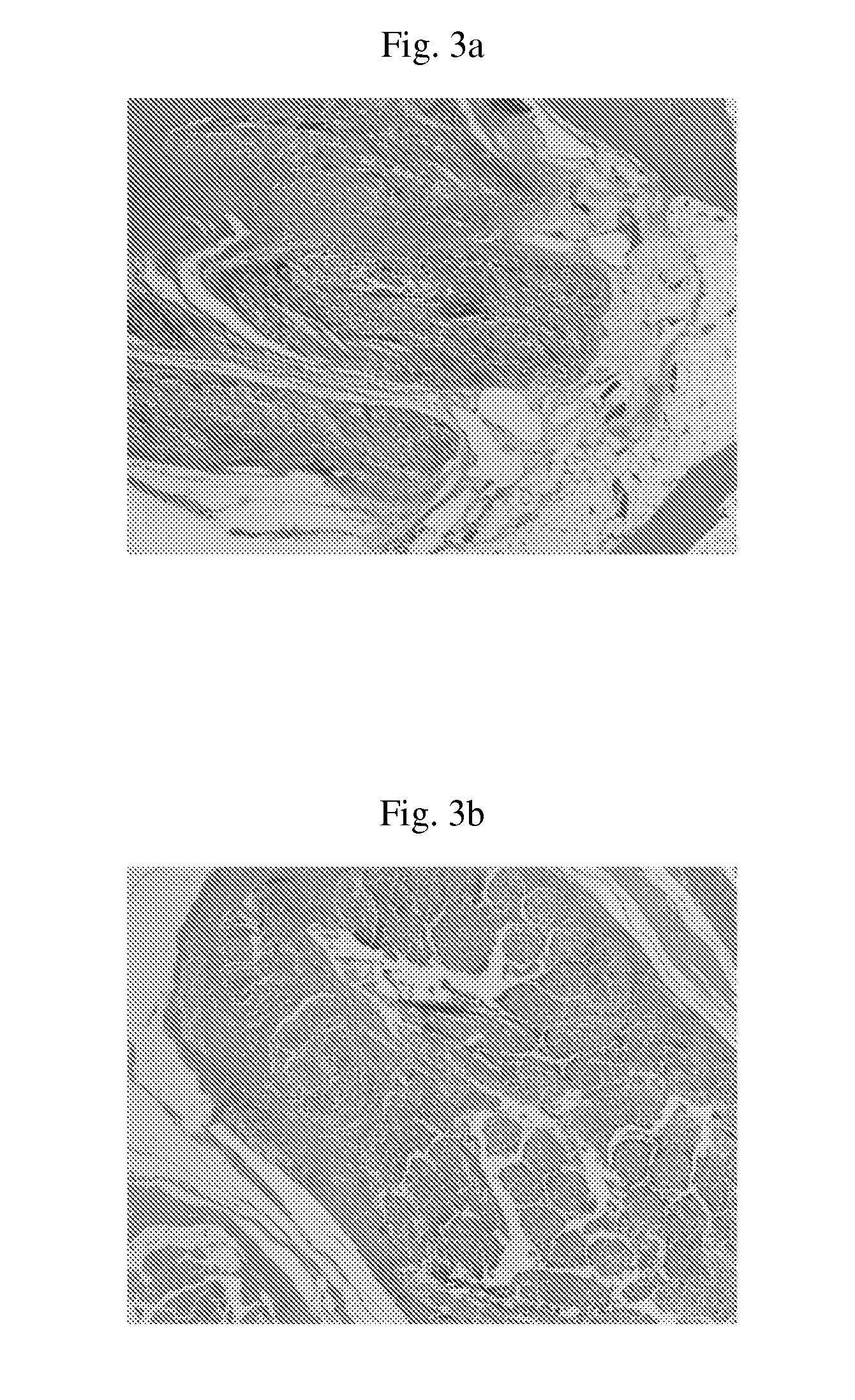
[0085]As shown in FIG. 2, a patellar tendon site was removed from rabbits (FIG. 2a), after which the collateral ligament of the knee was removed and then a tunnel was formed in the condyle portion using a 2.5-mm drill (FIG. 2b). A scaffold, prepared by coating a collagen on the outside of the silk tube of Preparation Example 1, or the composite scaffold of the present invention, was inserted into the formed tunnel in a ‘’ shape (FIG. 2c), and then the tunnel portion was sutured in a ‘’ shape (FIG. 2d). After the surgical operation, an antibiotic was administered to the rabbits for about 10 days.

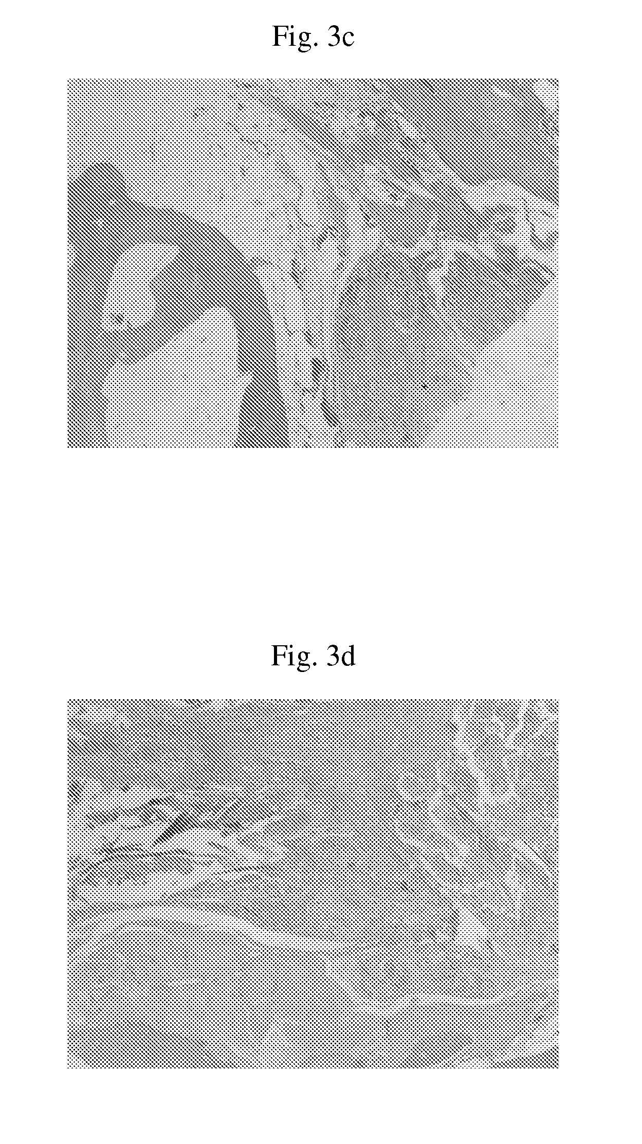
[0086]6 months after the implantation, the animal model with collateral ligament injury was subjected to histological examination in order to examine the biocompatibilities of the scaffold, comprising the collag...


PUM

PropertyMeasurementUnit
thicknessaaaaaaaaaa
thicknessaaaaaaaaaa
diameteraaaaaaaaaa

Abstract

Embodiments of the present invention relate to a biodegradable scaffold for replacing tissue or inducing tissue regeneration and a preparation method thereof, wherein the scaffold comprises at least one woven silk tube layer and a collagen layer inside the tube layer. The scaffold is excellent in terms of tissue regeneration and mechanical properties and causes little or no immune response after implantation. Thus, the scaffold can be effectively used as a matrix for the regeneration of ligaments and tendons and the repair of injured muscles.

Description

TECHNICAL FIELD[0001]The present invention relates to a biodegradable scaffold for replacing tissue or inducing tissue regeneration and a preparation method thereof.BACKGROUND ART[0002]In the United States, about 50 billion dollars are spent each year to treat injured ligaments of 130,000 persons or more. As various sports, including football, handball and ice hockey, have been spread, ligament injury patients have increased. In Europe, 75% of ligament injury patients required physical therapy or surgery to treat the anterior cruciate ligaments.[0003]For the repair of injured ligaments and tendons, methods, including xenograft, allograft and autograft methods, are generally used.[0004]The xenograft method comprises treating a bovine ligament with a chemical agent to remove the cells and grafting the treated ligament. This method was not approved by the US Food and Drug Administration (FDA), because it causes exudates, graft failure and synovitis.[0005]Moreover, the allograft method ...

Claims


Application Information

Patent Timeline
Patent Type & Authority Applications(United States)
IPC IPC(8): A61F2/02D06B1/00D03D15/00
CPCA61F2/08A61L27/24A61L27/32D06B1/00A61F2/02D03D15/00A61L27/54A61F2240/001A61L2300/414A61L2430/10A61L2430/30
Inventor PARK, JUNG-KEUGSEO, YOUNG KWONHAN, MI JUNGLEE, HWA SUNGEO, SU RAKYOON, HEE HOONKWON, SOON YONGPARK, HEE JUNG
Owner DONGGUK UNIV IND ACADEMIC COOPERATION FOUND
Why Eureka
Try Eureka