

Interventional photoacoustic imaging system
a photoacoustic imaging and imaging system technology, applied in the field of interventional photoacoustic imaging systems, can solve the problems of insufficient dosing to the cancer site, major technical limitation, and inability to localize or precisely place the implanted brachytherapy seeds,
- Summary
- Abstract
- Description
- Claims
- Application Information
AI Technical Summary
Benefits of technology
Problems solved by technology
Method used
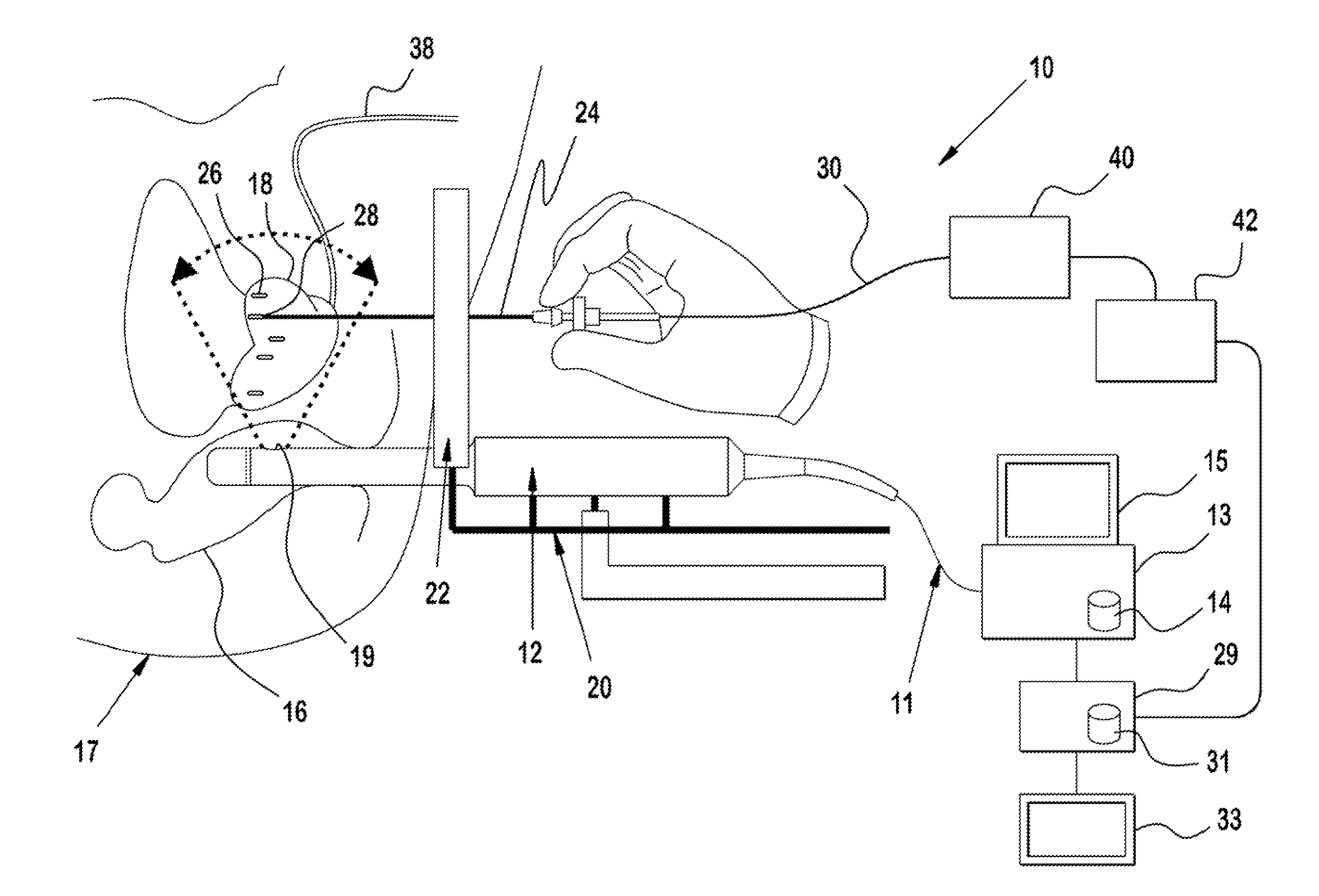
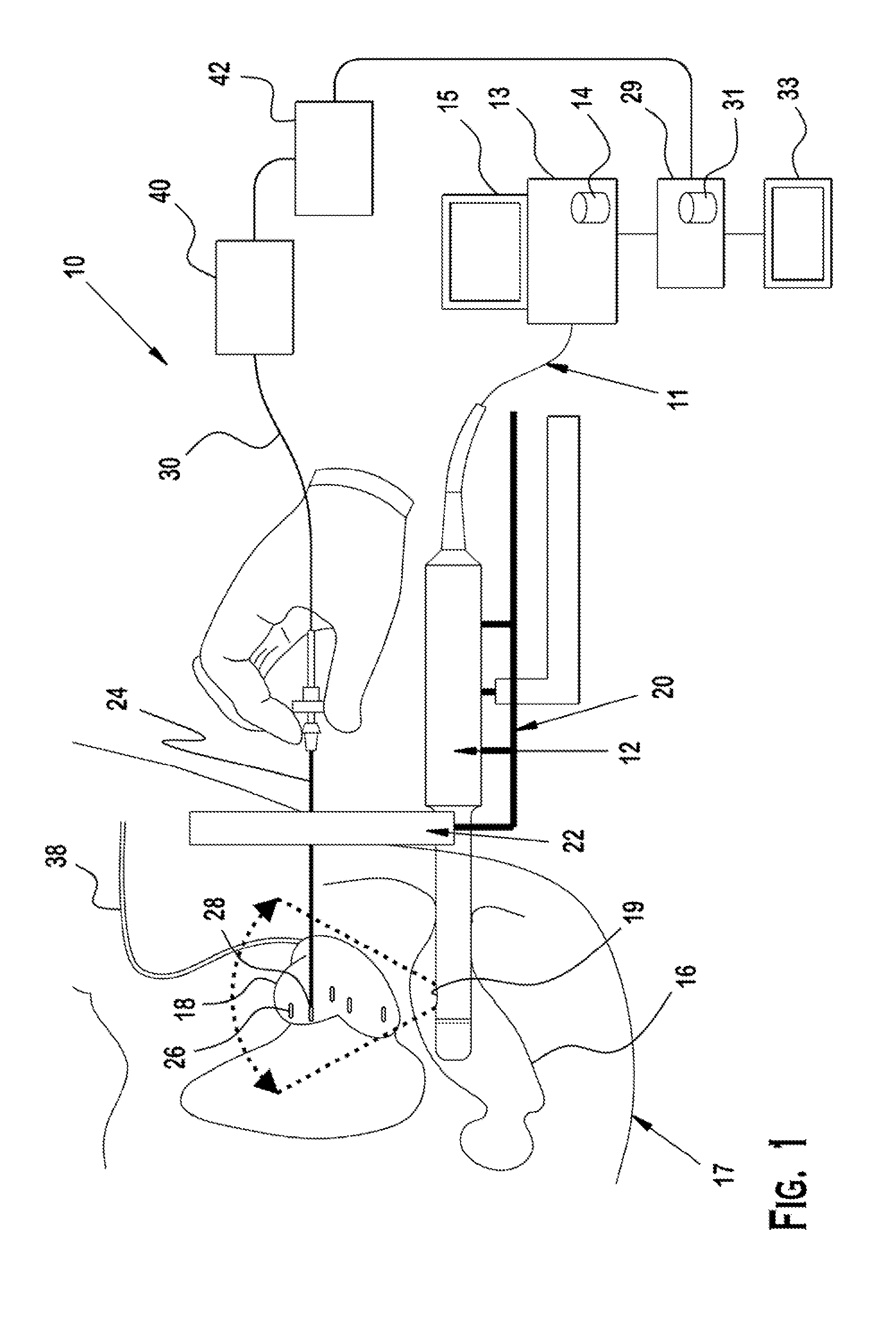
Image

Examples
example 1
[0065]An experimental system was developed to determine, through photoacoustic imaging, seed location in several test phantoms implanted with brachytherapy seeds. During the experiment, pulsed laser light from a Nd:YAG (neodymium-doped yttrium aluminum garnet) laser was directed towards the phantoms. Due to the intense nature of the generated laser beam, it was necessary to reduce the beam intensity. This was achieved through the use of 2, 45° dielectric mirrors, two black holes (to absorb the laser beam), and an adjustable aperture. The beam was passed through the first 45° dielectric mirror with a significant portion of the beam being deflected into the first black bole. This process was repeated and the resultant beam was passed through an adjustable aperture to further modify beam intensity. As such, the beam intensity could be adjusted to a value of approximately 10 mJ / cm2.
[0066]The phantoms used were made of two layers having different optical absorption coefficients (similar ...
example 2
[0068]With reference to FIG. 6, a more detailed experimental setup is shown therein, which included a pulsed Nd:YAG laser system 100 (Surelite II, developed by Continuum, Inc. in Santa Clara, Calif.). The laser system 100 was operated at a wavelength of 1064 nm, providing good contrast between the metallic seeds which absorb such light and the soft tissue of the prostate which does not. The Nd:YAG laser 100 operated within an energy density of 40 mJ / cm2 (roughly, energy of 40 mJ and a spot size of 1 cm2).
[0069]The laser 100 was incorporated into ultrasound system 102. The ultrasound system 102, including transducer 103, was used to detect sound waves generated by the photoacoustic effect. In particular, the ultrasound system 103 was an ultrasonic open research platform known as SONIXCEP manufactured by Ultrasonix Medical Corporation (“Ultrasonic”) located in Richmond, BC, Canada. For faster acquisition, a separately developed data acquisition hardware (DAQ) 104 known as SonixDAQ was...
PUM
 Abstract
Description
Claims
Application Information