Methods And Compositions For Cellular Imaging And Cancer Cell Detection Using Light Harvesting Conjugated Polymer-Biomolecular Conjugates
a technology of biomolecular conjugates and cellular imaging, applied in the direction of fluorescence/phosphorescence, instruments, group 3/13 element organic compounds, etc., can solve the problems of severe cytotoxicity, low photobleaching threshold, and difficulty in implementing such a strategy using cpes
- Summary
- Abstract
- Description
- Claims
- Application Information
AI Technical Summary
Benefits of technology
Problems solved by technology
Method used
Image

Examples
example 1
Synthesis of Biomolecule-Functionalized HCPEs
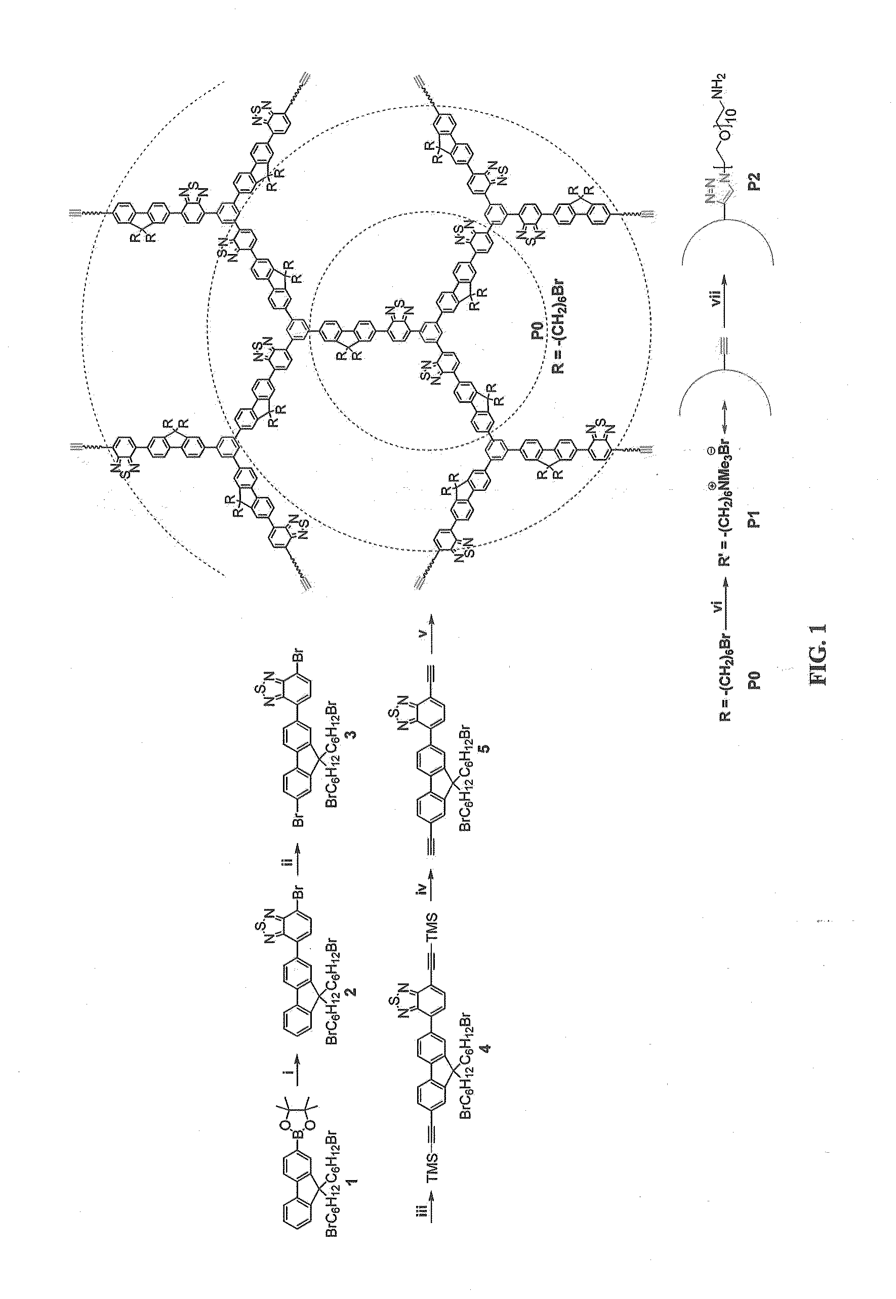
[0216]An affibody-attached hyperbranched conjugated polyelectrolyte (HCPE) was used for targeted fluorescence imaging of human epidermal growth factor receptor 2 (HER2) positive cancer cells. Early-stage detection of HER2 is of clinical significance in personalizing cancer treatment, because HER2 expression levels are closely related to tumor behavior and clinical outcome. Anti-HER2 affibody instead of commonly-used HER2-specific antibody (herceptin) was chosen as the recognition element, in view of its higher affinity for HER2 and smaller size (approximately 7 kDa) compared to herceptin (approximately 150 KDa). The HCPE (P2) used for bioconjugation was endowed with a unique core-shell molecular architecture to minimize nonspecific interactions with biomolecules and to facilitate bioconjugation and targeted cellular imaging.
[0217]The core-shell HCPE (P2) had a hyperbranched conjugated polymer as the fluorescent core and linear poly(ethyle...
example 2
Synthesis of a Folid Acid-Functionalized Molecular Brush
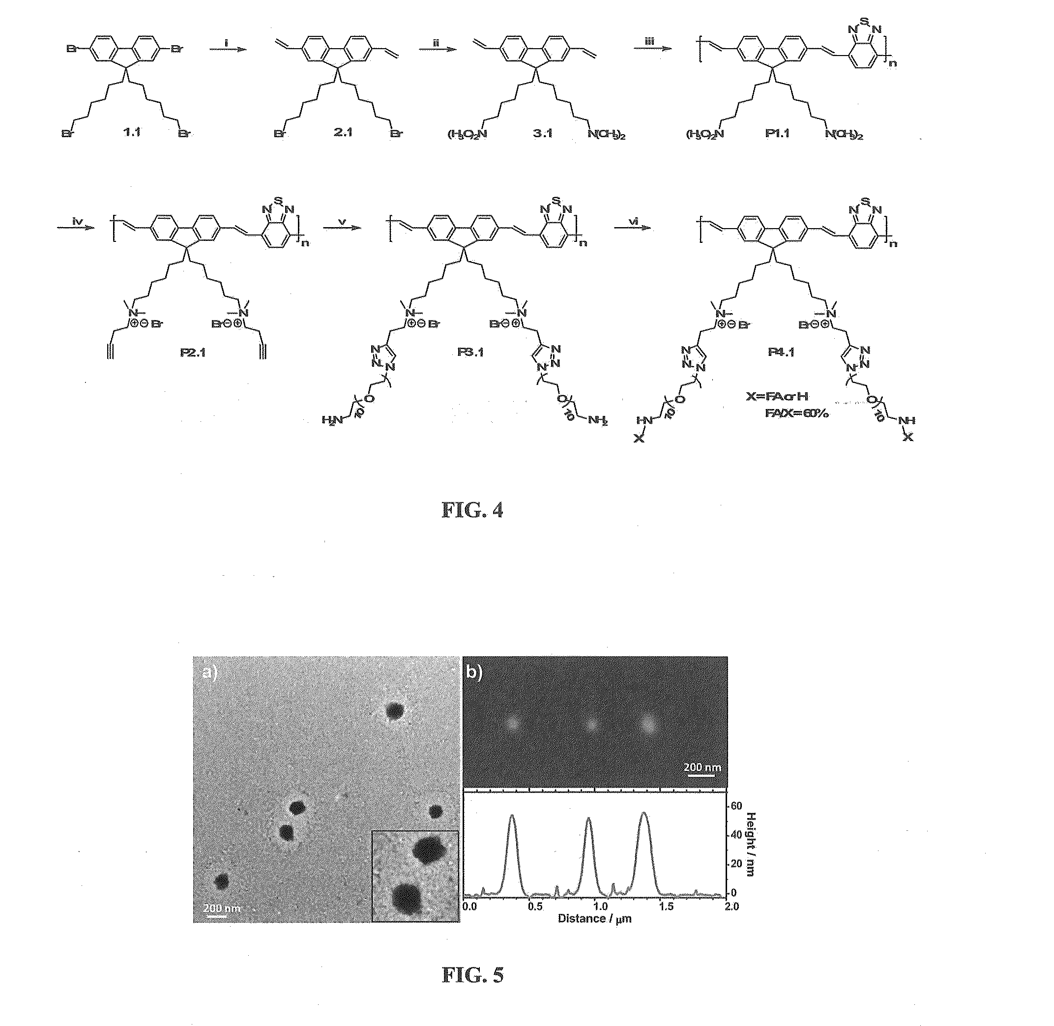
[0233]The molecular brush (P4.1) was synthesized via a stepwise “grafting onto” method involving click chemistry. P4.1 formed core-shell spherical nanoparticles in aqueous solution, wherein the PEG grafting chains constituted the shell layer encapsulating the charged, conjugated backbones. Such a self-assembled nanostructure not only resulted in a high PL quantum yield in aqueous solution (11%), but also led to minimal nonspecific interactions with biomolecules and suppressed nonspecific cellular uptake. These desirable biochemical and optical properties make P4.1 an effective FR / NIR cellular probe for discrimination and visualization of MCF-7 cancer cells from NIH-3T3 normal cells in a high contrast and selective manner. In view of its high photostability and low cytotoxicity, such a molecular brush based cellular nanoprobe holds great promises as an alternative to current stains such as QDs and silica nanoparticles for clinic...
example 3
Self-Assembly Properties of P2
[0243]High-resolution transmission electron microscopy (HR-TEM) shows that P2 self-assembles into spherical nanoparticles with an average diameter of 30 nm in aqueous solution. Moreover, these nanospheres possess a core-shell nanostructure, wherein the dark interior and the gray exterior correspond to the domains enriched with electron-rich conjugated segments and saturated PEG chains, respectively. Such a core-shell nanostructure is beneficial to both bioconjugation and cell imaging, as PEG shells could serve as a protective layer.
PUM
| Property | Measurement | Unit |
|---|---|---|
| Wavelength | aaaaa | aaaaa |
| Fluorescence | aaaaa | aaaaa |
Abstract
Description
Claims
Application Information
 - R&D
- Intellectual Property
- Life Sciences
- Materials
- Tech Scout
- Unparalleled Data Quality
- Higher Quality Content
- 60% Fewer Hallucinations
Browse by: Latest US Patents, China's latest patents, Technical Efficacy Thesaurus, Application Domain, Technology Topic, Popular Technical Reports.
© 2025 PatSnap. All rights reserved.Legal|Privacy policy|Modern Slavery Act Transparency Statement|Sitemap|About US| Contact US: help@patsnap.com