Integrated microfluidic radioassay and imaging platform for small sample analysis
a microfluidic and imaging platform technology, applied in the field of microfluidic devices and diagnostic schemes, can solve the problems of high cost, high cost, and difficult to study kinase activity on samples with a small number of cells, and achieve the effects of limiting the desired work pace, assessing kinase activity, and high cos
- Summary
- Abstract
- Description
- Claims
- Application Information
AI Technical Summary
Benefits of technology
Problems solved by technology
Method used
Image
Examples
example 1
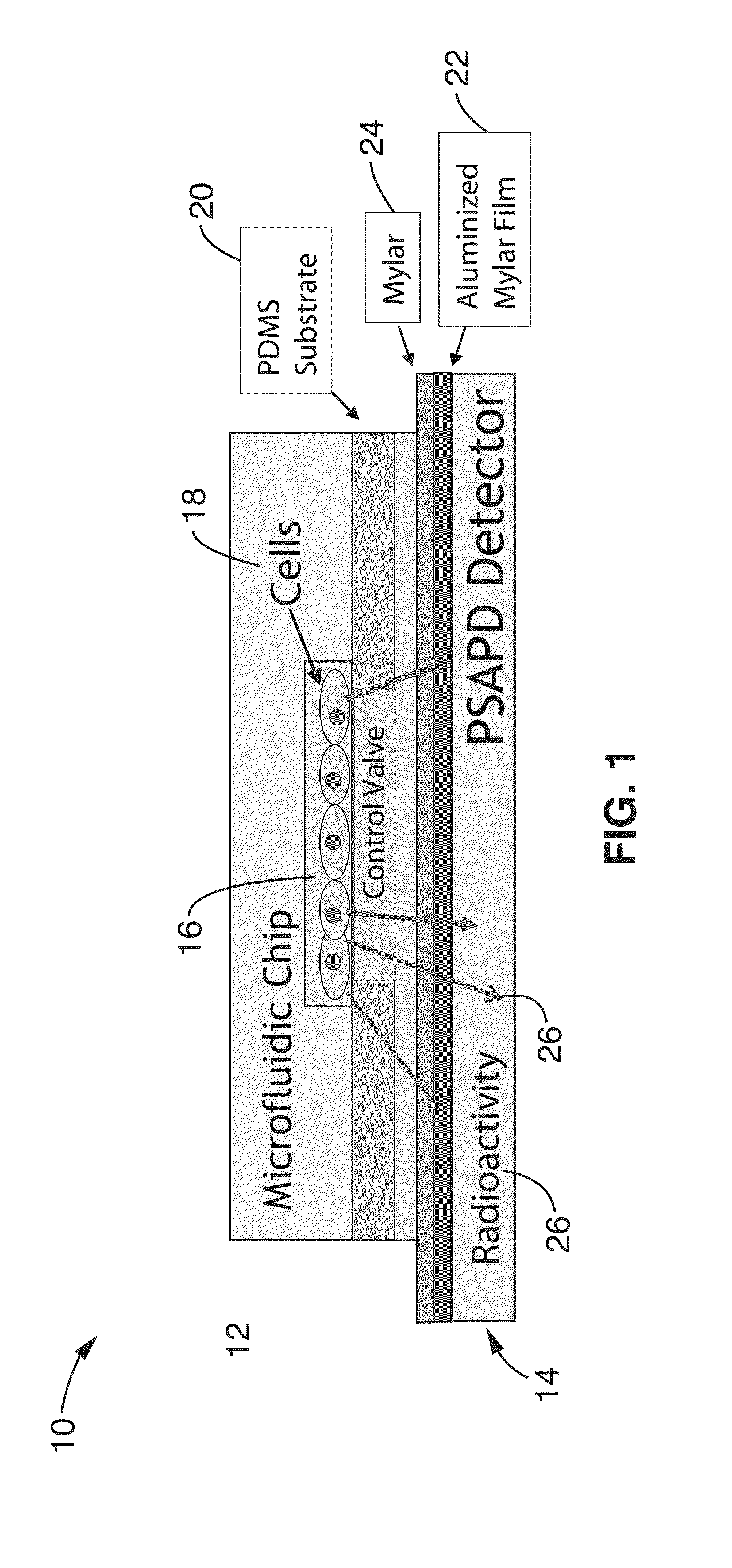
[0046]In order to demonstrate the invention, a microfluidic chip and detector were constructed. The microfluidic chip was fabricated using two-layer soft lithography. Two different molds were first fabricated by a photolithographic process to create the fluidic channels and the control channels that actuate the values located in the top and bottom layers, respectively, of the PDMS-based chip. The fluidic channel mold was made by a three-step photolithographic process. Both round-profile and square-profile channels were created to interface with fully closing regular valves and sieve values, respectively. Upon closure of the valve, round-profile channels close completely while square-profile values leave a small gap near the right-angle.
[0047]In the first photolithographic step, a 45-μm thick negative photoresist (SU8-2025) was spin coated on to a silicon wafer (Silicon Quest, San Jose, USA). After UV exposure and development, a square-profile channel pattern was obtained to generate...
example 2
[0054]In order to prove the concepts of the invention, a microfluidic chip and detector were constructed and an assay scheme was selected to measure small molecule kinases enzymatically phosphorylating small molecule substrates, such as hexokinase phosphorylating glucose or its derivatives. The first application was for nucleoside analog phosphorylation, specifically the phosphorylation of the deoxycytidine analog 18F-FAC by deoxycytidine kinase (dCK). Nucleoside kinases were selected to illustrate the methods because they are important biomarkers in cancer diagnostics and treatment. One important function of these kinases is the activation of certain classes of anticancer and antiviral prodrugs. They can be also useful in phenotyping cancers based on state of proliferation (thymidine kinase 1, TK1), as well as, their potential resistance to treatment (deoxycytidine kinase, dCK). One method to determine the activity of these enzymes is though a kinase assay, where the rate of phosph...
example 3
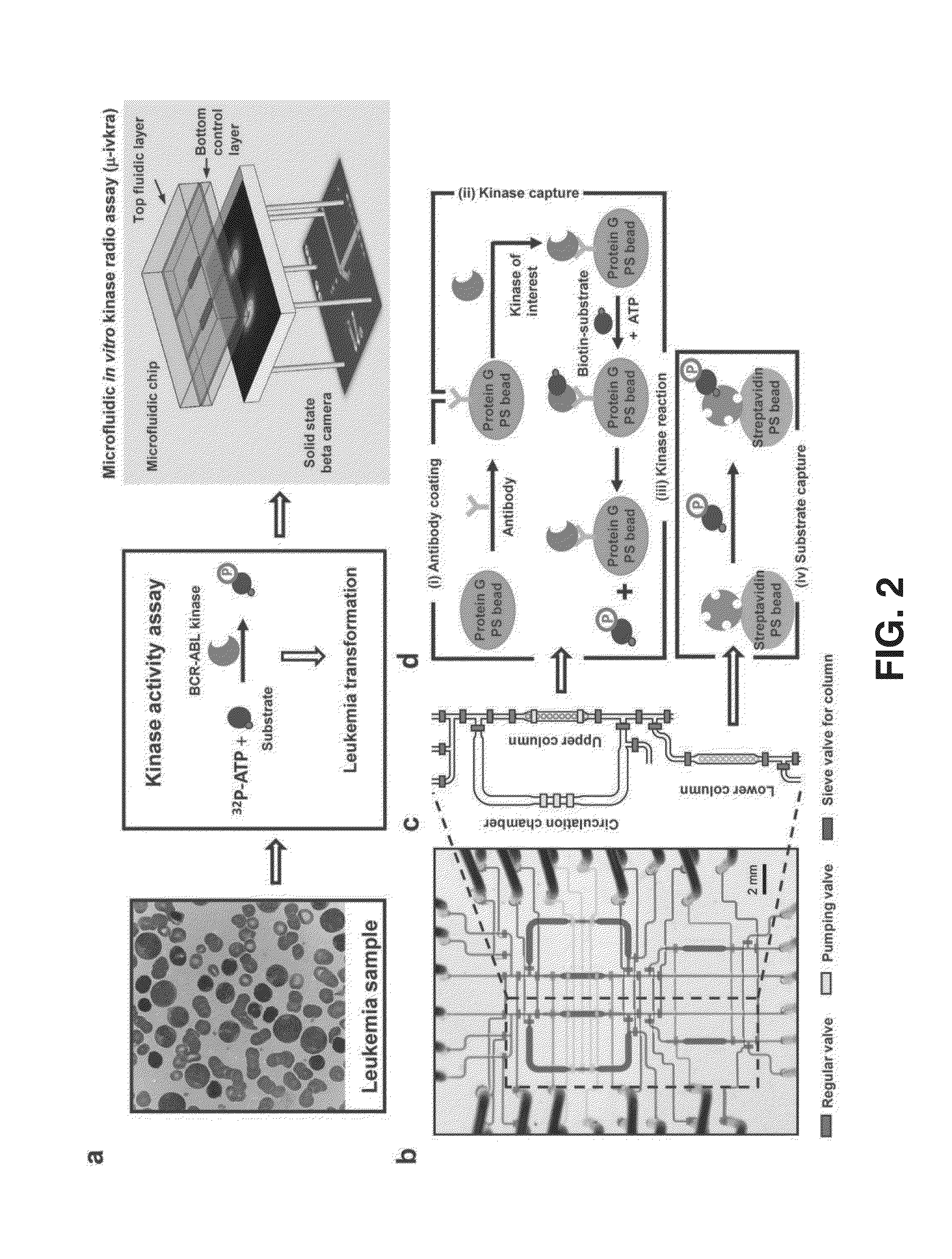
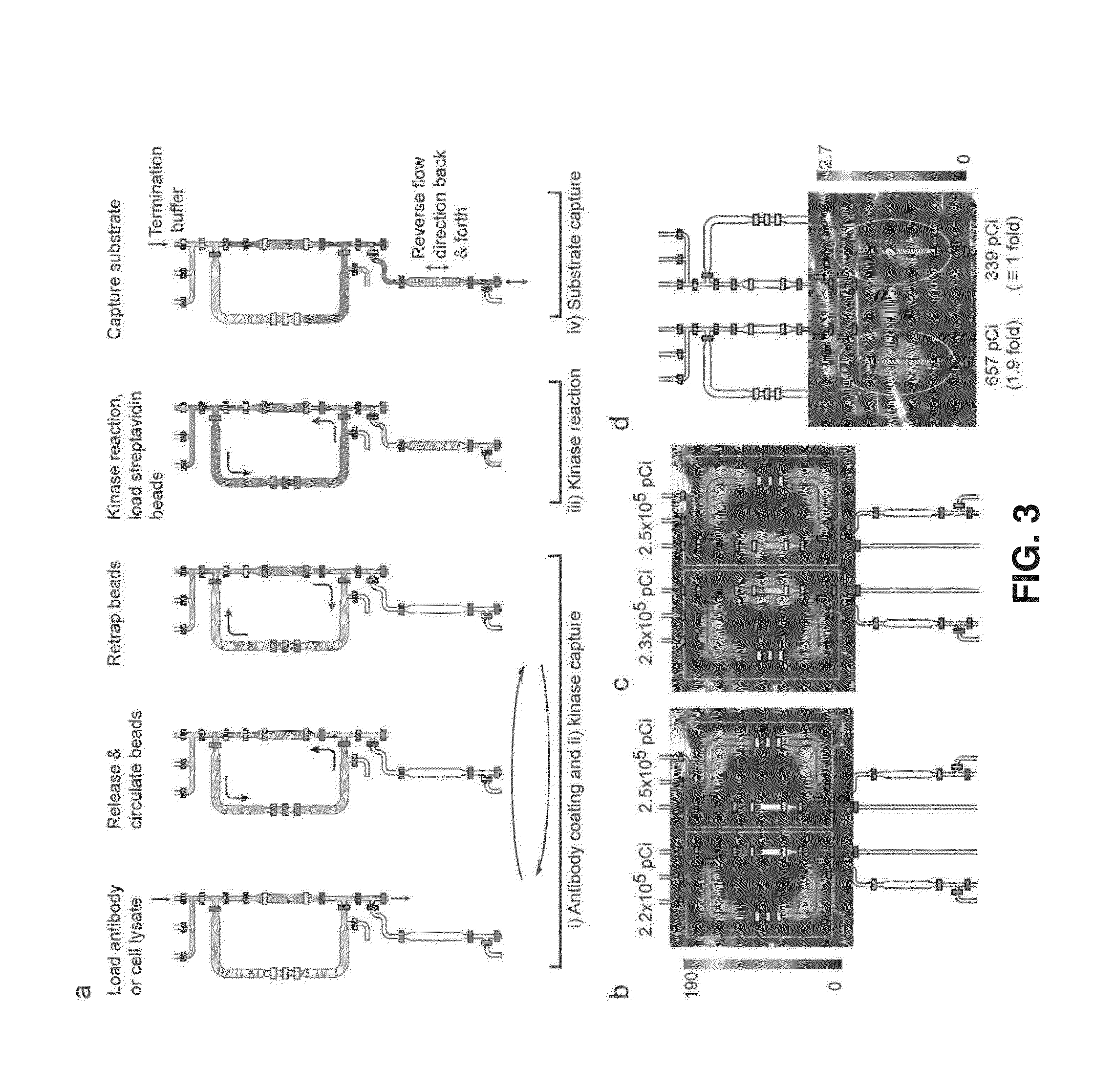
[0060]In order to further prove the concepts of the invention, microfluidic in vitro kinase radio assay (μ-ivkra) using BCR-ABL oncogenic kinase-positive leukemia samples was produced. A polydimethylsiloxane (PDMS)-based integrated microfluidic chip that performs an immunocapture-based kinase assay was fabricated with an integrated position sensitive avalanche photodiode detector (PSAPD) to function as a camera for imaging charged beta particles as a radioactivity-based readout for the assay. The beta camera allowed real-time monitoring of the radioactivity distribution during the assay, and quantified the final amount of radioactivity incorporated into the substrate. Control and readout of the microfluidic device and beta camera were performed via custom electronics and a personal computer. The resulting device performs an automated multi-step kinase reaction assay with coupled readout in a single unit, complete from sample loading to final quantitative data.
[0061]The cell lines th...
PUM
 Abstract
Description
Claims
Application Information
 - R&D
- Intellectual Property
- Life Sciences
- Materials
- Tech Scout
- Unparalleled Data Quality
- Higher Quality Content
- 60% Fewer Hallucinations
Browse by: Latest US Patents, China's latest patents, Technical Efficacy Thesaurus, Application Domain, Technology Topic, Popular Technical Reports.
© 2025 PatSnap. All rights reserved.Legal|Privacy policy|Modern Slavery Act Transparency Statement|Sitemap|About US| Contact US: help@patsnap.com


