
Microfluidic flow assay and methods of use
a technology of microfluidics and flow assays, applied in the direction of analytical using chemical indicators, laboratory glassware, instruments, etc., can solve the problems of inability to diagnose the severity of bleeding disorders, serious morbidity in developing countries, and important medical issues such as excessive bleeding
- Summary
- Abstract
- Description
- Claims
- Application Information
AI Technical Summary
Benefits of technology
Problems solved by technology
Method used
Image

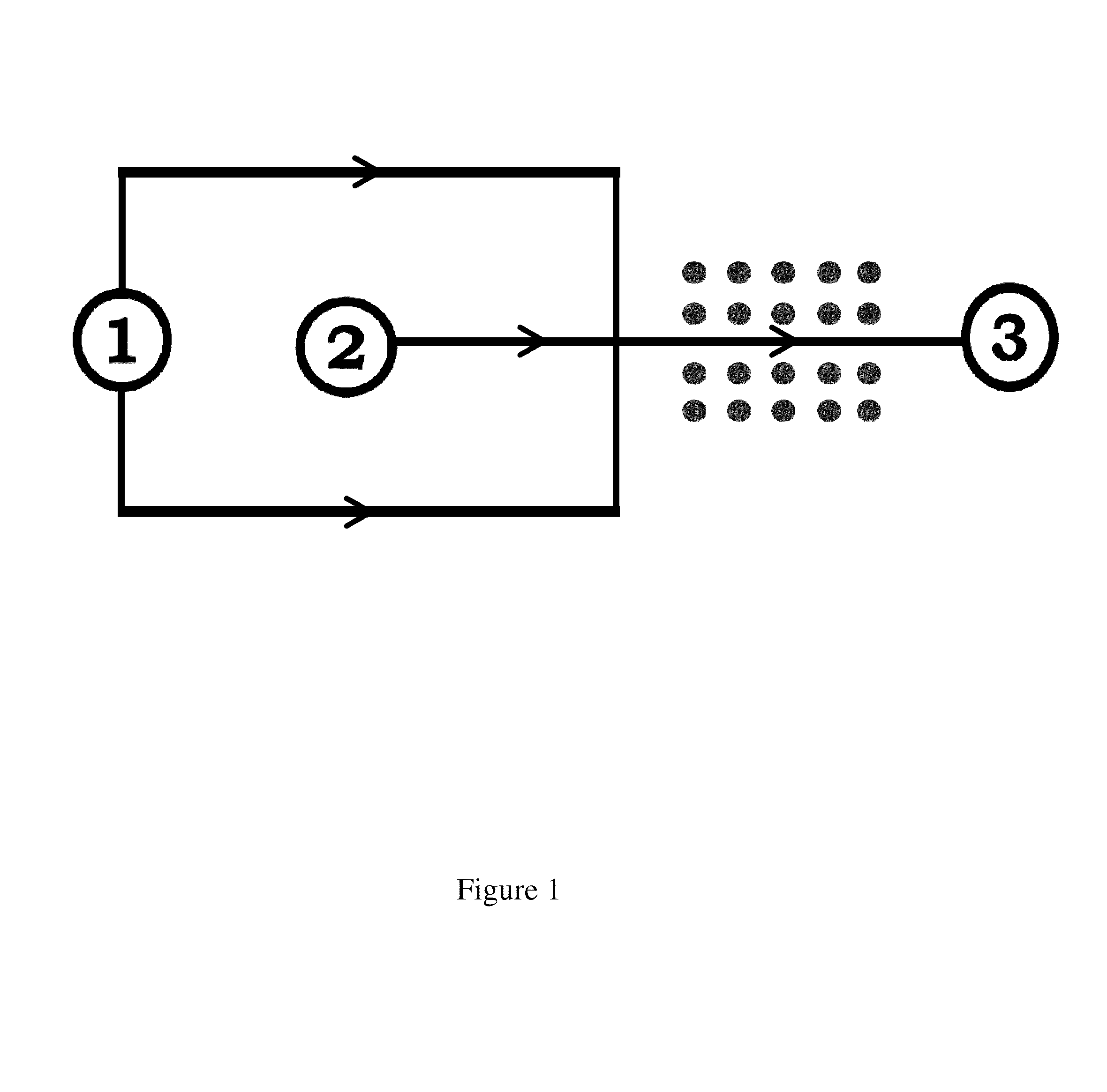
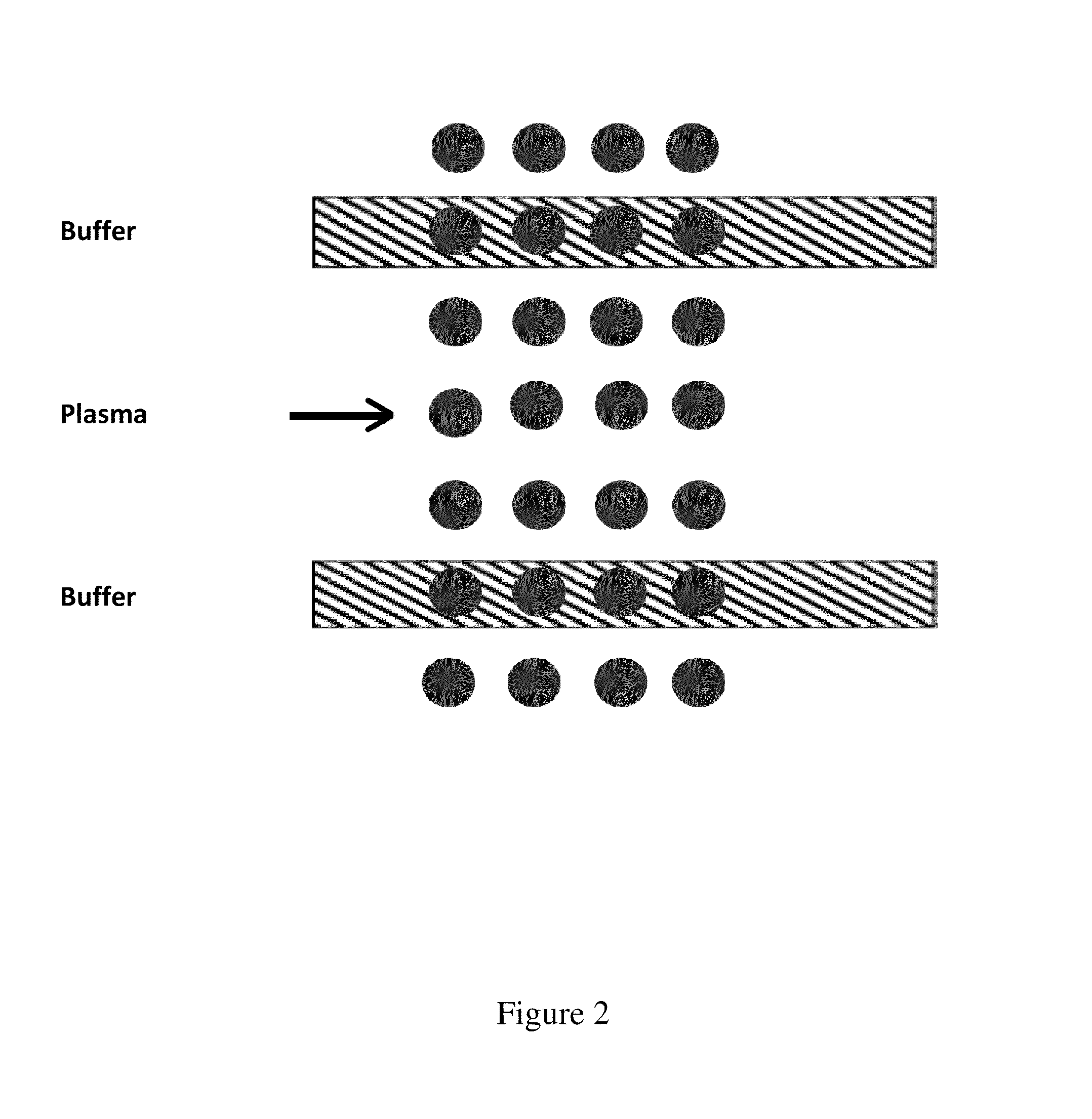
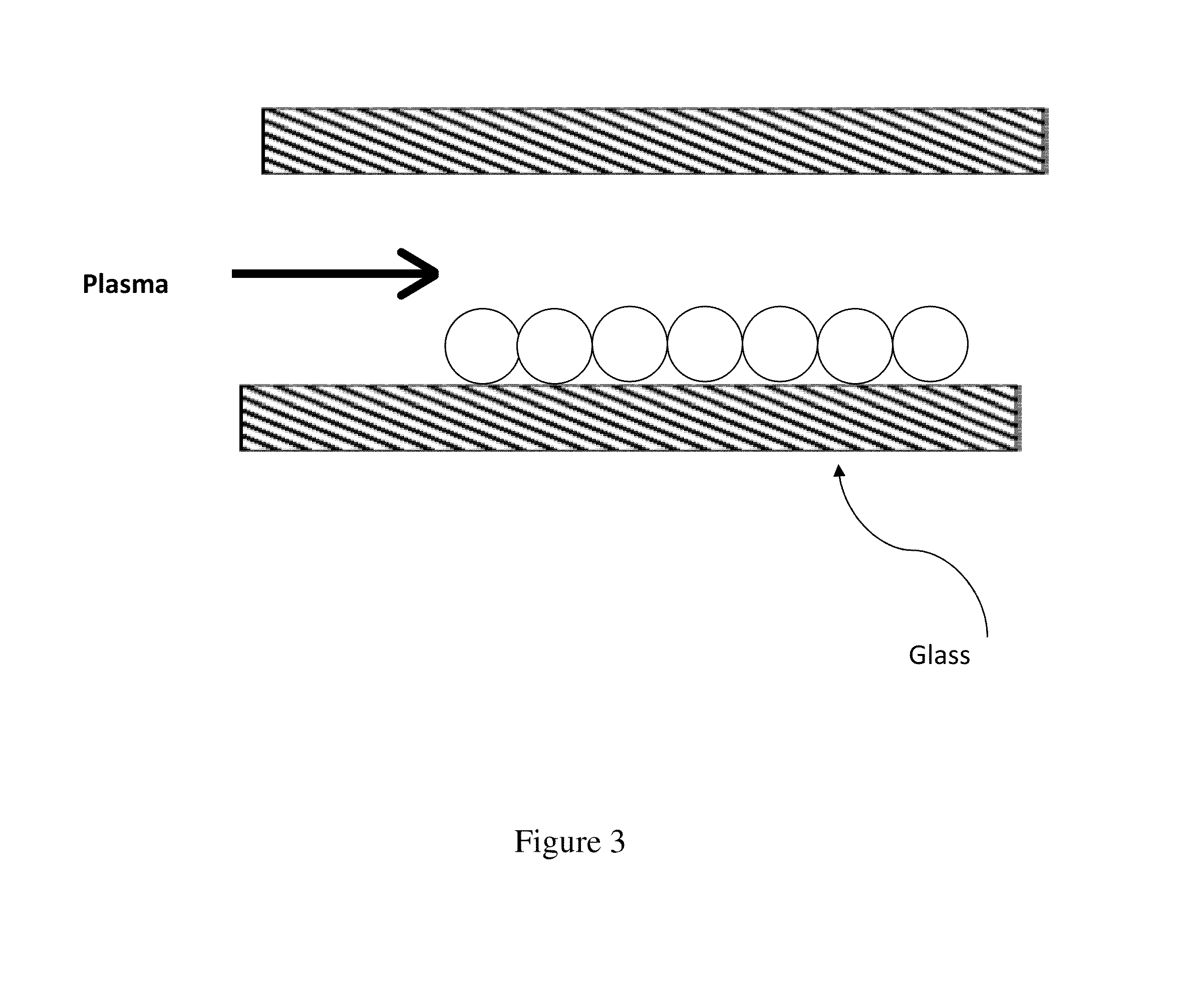
Examples
example 1
[0069]This example demonstrates the fabrication of lipid coated particles of the present invention to detect fibrin formation.
[0070]In order to initiate fibrin formation in the plasma-based model for coagulation using the microfluidic device of the present invention, silica microbeads are used to simulate the catalytic activity of platelets. Once the silica beads are made hydrophilic, their surfaces are coated with lipid bilayers, which contain varied concentrations of functional molecules such as tissue factor (TF) and thrombomodulin (TM) that initiate and inhibit coagulation. The composition of the lipid bilayer can be manipulated to mimic the surface of various cell types. This is accomplished by mixing different ratios of phospholipids such as phosphatidylserine (PS), phosphatidylcholine (PS), and phosphatidylethanolamine (PE). A description of the fabrication of silica beads and coating them with phospholipids is found below:[0071]1. Weigh or measure out 2.1 mL of deionized wat...
example 2
[0086]This example demonstrates an example of the patterning of lipid coated particles of the present invention.
[0087]Once the lipid coated particles are made as described in Example 1, they need to be immobilized and patterned onto a substrate. The immobilization of the particles can rely on methods such as covalent bonds (e.g. streptavidin-biotin), electrostatic interactions, or hydrogen bonding. The patterning may be achieved either by a subtractive technique such as microblotting or a lift-off process such as a microstencil. A microblotting technique is described below:[0088]1. Obtain a 100 μg / mL solution of silica beads functionalized with lipid bilayer as explained in Example 1.[0089]2. Incubate a 100 μg / mL solution of the lipid coated particles in HBS buffer onto and APTES functionalized glass slide for 1 hour at room temperature. Wash substrates three times in HBS buffer to remove unbound beads.[0090]3. Place glass slide into a 6-inch petri dish with 100 mL of deionized 18 M...
example 3
[0093]This example demonstrates an example of the integration of the lipid coated particles into microfluidic channels of the present invention.
[0094]After immobilizing and / or patterning the lipid coated particles, the microfluidic device of the present invention is aligned and bonded to the substrate. Because the lipid bilayers on the lipid coated particles should remain hydrated, this step should be done while the substrate is immersed or covered in buffer solution. The device may be aligned either manually or using alignment marks on the substrate. Once in contact, the device may be bonded to the substrate by vacuum assisted bonding.
PUM
 Abstract
Description
Claims
Application Information