
Drug coated balloon catheter and pharmacokinetic profile

a technology of angioplasty balloon and pharmacokinetic profile, which is applied in the direction of catheters, coatings, surgery, etc., can solve the problems of blood clots inside the stent, blood vessel collapse, and drug eluting balloons

Inactive Publication Date: 2014-02-13
ABBOTT CARDIOVASCULAR
View PDF1 Cites 10 Cited by
  • Summary
  • Abstract
  • Description
  • Claims
  • Application Information

AI Technical Summary

Benefits of technology

The invention is a drug delivery balloon that is designed to release the medication it contains in a controlled and targeted way to treat a specific area of the body. The coating of the balloon is made to have a specific concentration of the medication that can inhibit the growth of muscle cells in the blood vessels, reducing the likelihood of restenosis. The medication is detectable in the tissue for at least one week after the balloon is inflated. The concentration of the medication in the body increases over time, with a maximum concentration of 40 ng / ml or 0.2 ng / ml / ug. This ensures that the medication is not present in the blood in high concentrations for more than a few hours after treatment, minimizing the risk of toxicity.

Problems solved by technology

It was found that following angioplasty, although a blood vessel would be successfully widened, sometimes the treated wall of the blood vessel becomes weakened after balloon inflation or dilatation, causing the blood vessel to collapse after the balloon is deflated or later.
One drawback of drug eluting stents is a condition known as late stent thrombosis, an event in which blood clots inside the stent.
Although drug eluting balloons are a viable alternative and in some cases appear to have greater efficacy than drug eluting stents as suggested by the PEPCAD II study, drug eluting balloons present challenges due to the very short period of contact between the drug coated balloon surface and the blood vessel wall.
In particular, a non-perfusion balloon can only be inflated for less than one minute, and is often inflated for only thirty seconds which would otherwise starve distal regions of oxygenated blood.
Thus, there are challenges specific to drug delivery via a drug coated balloon because of the necessity of a short inflation time, and therefore time for drug or coating transfer—a challenge not presented by a drug eluting stent, which remains in the patient's vasculature once implanted.
However, not all embodiments result in an efficacious response in reducing restenosis after balloon and bare metal stent trauma.

Method used


Image

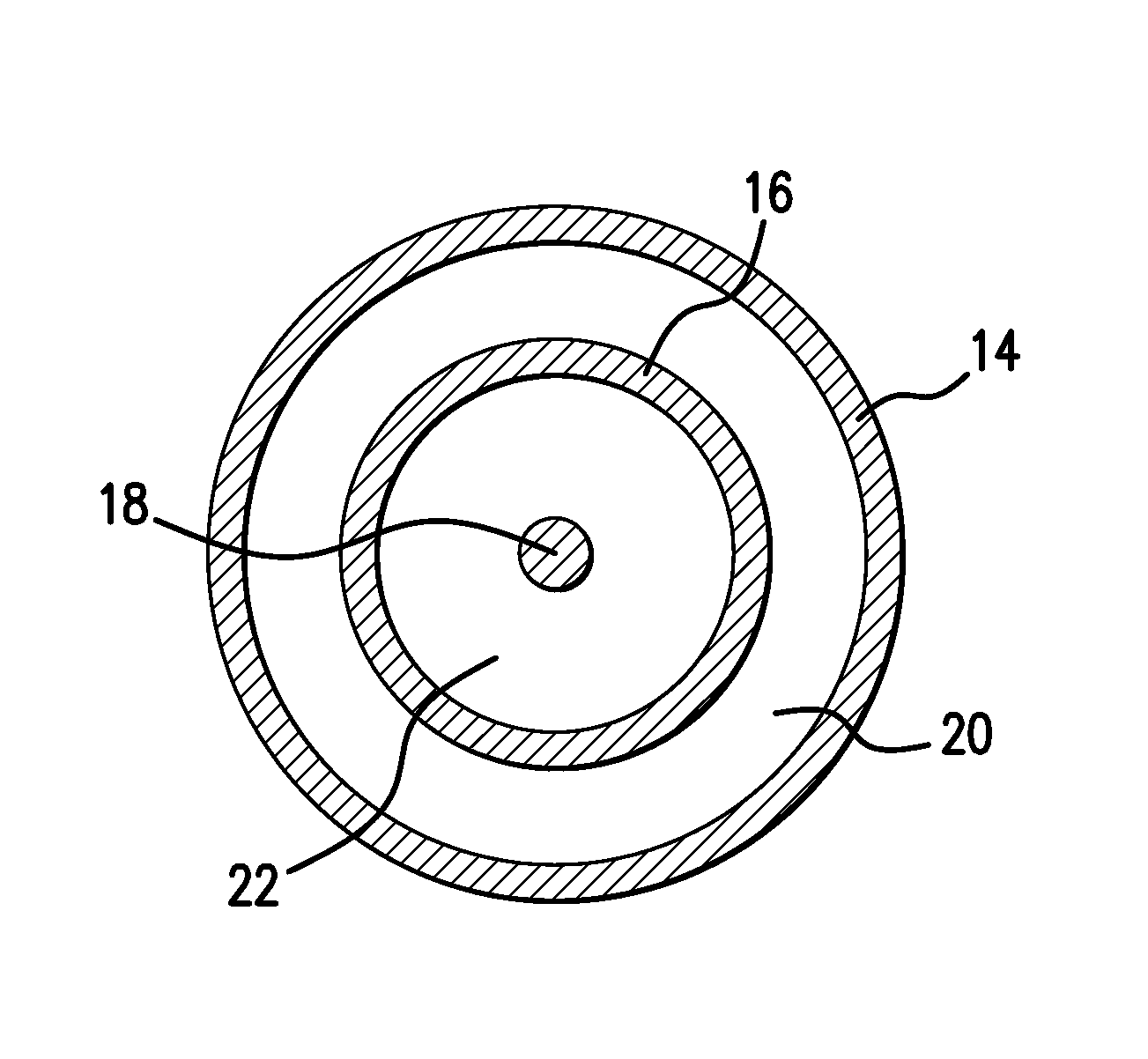
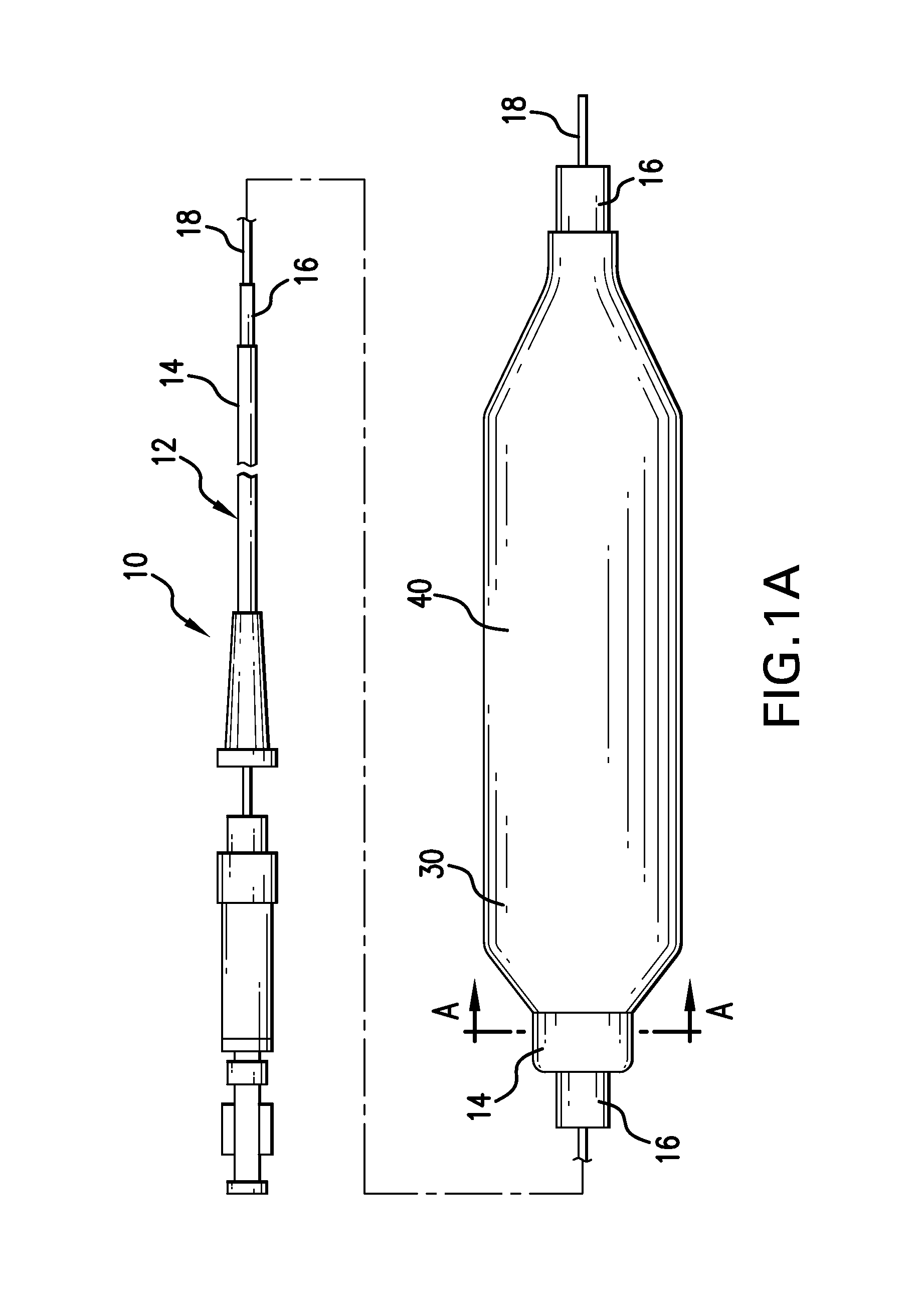
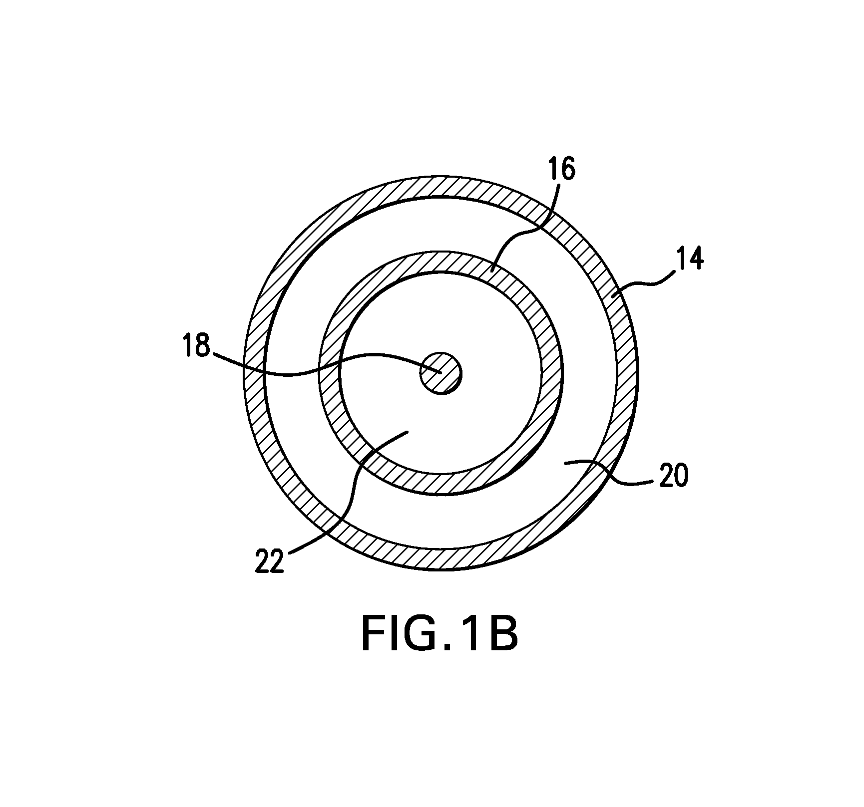
  • Drug coated balloon catheter and pharmacokinetic profile
  • Drug coated balloon catheter and pharmacokinetic profile
  • Drug coated balloon catheter and pharmacokinetic profile

Examples


example 1

Zotarolimus Coated Balloon

[0100]Zotarolimus was coated onto deflated 17×3.0 mm angioplasty balloons by syringing a solution of the drug dissolved in a mixture of Ultravist contrast agent, acetone, and ethanol onto the balloon surface. The average amount of zotarolimus coated was 662 μg per balloon. Balloon expansion was performed at 20% overstretch in porcine coronary arteries. The balloons were maintained expanded in position for 1 minute. Animals were sacrificed after 20 minutes, and the concentration of zotarolimus in the arterial tissue at the expansion sites was measured. The mean dose delivered was 6% of the total, which corresponded to a local concentration of 800 μM, at this early time point.

example 2

Everolimus Coated Balloon

[0101]Everolimus was coated onto inflated 3.0 mm×21 mm diameter Pebax angioplasty balloons using a custom designed Sonotek ultrasonic balloon coater. Three coating foimulations were evaluated including 1) everolimus alone (1025 m / balloon); 2) everolimus with hydrophilic Ultravist contrast agent at a 1:1 (w / w) ratio; and 3) everolimus with hydrophilic non-ionic polyvinylpyrrolidone polymer (Povidone C-30) and glycerol plasticizer at a 1:1:0.4 (w / w) ratio. The dosage of therapeutic agent coated on the balloons are 1) everolimus alone (1600 μg / balloon); 2) everolimus with hydrophilic Ultravist contrast agent at a 1:1 (w / w) ratio (1250 μg / balloon); and 3) everolimus with hydrophilic non-ionic polyvinylpyrrolidone polymer (Povidone C-30) at a 1:1 (w / w) ratio (1025 μg / balloon).

[0102]The coatings were sprayed and baked dry followed by balloon folding, 3.0 mm×18 mm Vision stent crimping, sheath placement, heat bonding to a full length catheter and hypotube seal, pac...

example 3

Zotarolimus Coated Balloon

[0105]In the following experiments, zotarolimus formulations were coated onto inflated 3.0 mm×12 mm Vision RX angioplasty balloons by air assisted spray atomization (zotarolimus only coatings at 88 ug / cm2 or 570 ug / cm2) or direct fluid volume application (zotarolimus:excipient coatings) of a solution of the drug dissolved neat in solvent or in a mixture of drug and excipient. The excipient formulations evaluated were zotarolimus-Ultravist 1.95-1 and zotarolimus-PVP-glycerol 2-1-0.4 with and without a bare metal stent and at either 88 ug / cm2 or 15 ug / cm2 zotarolimus dosages. Balloon expansion was performed at 20% overstretch in healthy domestic porcine coronary and / or mammary arteries. The balloons were maintained expanded in position for 30 seconds. Following balloon angioplasty, the animals were sacrificed after 30 minutes, 1 day (zotarolimus only), and 7 days and the concentration of zotarolimus in the arterial tissue at the expansion sites was measured v...


PUM

PropertyMeasurementUnit
concentration half lifeaaaaaaaaaa
time periodaaaaaaaaaa

Abstract

A drug delivery balloon is provided comprising a balloon having a surface, and a coating disposed on at least a portion of the balloon surface, the coating including an cytostatic therapeutic agent, an excipient, and a plasticizer. In accordance with the present subject matter, at least 30% of the coating transfers from the balloon surface within two minutes after inflation of the balloon. Alternatively, at least 30% of the coating transfers from the balloon surface within one minute after inflation. The coating results in an effective pharmacokinetic profile of an cytostatic therapeutic agent in a vasculature or target tissue.

Description

FIELD OF THE INVENTION[0001]The present invention is related to the delivery of drugs from an insertable medical device. More particularly, the present invention relates to a coated angioplasty balloon and the pharmacokinetic profile of the released drug from the tissue.BACKGROUND OF THE INVENTION[0002]Atherosclerosis is a syndrome affecting arterial blood vessels. It is a chronic inflammatory response in the walls of arteries, in large part due to the accumulation of blood cells and promoted by low density lipoproteins and the formation of plaque on the arterial wall. Atherosclerosis is commonly referred to as hardening of the arteries. Angioplasty is a vascular interventional technique involving mechanically widening an obstructed blood vessel, typically caused by atherosclerosis.[0003]During angioplasty, a catheter having a tightly folded balloon is inserted into the vasculature of the patient and is passed to the narrowed location of the blood vessel at which point the balloon i...

Claims


Application Information

Patent Timeline
Patent Type & Authority Applications(United States)
IPC IPC(8): A61L29/16A61L29/08
CPCA61L29/08A61L29/16A61L29/085A61L29/141A61L2300/416A61L2300/606
Inventor STANKUS, JOHNTROLLSAS, MIKAELHOSSAINY, SYEDLIANGXUAN, ZHANGBERGER, EDPACETTI, STEPHENTONER, JOHN L.
Owner ABBOTT CARDIOVASCULAR
Learn More