
Single molecule imaging techniques to aid crystallization

a single molecule and imaging technique technology, applied in the direction of fluorescence/phosphorescence, crystal growth process, instruments, etc., can solve the problems of slow and arduous process, unclear molecular basis of ribosome positioning of trnas in hybrid sites, and difficult to find suitable crystallization conditions from which to crystallize biomolecules into different conformational states. , to achieve the effect of high throughpu

Inactive Publication Date: 2014-04-03
CORNELL UNIVERSITY
View PDF0 Cites 4 Cited by
  • Summary
  • Abstract
  • Description
  • Claims
  • Application Information

AI Technical Summary

Benefits of technology

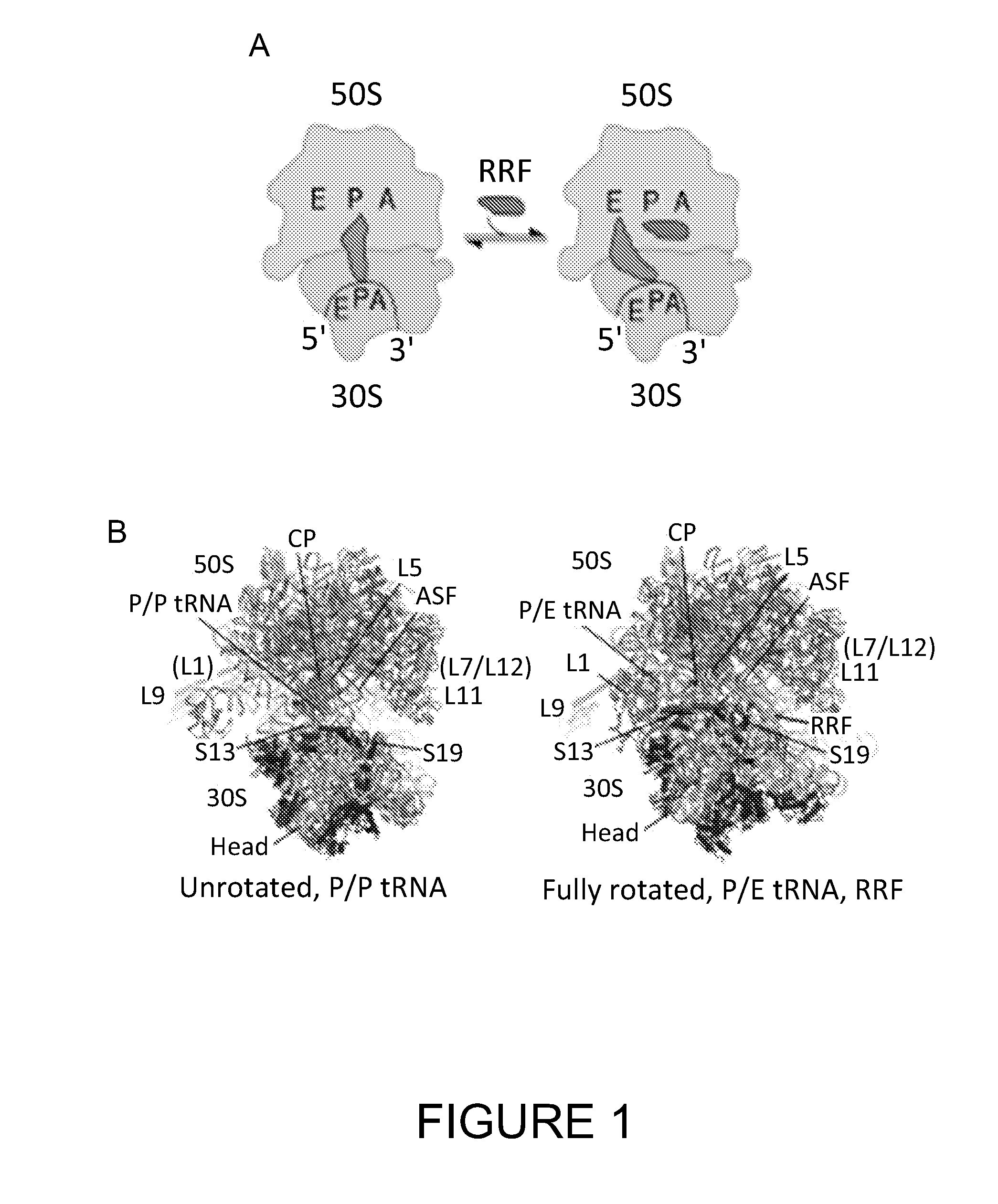
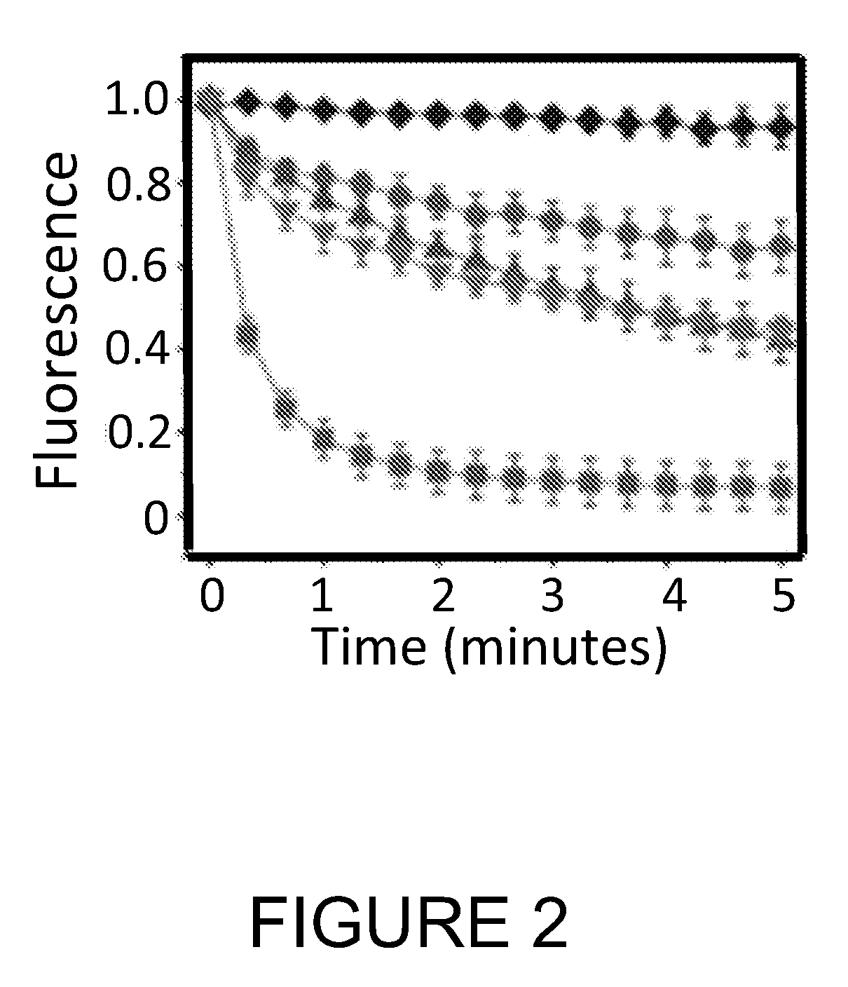
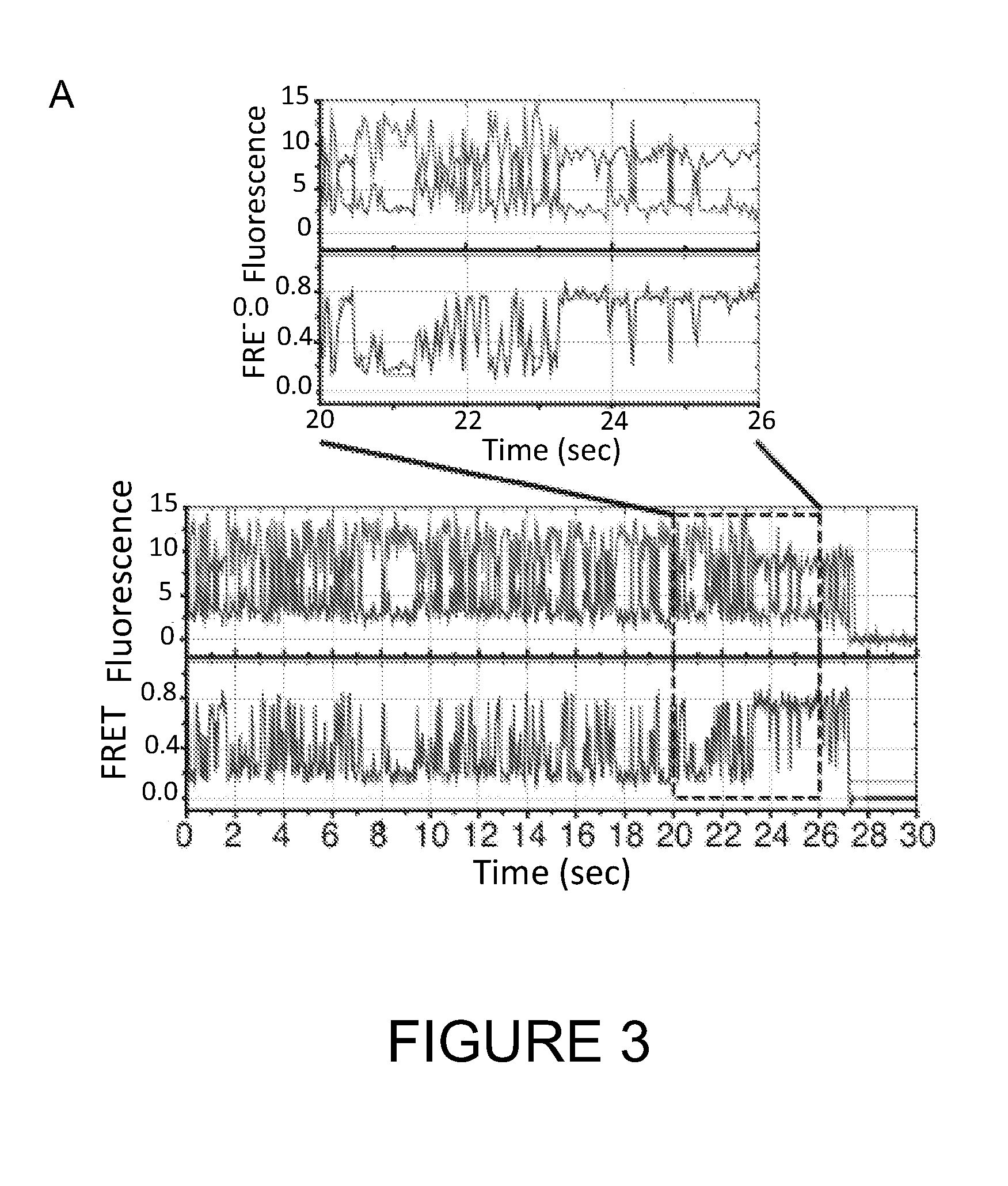
The present invention proposes a method to optimize the conditions for generating crystals of biomolecules in a desired conformation using single-molecule imaging techniques. This involves identifying the desired conformations of the biomolecule, preparing it with a fluorophore pair that can detect changes in conformation, and imaging it under various conditions while detecting the changes. The crystallization conditions are selected based on the increased distribution or time of the biomolecule in the desired conformation. This method can be applied to a variety of biomolecules, including proteins, peptides, nucleic acids, and complexes thereof. It can also be adapted for high throughput screening and analysis of potential crystallization conditions.

Problems solved by technology

However, the molecular basis for ribosome positioning of tRNAs in hybrid sites has been unclear.
Crystallization of biomolecules generally proceeds by empirical exploration of potential crystallization conditions and is a slow and arduous process, even when considerable structural information may already be available.
For example, if the biomolecule of interest is known to exist in, or to sample, multiple conformational states, as is the case with the ribosome during the defined phases of initiation, protein elongation, termination, and ribosome recycling, and one of those states is thermodynamically favored, it may be very difficult to find suitable crystallization conditions from which to crystallize the biomolecule into a different conformational state.

Method used


Image

  • Single molecule imaging techniques to aid crystallization
  • Single molecule imaging techniques to aid crystallization
  • Single molecule imaging techniques to aid crystallization

Examples


examples

[0039]Purification of Native E. Coli tRNAPhe

[0040]The purification protocol for tRNAPhe was adapted from a published protocol (Cayama 2000). Briefly, Escherichia coli (E. coli) cells (strain MRE600) harboring plasmid pBS-tRNAPhe, which overexpresses E. coli tRNAPhe, were cultured and harvested as previously described (Junemann 1996). The cell pellets were lysed by sonication in 20 mM Tris HCl, pH 7.5, 50 mM MgCl2 and 20 mM β-mercaptoethanol. The cell lysate was clarified by centrifugation at 35000 rpm in a Beckman Ti-70 rotor at 4° C. for 2 hours. Total cellular RNA was extracted from the supernatant by phenol extraction and ethanol precipitation. High molecular weight RNAs were removed by isopropanol precipitation (von Ehrenstein 1967). The soluble RNA fraction was then incubated for 15 min at 37° C. after adjusting the pH to 8 by addition of 0.5 M Tris HCl, pH 8.8 to deacylate tRNAs. As previously described (Blanchard 2004), tRNAPhe was specifically aminoacylated following brief ...


PUM

PropertyMeasurementUnit
pHaaaaaaaaaa
pHaaaaaaaaaa
pHaaaaaaaaaa

Abstract

This invention relates to a method to rapidly find crystallization conditions for a biomolecule in a desired conformation using single-molecule, fluorescent resonance energy transfer (smFRET) imaging techniques. The method provides significant cost and time advantages over the empricial exploration for crystallization conditions.

Description

CROSS-REFERENCE TO RELATED APPLICATIONS[0001]This application claims priority from U.S. Provisional Application No. 61 / 476,102, filed Apr. 15, 2011, which is herein incorporated by reference in its entirety.GOVERNMENT FUNDING[0002]This invention was made with Government support under Grant Numbers GM65050 and GM079238 awarded by the National Institutes of Health. The United States Government has certain rights in the invention.FIELD OF THE INVENTION[0003]This invention relates to a method to rapidly find crystallization conditions for a biomolecule in a desired conformation using single-molecule, fluorescent resonance energy transfer (smFRET) imaging techniques. The method provides significant cost and time advantages over the empirical exploration for crystallization conditions.BACKGROUND OF THE INVENTION[0004]Protein biosynthesis by the ribosome proceeds in defined phases of initiation, protein elongation, termination, and ribosome recycling (Schmeing 2009). Understanding the mole...

Claims


Application Information

Patent Timeline
Patent Type & Authority Applications(United States)
IPC IPC(8): G01N21/64
CPCG01N21/6486G01N33/542C30B29/58
Inventor BLANCHARD, SCOTT C.FELDMAN, MICHAEL BRIANWANG, LEYIALTMAN, ROGER
Owner CORNELL UNIVERSITY
Learn More