Device for reducing renal sympathetic nerve activity

a technology of renal sympathetic nerve and device, applied in the field of catheterassisted devices, can solve the problems of increased blood pressure, increased reflex pathway regulation, and considerable morbidity,

Inactive Publication Date: 2014-05-15
LAMBERT DAVID +1
View PDF3 Cites 50 Cited by
  • Summary
  • Abstract
  • Description
  • Claims
  • Application Information

AI Technical Summary

Benefits of technology

[0020]The probe can include one or more fiber optic cables in communication with the means for transmitting and receiving information. In certain embodiments the fiber optic cable(s) transmits and delivers imaging information and / or ablation energy.
[0021]The probe can include one or more reversibly inflatable balloons disposed on the distal end of the probe, which is in fluid communication with the at least one lumen. The balloon may be adapted for positioning and immobilizing the probe at the treatment site, such as by disposing the balloon opposite the ablation means. In other embodiments, the balloon is adapted for occluding blood flow. Typically, the balloon is inflated by means of a fluid such as saline or a gas such as CO2.

Problems solved by technology

While the underlying cause of this increased renal nerve activity is still unknown, the resulting increased blood pressure produces considerable morbidity including congestive heart failure (CHF) and kidney failure.
Although these drugs have proven beneficial, their use often results in up-regulation of reflex pathways that reduce their effectiveness over time.
The utility of such treatment can be compromised when the patient develops a fulminate hypertensive response to cyclosporine therapy.
In human patients, CsA administration following liver, kidney and heart transplants can cause an increase in blood pressure to pathological levels within days.
Compounding the situation, the increased afferent sympathetic nerve activity causes renal artery constriction leading to increased renin release and a worsening of kidney function.
This approach has not been used in human patients, however, due in part to the risks involved with denervation surgery.
Moreover, severed human renal nerves are able to regenerate, but often do so in an abnormal or pathological way.
Not only does this decrease the potential efficacy of surgical denervation, but may also lead to pain and other serious side effects.
Therefore, renal denervation is not considered a valid treatment for uncontrolled hypertension in human patients.
Radiofrequency energy is thus applied to the renal artery wall, heating the arterial wall, thereby damaging the underlying renal nerve.
However, the inability to direct the device to the precise position of the artery where the renal nerve lies makes it necessary to make repeated burns to ensure that the renal nerve is ablated.
Radiofrequency energy is thus applied in a pattern of discrete burns circumferentially around the lumen of the renal artery, resulting in damage to the renal nerve, but also resulting in significant damage unrelated to the therapeutic renal ablation effect.
A significant limitation of existing devices is the inability to visualize the renal nerve.
However, this method is limited by the skill of the operator without the aid of visual feedback, in making a sufficient number of burns to ensure that the nerve is damaged and in effect relying on chance.
Additionally, unnecessary damage to the wall of the renal artery occurs when energy is applied to areas of the renal artery wall not underlying the renal nerve.

Method used


Image

  • Device for reducing renal sympathetic nerve activity
  • Device for reducing renal sympathetic nerve activity
  • Device for reducing renal sympathetic nerve activity

Examples


example 1

Sonogram of Human Renal Vasculature to Investigate Anatomy and Best Approach to Renal Artery

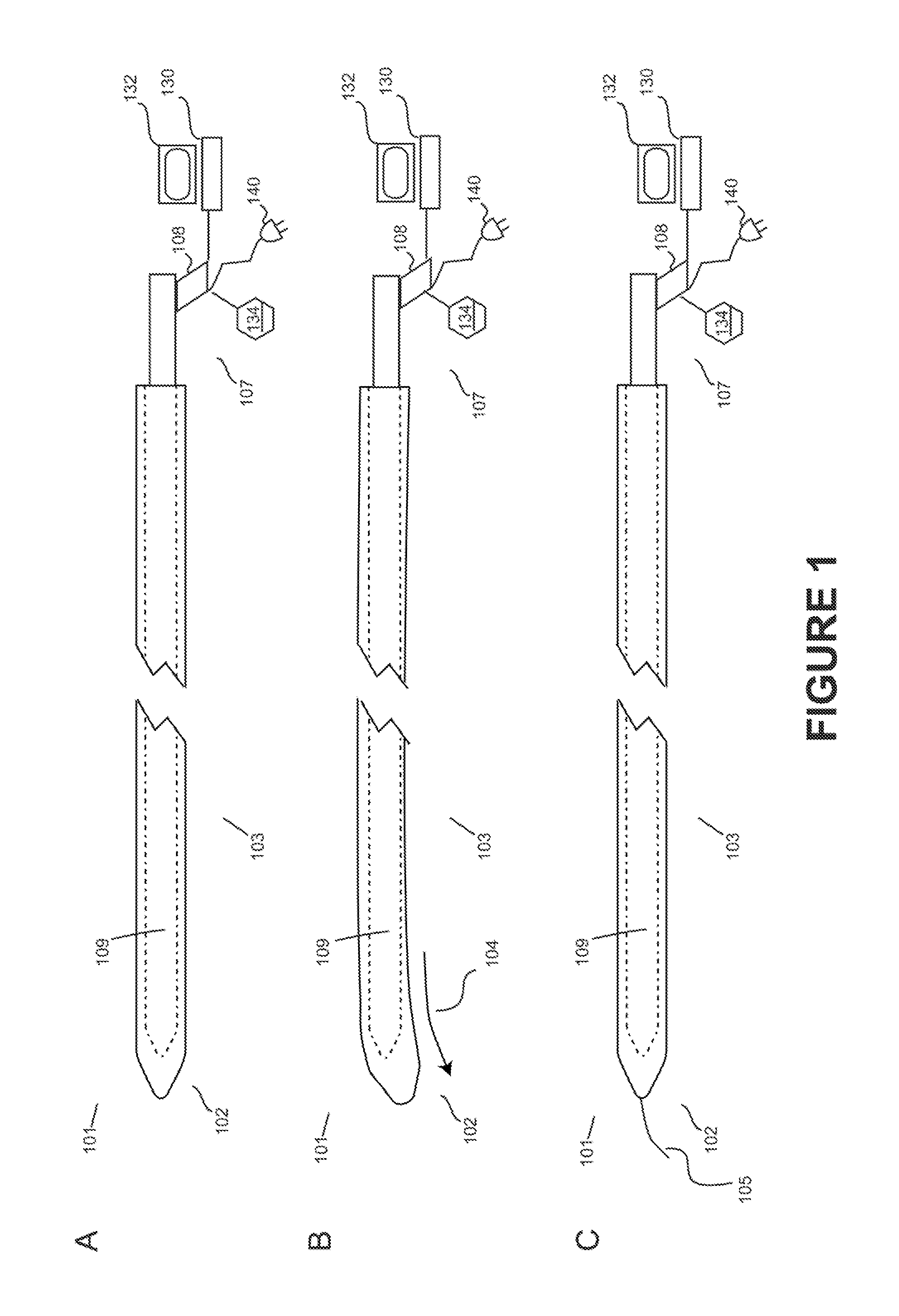
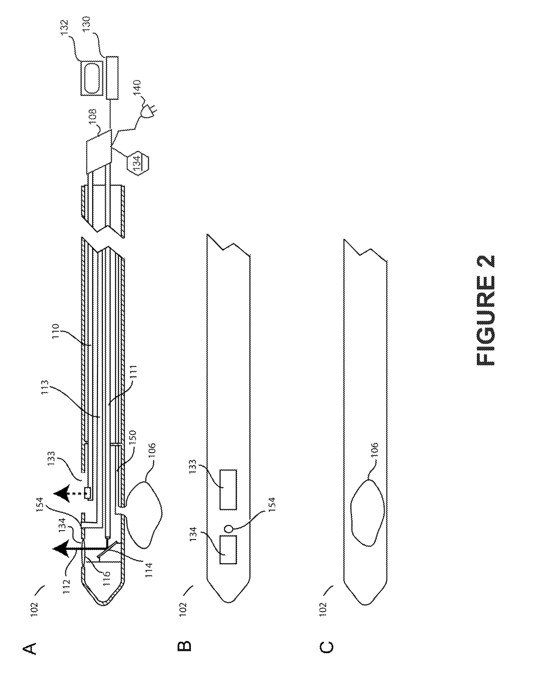
[0073]Increased renal sympathetic nerve activity is a major contributor to essential hypertension. However, the underlying cause of this centrally mediated increase in sympathetic nerve activity is unknown. The kidneys are highly sensitive to changes in blood pressure, and afferent sympathetic nerve traffic leading from the kidneys to the brain regulates reflex efferent sympathetic arterial constriction and vascular tone. The renal nerve branches from the renal plexus lying near the aorta and travels along the renal artery to the kidney. The renal nerve lies in close proximity to the renal artery and branches into a network, or plexus, of nerves nearer the kidney.

[0074]The main branch of the renal nerve lies immediately on top of the advential layer of the renal artery, encased in a layer of fat. Previous studies focused on determining whether a retroperitoneal approach, combined with imaging...

example 2

Histology of Pig Renal Artery Showing Renal Nerves

[0077]Materials and Methods.

[0078]The renal arteries were excised from Gottingen minipigs and placed in formalin for histological (H&E) staining. In FIG. 9, the renal artery is shown with distances from the lumen wall to the renal nerves.

Optical Coherence Tomography of Renal Nerves

[0079]To investigate the ability to visualize the renal nerve through the renal artery wall, freshly harvested pig renal arteries were obtained, still attached to the aorta and kidneys. The samples were placed on a dissection tray and excess fat was dissected from the renal artery. An incision was made in the abdominal aorta to facilitate placement of an OCT imaging catheter inside the renal artery. FIG. 11 is OCT imaging of the renal nerve, showing the location of the renal nerve in relation to the artery lumen. FIG. 10 shows H&E staining of the same section of renal artery verifying the ability to image the renal nerve through the renal artery wall.

example 3

Histology of Human Renal Artery Showing Renal Nerves

[0080]Materials and Methods.

[0081]The right renal artery from an 80 year old male cadaver is shown after histological (H&E) staining. Of significance to the usefulness of this device is the almost complete lack of intimal thicking or hyperplasia, FIG. 12 shows H&E staining of a section of the right renal artery. Renal nerves are shown. Separation of renal nerves from the renal artery is an artifact of tissue shrinkage and separation during formalin fixation.

Optical Coherence Tomography of Renal Nerves

[0082]To investigate the ability to visualize the renal nerve through the renal artery wall, the renal artery with renal nerves attached from an 80 year old male cadaver was thawed in room temperature saline and placed on a tray for optical coherence visualization. FIG. 13 is OCT imaging of the renal nerves, showing the location of the renal nerves in relation to the artery lumen.


PUM


Abstract

The present invention provides devices and methods for modulating or blocking renal sympathetic nerve activity by applying laser energy to the renal artery wall to effect ablation of the renal nerve or nerves. The devices and methods of the invention may be useful in the treatment of cardiovascular and renal disease resulting from hypertension. In addition the devices and methods of the invention may be useful for reducing reflex hypertension following cyclosporine administration after organ transplantation.

Description

FIELD OF THE INVENTION[0001]The present invention relates generally to catheter-assisted devices and specifically to devices for modulating renal sympathetic nerve activity.BACKGROUND[0002]Hypertension is now firmly linked with elevated renal sympathetic nerve activity in animal models and humans. While the underlying cause of this increased renal nerve activity is still unknown, the resulting increased blood pressure produces considerable morbidity including congestive heart failure (CHF) and kidney failure. Currently, the only approved method for treating hypertension is drug therapy, such as administration of diuretics, inhibitors of the rennin angiotensin system, and drugs that act on the heart to reduce cardiac workload. Although these drugs have proven beneficial, their use often results in up-regulation of reflex pathways that reduce their effectiveness over time.[0003]Pharmacologically mediated hypertension has been observed in the clinic with the use of the anti-rejection d...

Claims


Application Information

Patent Timeline
Patent Type & Authority Applications(United States)
IPC IPC(8): A61B18/24A61M5/158
CPCA61B18/1492A61B18/24A61B2018/00285A61B2018/00404A61B2018/00434A61B2018/00511A61B2090/3735A61B2018/00791A61B2018/00982A61B2018/2272A61B2218/002A61M5/158A61B2018/00577
Inventor LAMBERT, DAVIDIKENO, FUMIAKI
Owner LAMBERT DAVID
Why Eureka
Try Eureka