
Erythropoietin and fibronectin compositions for therapeutic and cosmetic applications

a technology of fibronectin and erythropoietin, which is applied in the direction of growth factors/regulators, animal/human proteins, non-active ingredients, etc., can solve the problems of skin maladies and conditions such as acne, actinic keratoses, photoaging skin, etc., and achieve the effect of promoting wound healing or connective tissue reconstruction

Inactive Publication Date: 2014-06-05
REMEDOR BIOMED
View PDF7 Cites 8 Cited by
  • Summary
  • Abstract
  • Description
  • Claims
  • Application Information

AI Technical Summary

Benefits of technology

The patent text describes a method for promoting wound healing or connective tissue reconstruction in a subject by topically applying a combination of Erythropoietin and Fibronectin to the wound. The method involves applying a certain amount of Erythropoietin and Fibronectin to the wound tissue, which can be calculated based on the size of the wound. The technical effect of this method is to accelerate the healing process and improve the quality of wound healing or connective tissue reconstruction.

Problems solved by technology

However, the breakdown of healthy collagen, the decline in collagen production (e.g. by deceleration in the division rate of skin cells) and the defective cross-linking of collagen and elastin fibers in the skin, leads to the development of skin maladies and conditions including acne, actinic keratoses, photoaged skin, unwanted wrinkles and the appearance of aged skin (e.g. sags, changes in tone and texture).
However, in patients with diabetes or with the classical skin maladies, wound healing is usually delayed or even impaired completely and consequently chronic wounds develop.
This impaired process represents a major health-care problem with considerable socioeconomic impact.
One contributing factor that may play a crucial role is poor subcutaneous blood supply.
Likewise, in diabetes, fibroblasts fail to produce extracellular matrix (ECM) and keratinocytes fail to induce reepithelialization.
Furthermore, decreased collagen deposit, chemotaxis and the inhibition of fibroblast proliferation may all be associated with impairment of wound healing in diabetes

Method used


Image

  • Erythropoietin and fibronectin compositions for therapeutic and cosmetic applications
  • Erythropoietin and fibronectin compositions for therapeutic and cosmetic applications
  • Erythropoietin and fibronectin compositions for therapeutic and cosmetic applications

Examples


example 1

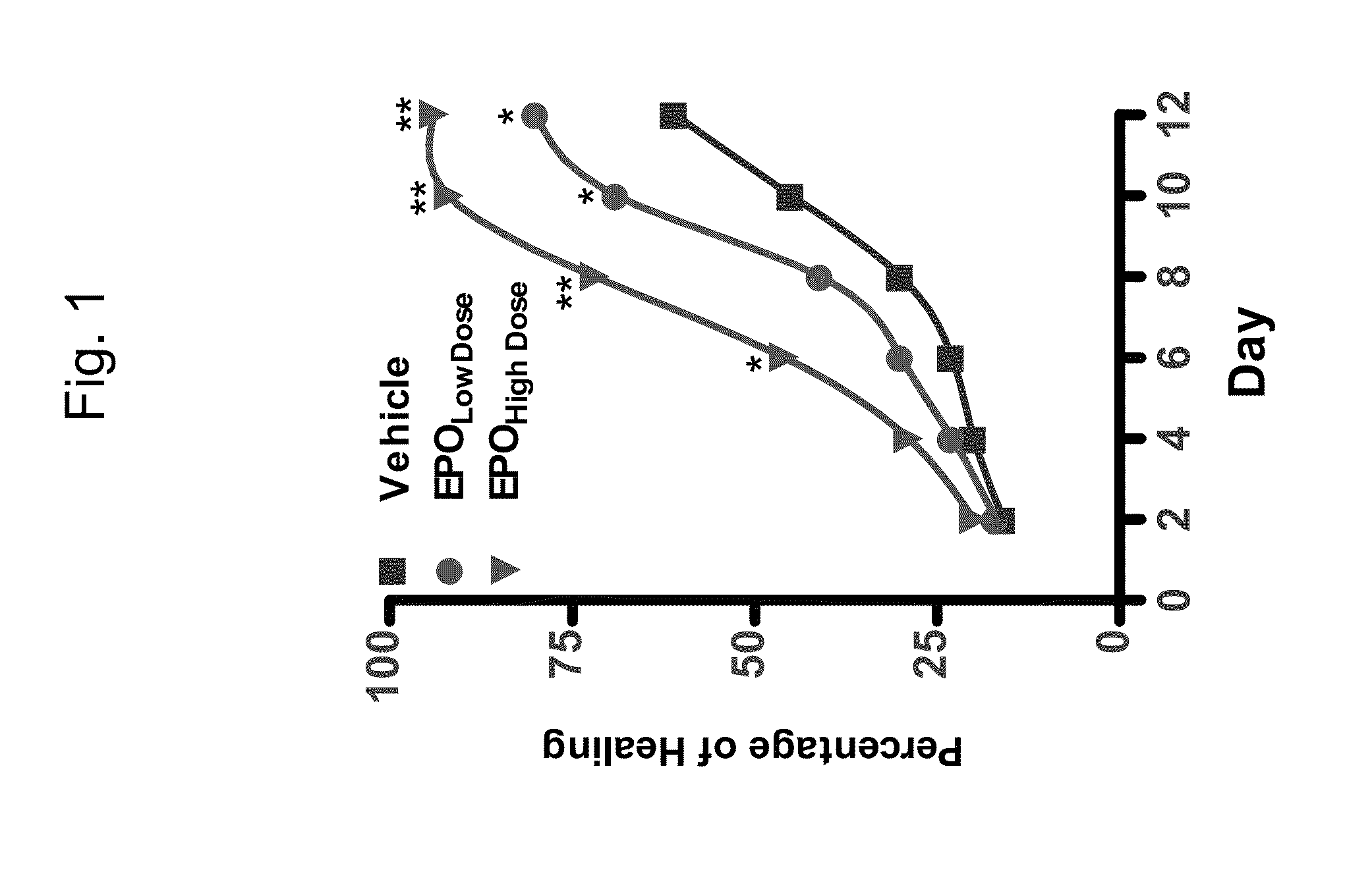
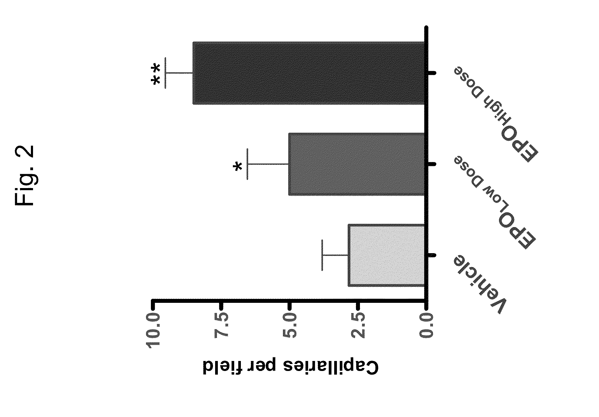
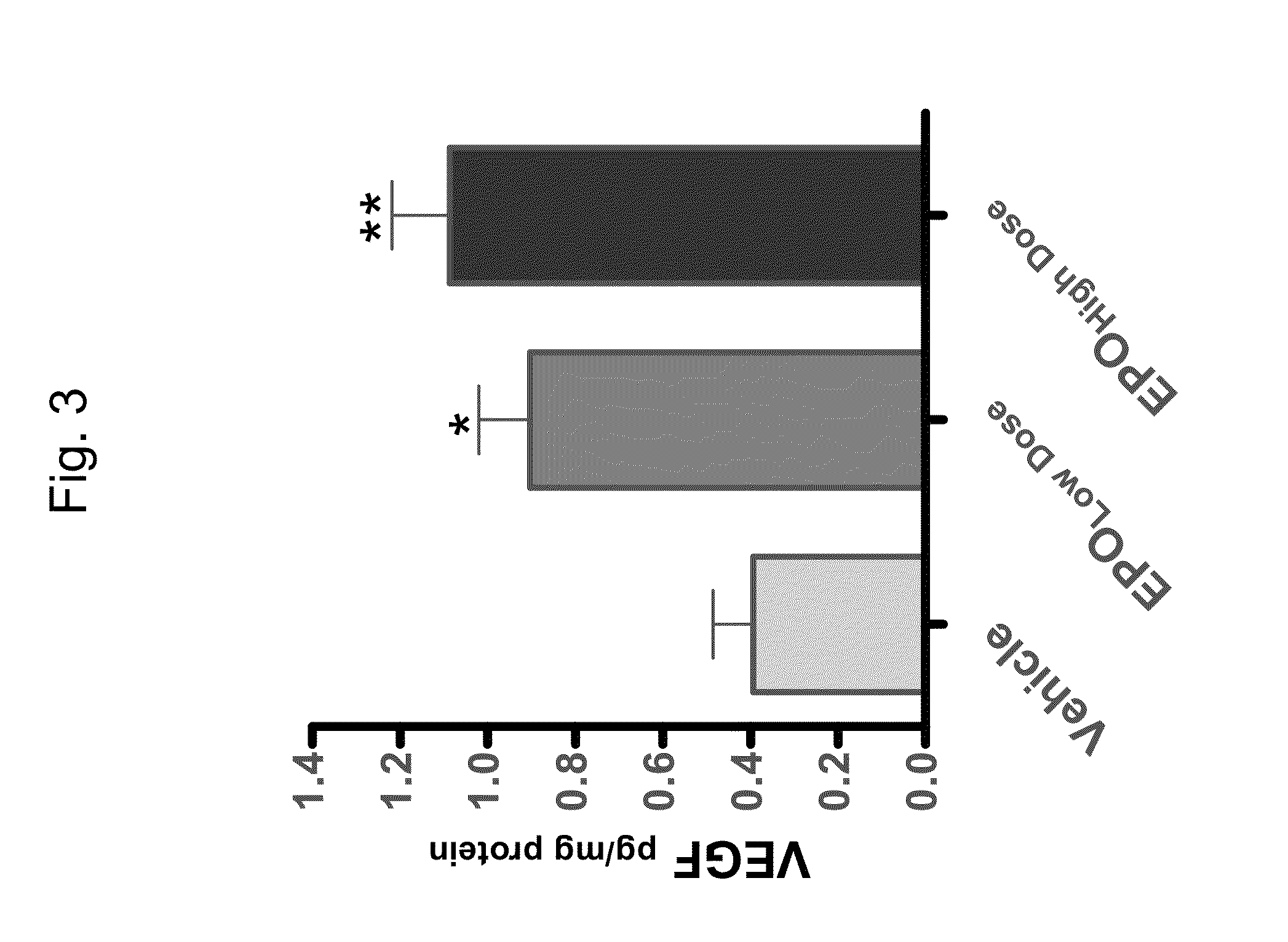
[0185]Topical treatment with Erythropoietin improves angiogenesis and wound healing in cutaneous wounds of diabetic rats

[0186]Materials and Experimental Procedures

[0187]Experimental Animals

[0188]Thirty Male Sprague-Dawley rats, aged 8 weeks (obtained from Harlan, Jerusalem, Israel) were kept in an environment comprising constant temperature and humidity and with an artificial 12-hour light / dark cycle. All rats were allowed free access to food and water. All experimental procedures followed the guidelines of the Animal Care and Use Committee of the Rappaport Faculty of Medicine—Technion Animal Center.

[0189]Induction of Diabetes Mellitus

[0190]Diabetes was induced by a single 60 mg / kg intraperitoneal injection of streptozotocin (STZ, Sigma Aldrich, St. Louis, Mo., USA), a toxin specific for insulin-producing cells, in saline-sodium citrate buffer (Sigma Aldrich, St. Louis, Mo., USA, pH 4.5). Blood glucose levels were measured using an acute glucometer (FreeStyle, Alameda, Calif., USA)....

example 2

[0218]Accelerated cutaneous wound healing in diabetic mice by topical treatment with Erythropoietin and Fibronectin

[0219]Materials and Experimental Procedures

[0220]Experimental Animals

[0221]Thirty two CD1 nude mice, aged 6 weeks (obtained from Harlan, Jerusalem, Israel) were kept in an environment comprising constant temperature and humidity and with an artificial 12-hour light / dark cycle. All mice were allowed free access to food and water. All experimental procedures followed the guidelines for Animal Care and Use Committee of the Rappaport Faculty of Medicine—Technion Animal Center.

[0222]Induction of Diabetes Mellitus

[0223]Diabetes was induced by a single 60 mg / kg intraperitoneal injection of streptozotocin (STZ; Sigma Aldrich, St Louis, Mo., USA), a toxin specific for insulin-producing cells, in saline-sodium citrate buffer (Sigma Aldrich, St Louis, Mo., USA, pH 4.5). Blood glucose levels were measured using an acute glucometer (FreeStyle, Alameda, Calif., USA). Five days after ...

example 3

EPO Upregulates β1-Integrin Expression in HEMCs

[0248]Materials and Experimental Procedures

[0249]Human Epidermal Microvascular Cell Culture and Experimental Conditions

[0250]Primary human epidermal microvascular cells (HEMCs) were purchased from PromoCell (GmbH, Heidelberg, Germany). HEMCs were maintained in human epidermal microvascular endothelial medium (PromoCell, GmbH, Heidelberg, Germany), a modified and optimized DMEM / F-12 (1:1) supplemented with 15 mM HEPES, 10% fetal bovine serum (FBS), growth factor (acidic FGF stabilized with Heparin) and 1% antibiotic solution containing streptomycin, neomycin and penicillin (Biological Industries, Beit Haemek, Israel). All experiments were performed in passages 3-6. HEMCs were seeded in culture dishes coated with fibronectin (10 μg / ml, Chemicon International, Temecula, Calif., USA). Cultured HEMCs were detached by trypsinization and reseeded in fibronectin coated 24× well plates (2.5×10⁵ cells / well) in triplicates. These cultured HEMCs we...


PUM

PropertyMeasurementUnit
concentrationaaaaaaaaaa
diameteraaaaaaaaaa

Abstract

A method of promoting wound healing or connective tissue reconstruction and a method of treating ischemia in a subject in need thereof are disclosed. The methods comprising topically administering to the subject about 10-30 mg per cm2 wound tissue of Erythropoietin and about 100-300 mg per cm2 wound tissue of Fibronectin, thereby promoting wound healing or connective tissue reconstruction or treating ischemia in the subject. Unit dosage forms, pharmaceutical compositions, cosmetic compositions and formulations comprising Erythropoietin and / or Fibronectin are also disclosed.

Description

RELATED APPLICATIONS[0001]This application is a continuation of a U.S. patent application Ser. No. 12 / 673,519 filed on Feb. 15, 2010, which is a National Phase of PCT Patent Application No. PCT / IL2008 / 001119 having International filing date of Aug. 13, 2008, which claims the benefit of priority of U.S. Provisional Patent Application Nos. 61 / 064,311 filed on Feb. 27, 2008 and 60 / 935,497 filed on Aug. 16, 2007. The contents of the above applications are all incorporated herein by reference.FIELD AND BACKGROUND OF THE INVENTION[0002]The present invention, in some embodiments thereof, relates to Erythropoietin and Fibronectin compositions and, more particularly, but not exclusively, to the use of same in therapeutic and cosmetic applications.[0003]The extracellular matrix (ECM), often referred to as the connective tissue, is a complex structural entity found within the mammalian tissue which surrounds and supports cells. The three major classes of ECM biomolecules are the structural pro...

Claims


Application Information

Patent Timeline
Patent Type & Authority Applications(United States)
IPC IPC(8): A61K38/18A61K38/43A61K38/36A61K38/39A61K31/21A61K31/198A61K38/22A61K38/19A61K31/70
CPCA61K38/1816A61K31/198A61K31/21A61K31/70A61K38/43A61K38/22A61K38/36A61K38/39A61K38/19A61K9/0014A61K9/06A61K47/32A61P17/02A61P3/02A61P31/00A61P39/04A61P43/00A61P7/04A61P9/10A61K2300/00
Inventor HAMED, SAHER
Owner REMEDOR BIOMED
Learn More