Optical-imaging probe for detecting sentinel lymph nodes which contains a composite of poly-gamma-glutamic acid and an optical-imaging die

a technology of optical imaging and sentinel lymph nodes, which is applied in the direction of instruments, diagnostic recording/meauring, ultrasonic/sonic/infrasonic diagnostics, etc., can solve the problems of serious problems in the disposal of samples after pathological analysis, the surgical technique is not preferable in terms of patient life quality, and the disposal of samples is not convenient. , to achieve the effect of reducing the tendency to migrate and being convenient and effectiv

Active Publication Date: 2014-06-12
BIOLEADERS CORP +2
View PDF2 Cites 2 Cited by
  • Summary
  • Abstract
  • Description
  • Claims
  • Application Information

AI Technical Summary

Benefits of technology

[0015]It is an object of the present invention to provide an optical imaging probe for detecting a sentinel lymph node, which contains a complex of poly-gamma-glutamic acid and an optical imaging dye, in which the optical imaging probe serving as a molecular imaging probe remains in the sentinel lymph node for an extended period of time and has a reduced tendency to migrate to other lymph nodes so that the sentinel lymph node can be detected in an easier and more effective manner.

Problems solved by technology

Thus, this surgical technique is not preferable in terms of the patient's life quality, even though it is successful.
However, conventional methods for detecting the sentinel lymph node using radiopharmaceuticals have many problems.
Specifically, the contamination of an analyzer and an analysis room during the pathological analysis of a lymph node labeled with a radiopharmaceutical, and the disposal of a sample after pathological analysis become serious problems.
Although ICG was used in studies on the detection of the sentinel lymph node, but there were problems in that ICG has a too small size, and thus when it is injected in vivo, it does not remain in the sentinel lymph node for a long period of time and migrates to other surrounding lymph nodes.
This makes it difficult to distinguish the sentinel lymph node from lymph nodes around cancer tissue during an actual surgical operation.
Such polymer particles are nano / micro-sized particles having a particle diameter of more than 100 nm can be used mainly for intravenous administration and oral administration, but are not suitable for the detection of the sentinel lymph node, which requires a particle diameter of 10-25 nm.
However, such polymer particles are not suitable for the detection of the sentinel lymph node, because they have a particle diameter of more than 10 μm for cell encapsulation.
Such polymer microcapsules have a particle diameter of 0.6-2 μm and are used in photodynamic therapy based on the characteristics of indocyanine green, but are not suitable for the detection of the sentinel lymph node.

Method used


Image

  • Optical-imaging probe for detecting sentinel lymph nodes which contains a composite of poly-gamma-glutamic acid and an optical-imaging die
  • Optical-imaging probe for detecting sentinel lymph nodes which contains a composite of poly-gamma-glutamic acid and an optical-imaging die
  • Optical-imaging probe for detecting sentinel lymph nodes which contains a composite of poly-gamma-glutamic acid and an optical-imaging die

Examples


example 1

Preparation of PGA / ICG Complex

[0041]Indocyanine green which is used as a fluorescent dye is an anionic amphipathic (hydrophobic and hydrophilic) material. The negative charge of indocyanine is hydrophobically (non-covalently) coupled to the hydrophobic moiety of poly-gamma-glutamic acid (γ-PGA) in an aqueous solution to form a complex.

[0042]A complex of indocyanine green (ICG) and γ-PGA was prepared in the following manner.

[0043]1.01 mg of ICG (Dongindanq Pharmaceutical Co., Ltd.) and each of 0.1, 1 and 10 mg of γ-PGA (Bioleaders Corp.) were dissolved in 1 ml of triple-distilled water to prepare a complex.

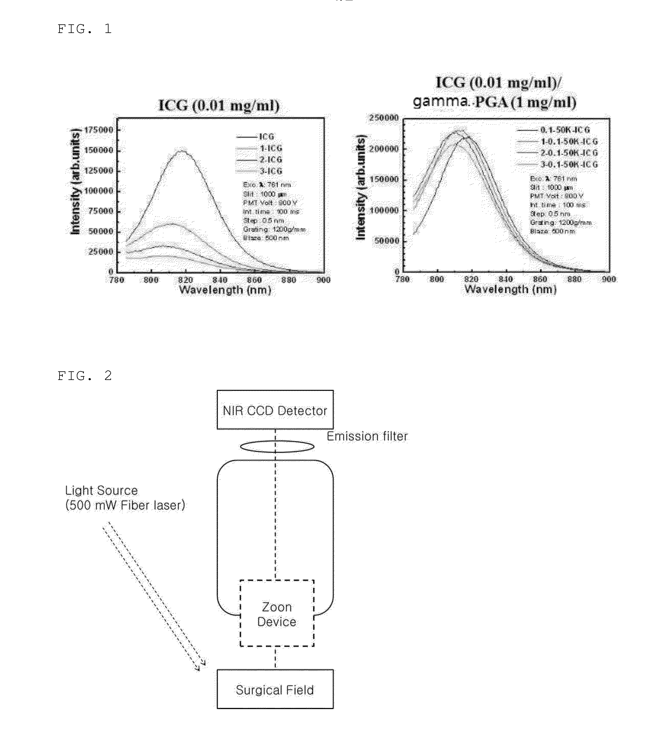
[0044]50 μl of the prepared ICG / γ-PGA complex solution was dispensed into a 1.5 Ml tube, and then the near-infrared fluorescence spectrum thereof was measured using a fluorescence spectrophotometer (FluoroMate FS-2) (see FIG. 1).

[0045]As can be seen in FIG. 1, the fluorescence intensity of ICG alone rapidly decreased with the passage of time, but the fluorescence intensity of the I...

example 2

In Vivo Imaging of ICG / γ-PGA Complex

[0046]In animal tests, 6-week-old female BALB / c nude mice (SLC Inc., Japan) with no pathogens were used. All the tests were performed under the approval of the Laboratory Animal Center, Chungnam National University.

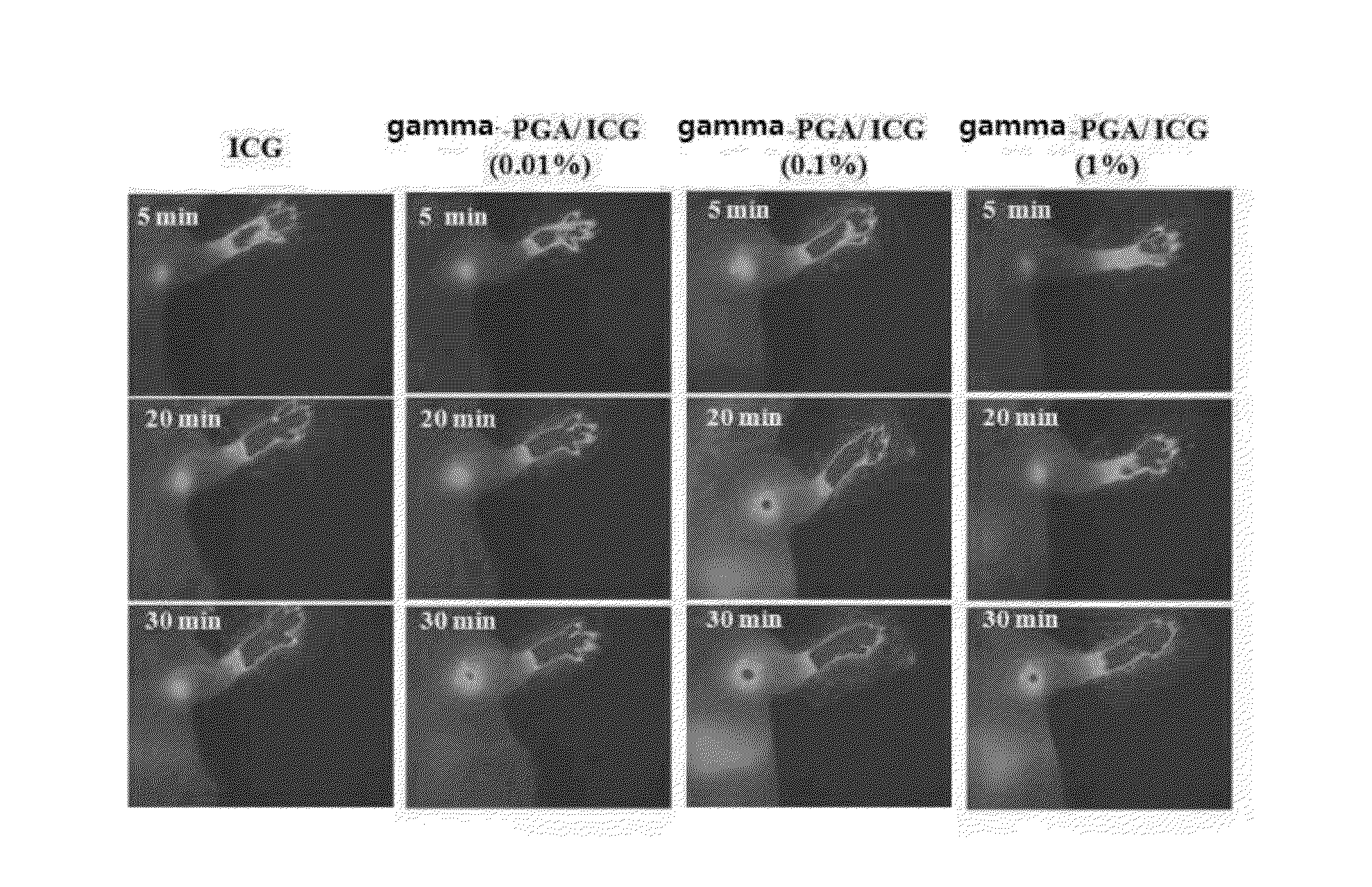
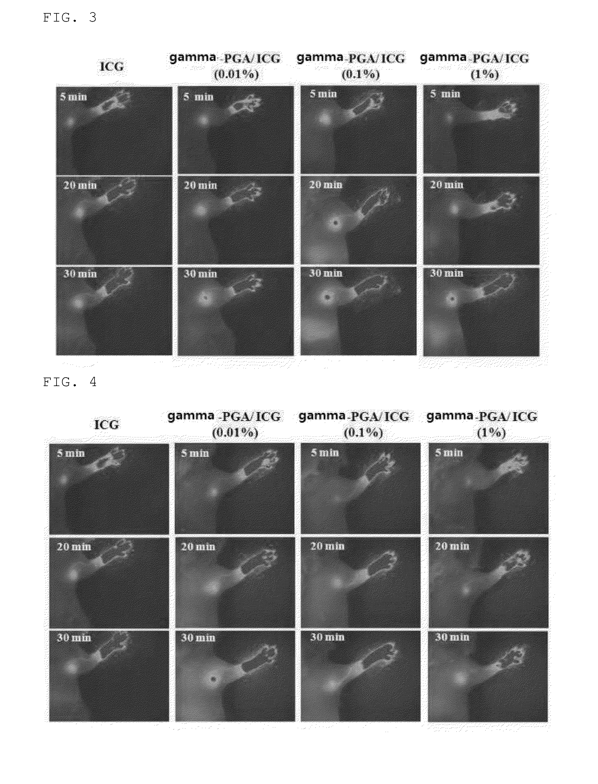
[0047]For data acquisition and analysis, images (see FIG. 3) were obtained using a home-made NIR optical imaging system (see FIG. 2) manufactured by the present inventors. Herein, the near-infrared optical imaging system comprises a light source consisting of a 500 mW fiber-type laser, an emission filter, a lens system equipped with a microzoom, and a near-infrared detector (NIR CCD detector) (see FIG. 2). In the near infrared imaging system, the intensity of fluorescence appears as a pseudo color, and more red fluorescence indicates stronger intensity.

[0048]Before imaging, mice were anesthetized by intraperitoneally injecting a 2.5% avertin (2,2,2-tribromoethanol-tert-amyl alcohol, Sigma) solution at a dose of 0.01 Ml / g weight. 50 μl o...


PUM

PropertyMeasurementUnit
molecular weightaaaaaaaaaa
particle diameteraaaaaaaaaa

Abstract

The present invention relates to an optical imaging probe for detecting a sentinel lymph node, which contains a complex of poly-gamma-glutamic acid and an optical imaging dye, and more particularly to an optical imaging probe for detecting a sentinel lymph node, which contains a poly-gamma-glutamic acid/optical imaging dye complex that, when injected subcutaneously in vivo, remains in the sentinel lymph node for a relatively long period of time and has a low tendency to migrate to other lymph nodes. The present invention provides a poly-gamma-glutamic acid/fluorescent dye complex, which contains no radiopharmaceutical and is harmless to the human body. The use of the poly-gamma-glutamic acid/fluorescent dye complex makes it possible to accurately detect the position of a sentinel lymph node in real time without concerns about radioactive contamination.

Description

TECHNICAL FIELD[0001]The present invention relates to an optical imaging probe for detecting a sentinel lymph node, which contains a complex of poly-gamma-glutamic acid and an optical imaging dye, and more particularly to an optical imaging probe for detecting a sentinel lymph node, which contains a poly-gamma-glutamic acid / optical imaging dye complex that, when injected subcutaneously in vivo, remains in the sentinel lymph node for a relatively long period of time and has a low tendency to migrate to other lymph nodes.BACKGROUND ART[0002]Anatomically, the sentinel lymph node can be defined as the first node that receives lymphatic drainage from a primary tumor. If whether tumor cells in the sentinel lymph node are benign or malignant is determined, whether other lymph nodes contain metastatic tumor can be determined. Thus, if whether the sentinel lymph node contains metastatic tumor cells is determined, the status of other lymph nodes can also be predicted.[0003]In other words, if ...

Claims


Application Information

Patent Timeline
Patent Type & Authority Applications(United States)
IPC IPC(8): A61K49/00
CPCA61K49/0054G01N21/6428G01N21/6456A61K49/0034A61K49/0056
Inventor SUNG, MOON-HEELIM, YONG TAIKNOH, YOUNG-WOOCKLEE, II HAN
Owner BIOLEADERS CORP
Why Eureka
Try Eureka