

Cell-Friendly Inverse Opal Hydrogels for Cell Encapsulation, Drug and Protein Delivery, and Functional Nanoparticle Encapsulation
- Summary
- Abstract
- Description
- Claims
- Application Information
AI Technical Summary
Benefits of technology
Problems solved by technology
Method used
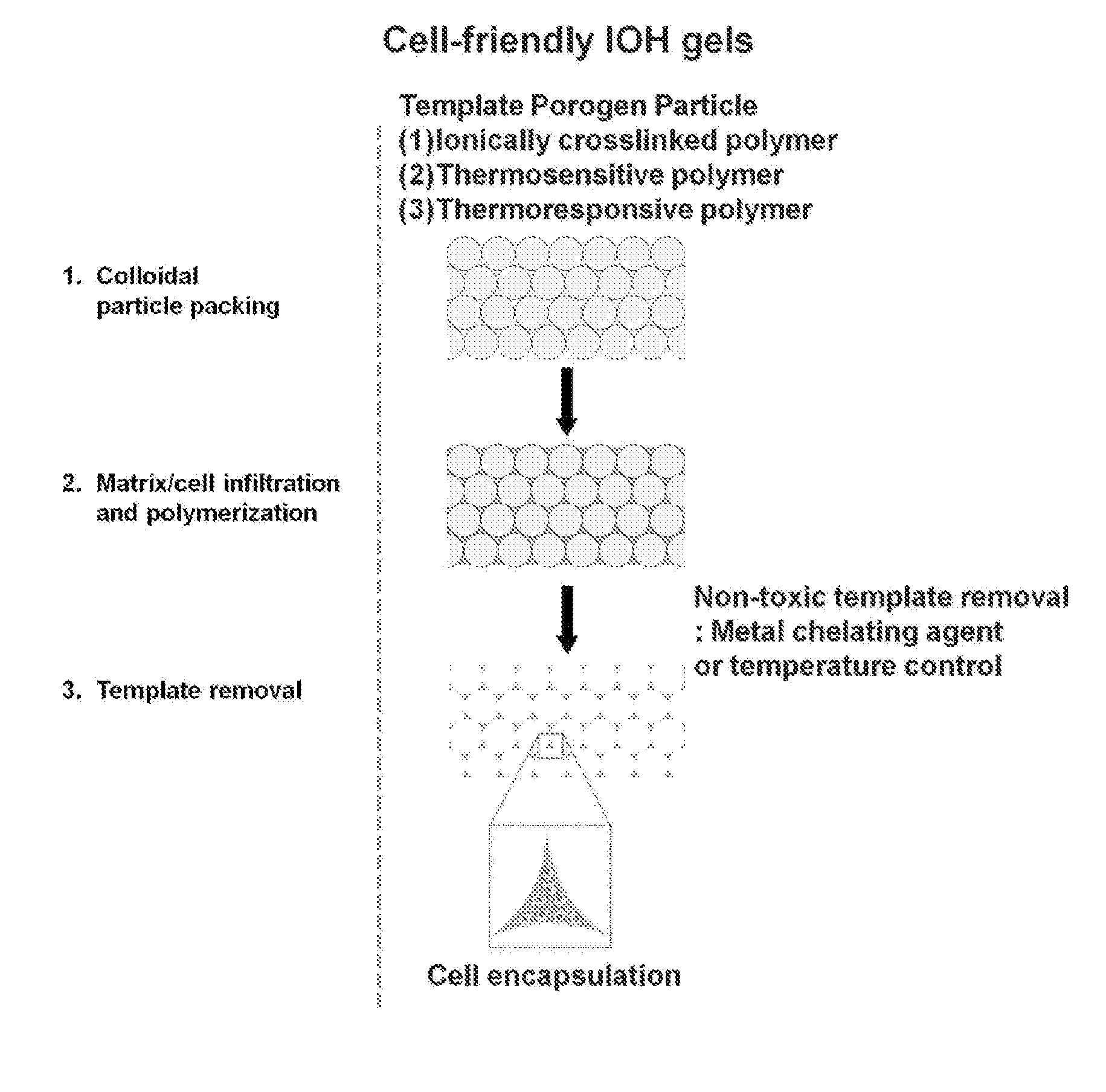
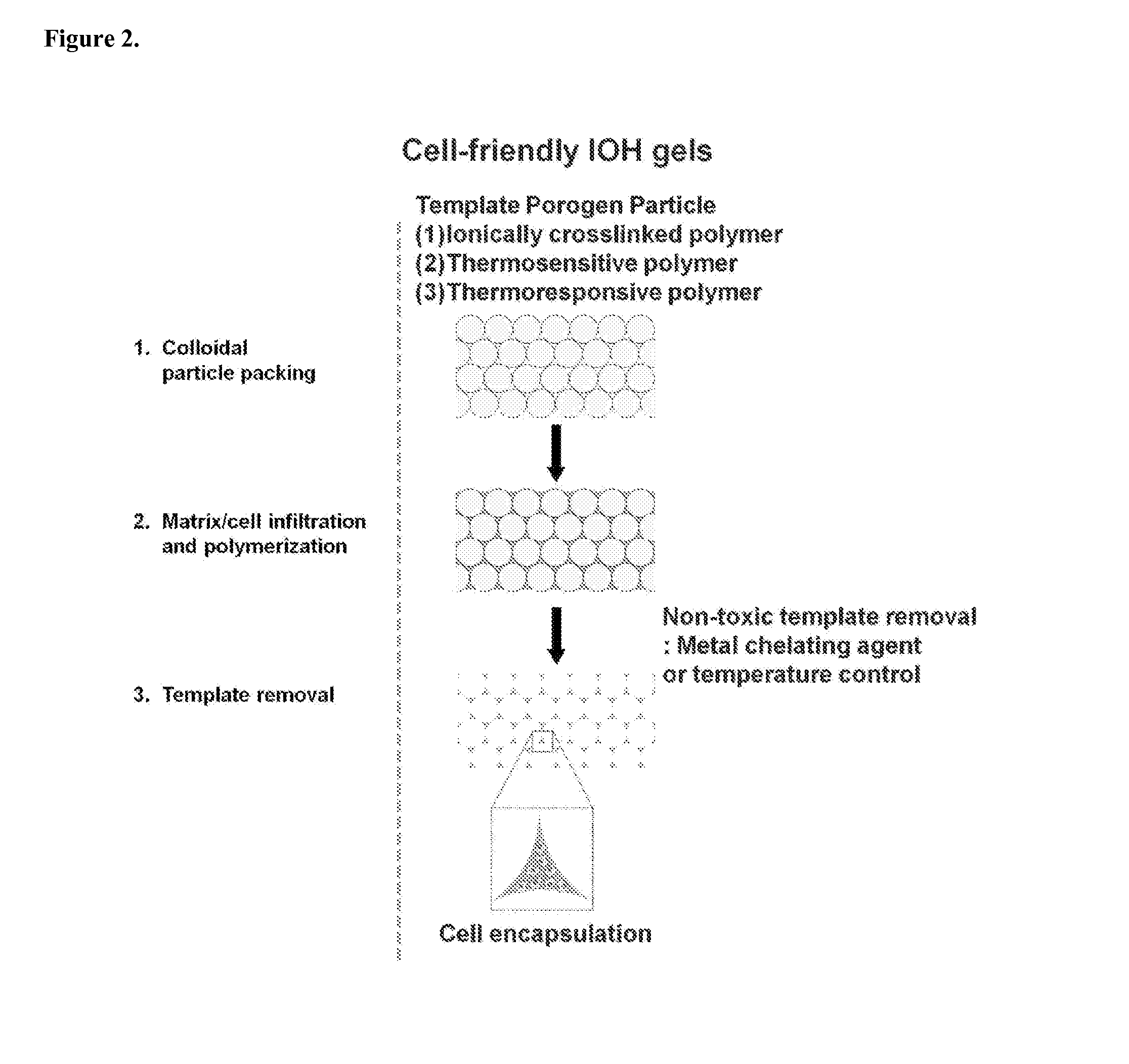
Image

Examples
example 1
Alginate Beads as a Sacrificial Template
[0046]Described herein is an example of the fabrication of a cell-friendly inverse opal hydrogel. Alginate beads, formed using Ca2+-crosslinking were used as the porogen, and
50 mM EDTA solution, a metal chelating agent, was used as the template removal solution. To evaluate if EDTA can dissolve the alginate beads efficiently, three different sized alginate beads were prepared using 2% alginate solution in 100 mM Ca2+solution (FIG. 3, upper row). Rhodamine-labeled bovine serum albumin (BSA) was encapsulated in alginate beads to visualize the beads and their dissolution. The resulting alginate beads were incubated in 50 mM EDTA solution under shaking. After 20 min, all alginate beads dissolved in EDTA solution and lost their spherical morphology, and this resulted in a pink solution due to released rhodamine-labeled BSA from alginate beads (FIG. 3, lower row). This demonstrates that the alginate beads were used as sacrificial template by using E...
example 2
Fabrication of Inverse Opal Hydrogels
[0047]Inverse opal hydrogels were fabricated using alginate beads as the template and methacrylated-gelatin as the hydrogel precursor (FIG. 4). Alginate beads were close-packed in molds with different shapes. Methacrylated-gelatin (10 wt %) solution was infiltrated by adding to the top of the packed alginate beads, and was subsequently polymerized under UV (365 nm) irradiation for 20 min. FIG. 4a shows alginate / gelatin composites with different shapes, such as disc, cubic, and cylinder. The interstitial space was filled with opaque gelatin hydrogels. Finally, the resulting alginate beads / hydrogel composites were incubated in 50 mM EDTA solution for 1 h at 37° C. under shaking to remove alginate beads. FIG. 4b shows disc-shaped porous IOHs with different pore sizes: 1500, 1000, and 600 um, respectively. The IOH maintained the original structure after removal of templates. The pore size was uniform and 3-dimensionally interconnected pores were clea...
example 3
EDTA is Non-Toxic to Cells Encapsulated in IOHs
[0048]To evaluate if incubation in EDTA solution is toxic to cells, cell viability was checked after incubation in 50 mM EDTA solution. Mouse mesenchymal stem cells (MSCs) were cultured in a flask, and incubated in a 50 mM EDTA solution for 10, 30, 60, 120, or 180 min. Subsequently, the viability of cells was measured with a live / dead cell assay by using calcein AM and ethidium homodimer-1. Although the cell morphology changed to a round shape, the representative fluorescent images of the live / dead assay showed that there was no significant toxicity of the EDTA solution to the cells for up to 3 h incubation (FIG. 5a). Quantitative analysis also showed ˜98% viability even after 3h incubation in EDTA solution (FIG. 5b). Based on these observations, incubation of the gels in 50 mM EDTA solution for up to 3 h to remove alginate beads was determined as a nontoxic process to cells encapsulated in IOHs.
PUM
| Property | Measurement | Unit |
|---|---|---|
| Length | aaaaa | aaaaa |
| Length | aaaaa | aaaaa |
| Length | aaaaa | aaaaa |
Abstract
Description
Claims
Application Information