Molecular diagnostic panel of eosinophilic gastrointestinal disorders

a gastrointestinal disorder and molecular diagnostic panel technology, applied in the field of diagnosis and treatment of an eosinophilic gastritis (eg) disease state, can solve the problems of less well-studied and less well-established diagnostic criteria

Active Publication Date: 2014-09-25
CHILDRENS HOSPITAL MEDICAL CENT CINCINNATI
View PDF1 Cites 10 Cited by
  • Summary
  • Abstract
  • Description
  • Claims
  • Application Information

AI Technical Summary

Benefits of technology

[0026]Embodiments of the invention also encompass kits for the detection of a level of one or more genes associated with EG, including: one or more oligonucleotide probes complementary to subsequences of said one or more markers or genes, wherein t

Problems solved by technology

Other EGIDs, such as EG, have been less well-studied than EoE,

Method used


Image

  • Molecular diagnostic panel of eosinophilic gastrointestinal disorders
  • Molecular diagnostic panel of eosinophilic gastrointestinal disorders
  • Molecular diagnostic panel of eosinophilic gastrointestinal disorders

Examples


example 1

Patient Selection

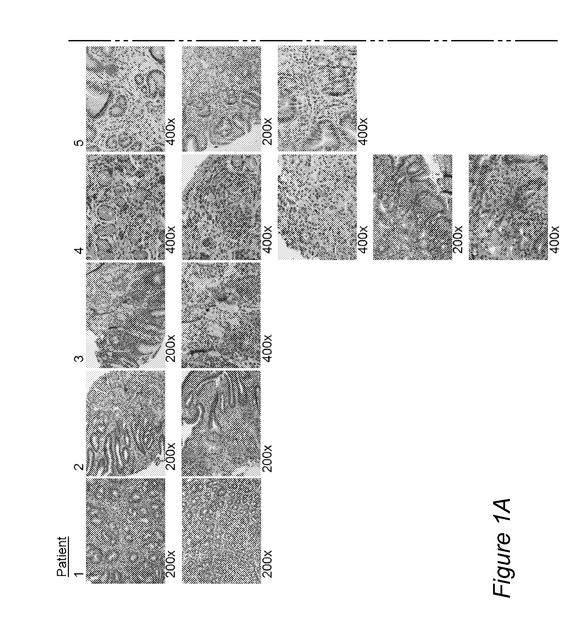
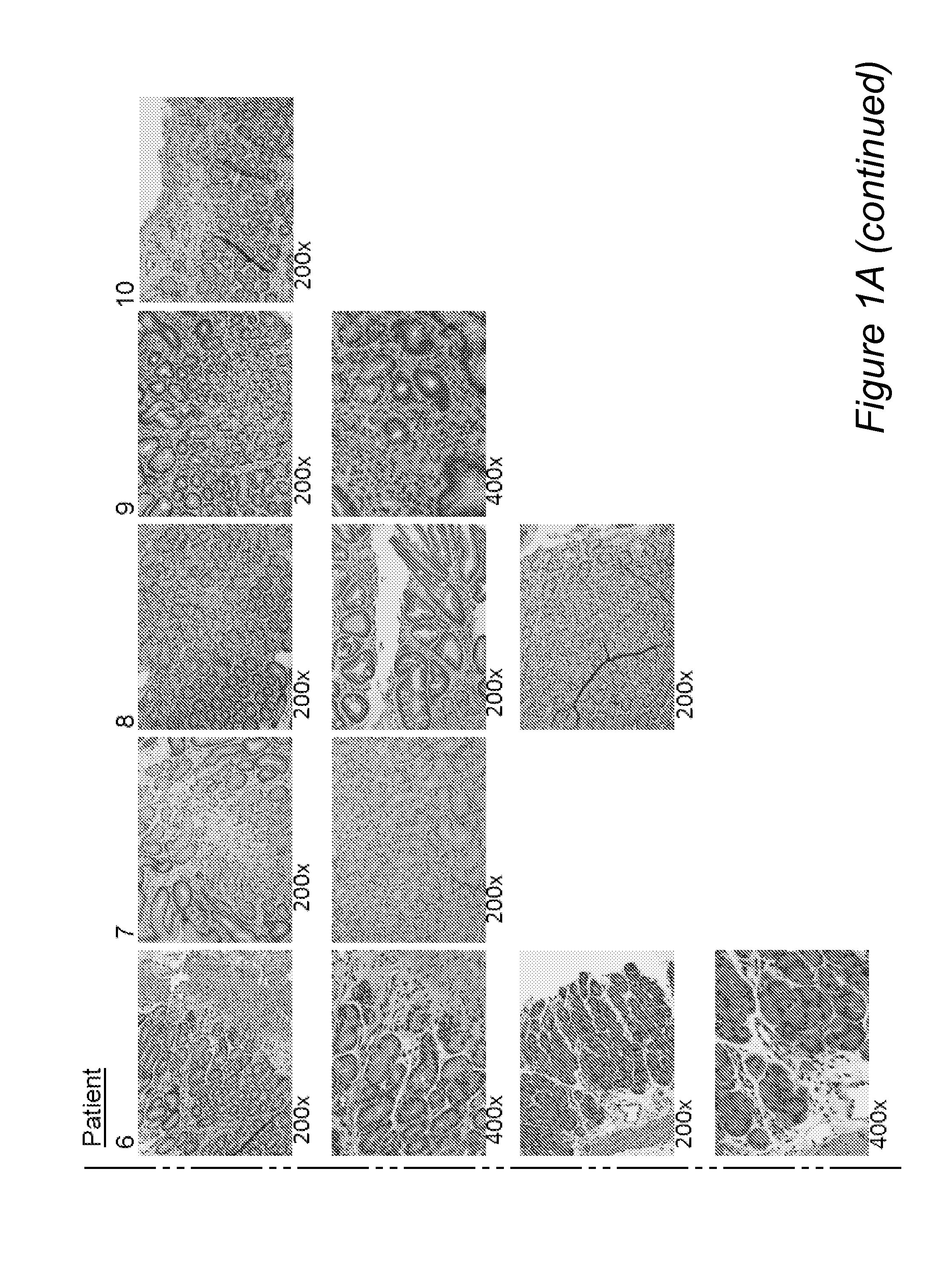
[0105]Entry criteria were that patients who had signs and symptoms consistent with upper GI tract disease and had biopsies that documented active eosinophilic gastritis (EG), with gastric tissue samples preserved for genetic and molecular analyses. The endoscopic procedure at which samples for histopathology, gene microarray, and PCR analyses were obtained was designated the incident endoscopy. All gastric samples used for ancillary studies were obtained from the antrum, and all samples obtained for routine histology and immunohistochemistry were obtained from the antrum or antrum / body.

[0106]Active EG was defined as increased numbers of eosinophils, which were the predominant inflammatory cells in some areas, in a gastric biopsy that showed architectural abnormalities, including excessively branched and / or coiled glands. Similar criteria were used to diagnose eosinophilic duodenitis (ED), eosinophilic jejunitis (EJ), and eosinophilic colitis (EC). Eosinophilic esoph...

example 2

Histopathology and Genetic and Molecular Analysis

Biopsy Preparation

[0108]Samples were obtained at the incident endoscopy from the duodenum, stomach, and esophagus of all patients. Some patients also had colonoscopy. Biopsies for histologic evaluation were fixed in 10% formalin, routinely processed, and embedded in paraffin. Sections were cut at 5 microns thickness and stained with hematoxylin and eosin or antibody. Alcian blue / PAS stain (Polyscientific, Bay Shore, N.Y.) was also performed on all biopsies.

Immunohistochemistry

[0109]Antibody information is provided in Table 1. The antibodies used for immunohistochemical staining in this study, their source, the dilution at which they were used, and the antigen retrieval method applied to the tissue sections are listed. Immunohistochemical stains were performed using a Ventana Benchmark XT automated immunostainer (Ventana, Tucson, Ariz.).

Quantitative Microscopy

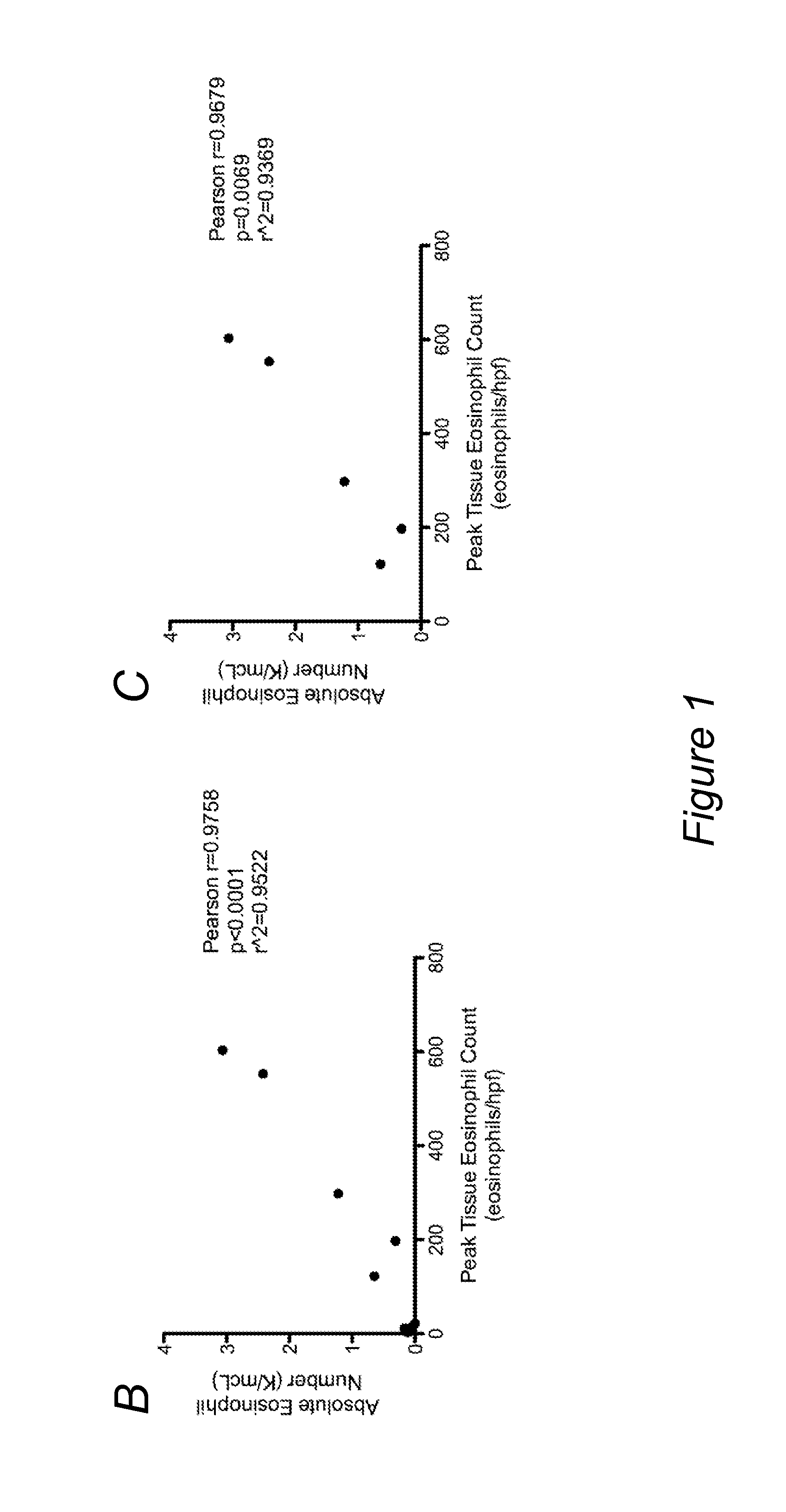
[0110]Multiple levels of gastric biopsies were surveyed, the area containing ...

example 3

Cell Culture and Treatment

Culture of Primary Esophageal Epithelial Cells

[0115]A sample of distal esophageal epithelium was collected during incident or other endoscopy. Following digestion with trypsin / EDTA, biopsy samples were cultured in modified F-media (3:1 F-12 / Dulbecco modified Eagle's medium) supplemented with FBS (5%), adenine (24.2 μg / ml), cholera toxin (10-4 μmol / L), insulin (5 μg / ml), hydrocortisone (0.4 μg / ml), and epidermal growth factor (10 ng / ml) in the presence of penicillin, streptomycin, and amphotericin (Invitrogen). Cultures were supplemented with 1 to 5×10⁵ feeders (NIH 3T3 J2 cells irradiated 6000 rad). Media were changed twice weekly. Upon reaching confluency, cells were trypsinized and re-plated for experiments.

Culture of Cell Lines and Cytokine Treatment

[0116]Cells from human esophageal cell line TE-7 (IARC, Lyon, France) were maintained in RPMI medium (Invitrogen) supplemented with 5% FBS (Atlanta Biologicals, Lawrenceville, Ga.) and 1% penicillin / streptomy...


PUM

PropertyMeasurementUnit
Levelaaaaaaaaaa
Exposure limitaaaaaaaaaa

Abstract

Embodiments of the invention are directed to methods of diagnosing eosinophilic gastritis (EG), or remission therefrom in a subject, wherein the methods include applying a sample from the subject to a diagnostic panel that contains selected markers for EG, analyzing the results thereof, and making a determination as to the EG status of the subject. Embodiments of the invention are also directed to methods of monitoring the pathological development or medical prognosis of EG in a subject. Embodiments of the invention are also directed to use of CDH26 as a marker for EG, eosinophilic esophagitis, or allergic inflammatory conditions. Embodiments of the invention also relate to the use of anti-CDH26-based therapeutics to treat allergic inflammatory conditions.

Description

CROSS REFERENCE TO RELATED APPLICATION[0001]The present application claims the benefit of priority under 35 U.S.C. §119(e) to U.S. Provisional Application No. 61 / 500,508, MOLECULAR DIAGNOSTIC PANEL OF EOSINOPHILIC GASTROINTESTINAL DISORDERS, filed on Jun. 23, 2011, which is currently co-pending herewith and which is incorporated by reference in its entirety.STATEMENT REGARDING FEDERALLY-SPONSORED RESEARCH[0002]This invention was made with U.S. Government support on behalf of National Institute of Health (NIH) Grant Nos. R01 DK76893 and U19 AI070235. The U.S. Government has certain rights in this invention.SEQUENCE LISTING[0003]The instant application contains a Sequence Listing which has been submitted in ASCII format via EFS-Web and is hereby incorporated by reference in its entirety. Said ASCII copy, created on Jun. 25, 2012, is named 88544023.txt and is 81,136 bytes in size.FIELD OF THE INVENTION[0004]The invention disclosed herein generally relates to methods and compositions fo...

Claims


Application Information

Patent Timeline
IPC IPC(8): C12Q1/68G01N33/68
CPCG01N33/6893C12Q1/6881C12Q2600/158C12N15/113C12N2310/11C12N2310/141C12N2310/531A61K31/7088A61K31/713C12Q1/6883Y02A90/10A61K39/395A61K48/00C07K16/18C07K2319/30C12N15/11C12Q2600/118C12Q2600/16G01N2800/06G01N2800/52
Inventor ROTHENBERG, MARC E.CALDWELL, JULIE
Owner CHILDRENS HOSPITAL MEDICAL CENT CINCINNATI
Why Eureka
Try Eureka