
System, device and method for measurement of esophageal wall blood perfusion

a technology of esophageal wall and blood perfusion, which is applied in the field of gastroenterology and esophageal biology, can solve the problems of unsatisfactory therapies, inability to identify abnormal motor events at the time of pain, and billions of dollars in costs, so as to reduce the association of esophageal wall blood perfusion, reduce swallowing-induced esophageal contraction amplitude, and reduce the effect of esophag

Inactive Publication Date: 2015-02-19
RGT UNIV OF CALIFORNIA
View PDF33 Cites 3 Cited by
  • Summary
  • Abstract
  • Description
  • Claims
  • Application Information

AI Technical Summary

Benefits of technology

The present patent describes a new laser Doppler probe that can be used to measure the blood flow in the esophageal wall. The probe is placed on the esophageal wall and stays in place during peristalsis and other movements. The device also includes a high-resolution manometry catheter to record pressure and reflux events. By monitoring these movements, the device can help identify the cause of esophageal pain. The laser Doppler probe can successfully measure esophageal wall perfusion and provide valuable information on the health of the esophageal tissue.

Problems solved by technology

Non-cardiac “angina like pain” of esophageal origin and heartburn, nonresponsive to potent acid inhibition therapy are major health problems for which there are no satisfactory therapies.
Angina like esophageal pain accounts for 26% of all emergency room visits in the United States and costs billions of dollars to the healthcare system.
However, some studies that utilized prolonged ambulatory manometry and pH recordings, conducted in the 1980's and early 1990s, failed to identify abnormal motor events at the time of pain.
Intraluminal manometry cannot monitor longitudinal muscle contraction or sustained esophageal contraction.
However, recording and analysis of US images, especially over extended time is expensive, tedious and labor intensive.
As a result, other laboratories have not studied longitudinal muscle and its relationship to esophageal pain.
In animal studies, one can anchor the laser Doppler probe to esophageal well using surgically placed sutures, which is obviously not practical for human studies.

Method used


Image

  • System, device and method for measurement of esophageal wall blood perfusion
  • System, device and method for measurement of esophageal wall blood perfusion
  • System, device and method for measurement of esophageal wall blood perfusion

Examples


example 1

Experimental Design

[0035]Studies were conducted in the 12 healthy volunteers (mean age 32.5 years, range 18-51, 11 males). Protocol for the studies was approved by the “University of California San Diego Institutional Review Board for the Protection of Humans” and each individual gave a written consent prior to enrollment in the study. Subjects fasted for at least 6 hours prior to the commencement of the study.

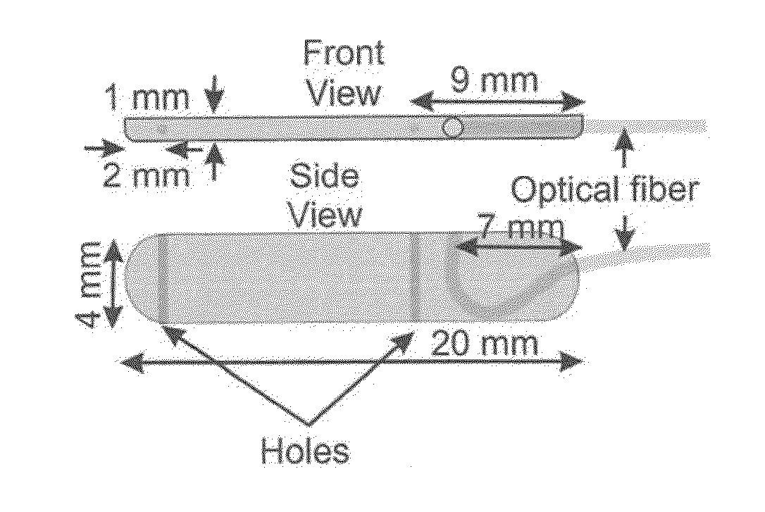
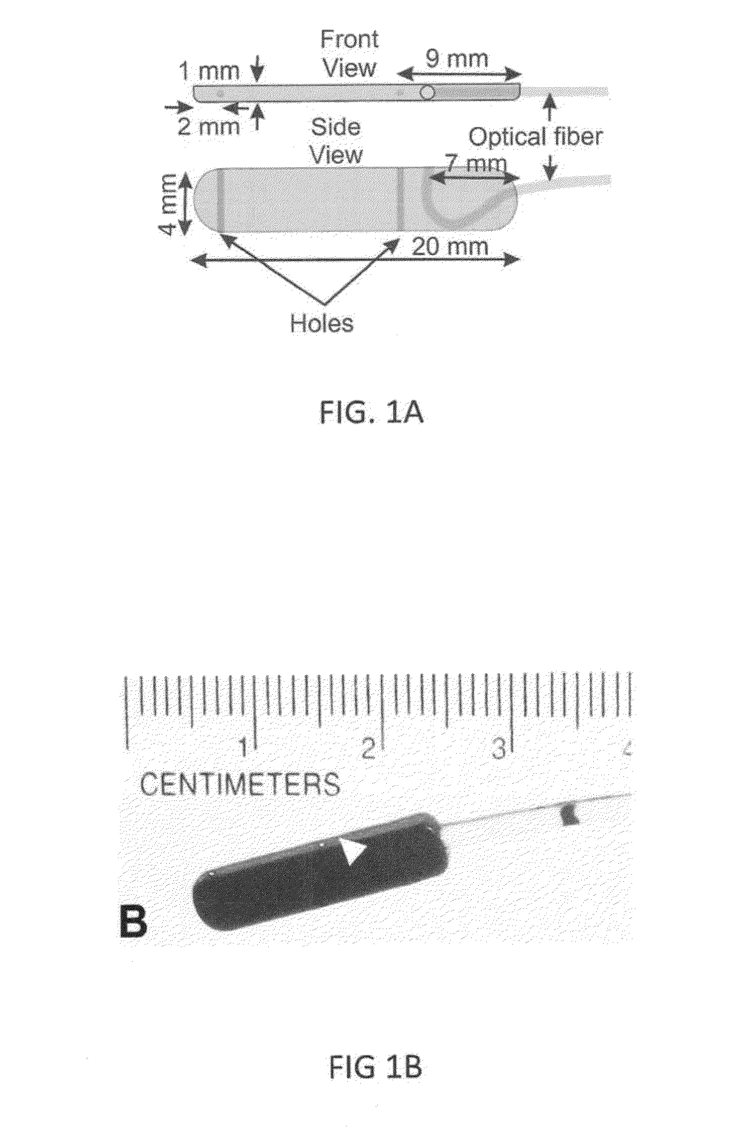
[0036]Esophageal wall perfusion can be monitored using a custom designed laser Doppler probe (FIGS. 1A and 18) that can be taped to a Bravo pH monitoring system and the two together can be anchored to the esophageal wall. A wireless Bravo pH monitoring technique makes it possible to anchor the pH capsule to the esophageal mucosa. Through a vacuum, connected to the cup of a Bravo pH capsule by the delivery system, a small volume of mucosa can be sucked into the cup of Bravo pH capsule. Using a mechanical system located in the handle of delivery system, a pin can be inserted thr...

example 2

Laser Doppler Probe & Catheter

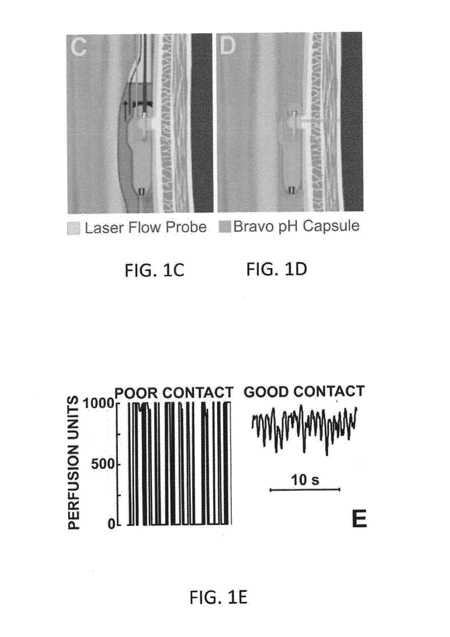
[0037]A unique aspect of the laser Doppler probe of the present disclosure is its relatively thinness, i.e., 1 mm wide. The probe can be 4 mm deep and 20 mm in length (FIGS. 1A and 1B) and can be connected to the laser Doppler perfusion monitor via a 1.5 mm diameter, 150 cm long fiber optic cable. The laser Doppler probe can be firmly taped to the Bravo pH capsule using paraffin film (FIGS. 1C and 1D) in a fashion so that when Bravo pH capsule is anchored to the esophageal wall so is the laser Doppler probe. The laser beam exits from the laser Doppler probe in the direction of esophageal wall, and at the level of the suction cup in the Bravo pH capsule. The combined dimensions of the Bravo capsule and laser probe can be as small as 5×4 mm or less, approximately the size of manometry catheters use in clinical practice (4-5 mm in diameter).

[0038]The laser Doppler probe taped to the Bravo capsule and delivery system can be passed through the nose in patien...

example 3

[0041]Data Analysis & Statistical Methods

[0042]In addition to the laser Doppler perfusion units (PU) signal, the Doppler monitor can also provide continuous record of the Total Backscatter (TB) of the laser light signal. Total Backscatter can be an indicator of the relative movements between the esophageal mucosa / wall and laser Doppler probe. Total backscatter can be essentially a reading of how much light is reflected back to the probe / instrument. Total backscatter signal reading of greater than 1 and a relatively flat line can indicate that the measurement conditions were stable and there is minimal to no relative movement between the tissue and the probe. An esophageal blood perfusion signal can be temporally filtered using a 2 seconds moving time average filter. Duration of esophageal blood flow reduction can be determined by the interval over which the perfusion drops by more than 10% of the baseline values. Baseline pressures and esophageal blood flow can be averaged over a 10...


PUM


Abstract

The present invention provides a device and system useful as a new diagnostic modality to differentiate the etiology of heartburn / chest pain by measuring esophageal blood flow. Furthermore, the device and system of the present invention can be used as a new diagnostic modality to differentiate the etiology of abdominal pain by measuring blood in the wall of gastrointestinal tract including rectum and colon blood flow.

Description

FEDERAL FUNDING LEGEND[0001]This invention was made with governmental support under a Grant Number RO-1 awarded by the National Institutes of Health. The government has certain rights in the invention.BACKGROUND OF THE INVENTION[0002]1. Field of the Invention[0003]The present invention relates generally to the fields of gastroenterology and esophageal biology. More specifically, the invention relates to the monitoring of the esophageal gastrointestinal tract wall blood perfusion and uses thereof.[0004]2. Background Information[0005]Non-cardiac “angina like pain” of esophageal origin and heartburn, nonresponsive to potent acid inhibition therapy are major health problems for which there are no satisfactory therapies. Angina like esophageal pain accounts for 26% of all emergency room visits in the United States and costs billions of dollars to the healthcare system. With the availability of potent acid inhibitors (proton pump inhibitors), it is clear that in 50-70% of patients with ga...

Claims


Application Information

Patent Timeline
IPC IPC(8): A61B5/00A61B17/34A61B5/145A61B8/12A61B5/026A61B5/0205
CPCA61B5/6852A61B5/0261A61B5/0205A61B5/14539A61B5/0538A61B8/12A61B17/3468A61B5/7475A61B5/14542A61B5/4233
Inventor MITTAL, RAVINDERBHARGAVA, VALMIK
Owner RGT UNIV OF CALIFORNIA
Learn More