Composition for detecting nucleic acid and method for detecting nucleic acid using same

Active Publication Date: 2015-03-19
SEOUL NAT UNIV R&DB FOUND
View PDF1 Cites 13 Cited by
  • Summary
  • Abstract
  • Description
  • Claims
  • Application Information

AI Technical Summary

Benefits of technology

[0010]By using the composition for detecting a nucleic acid and the method for detecting a nucleic acid using the composition in accordance with the example embodiments, presence or absence of a target nucleic acid in a sample or cell and an expression pattern of the target nucleic acid can be observed in a real time as well as a multiplexed-detection of plural target nucleic acids is enabled. Further, candidates of medicinal substances and low-molecular substances capable of adjusting expression of a specific miRNA positively or negatively can be derived by performing high-throughput screening (HIS). Further, a mechanism of action of the substances derived through this process and an influence of such substances upon the control of the miRNA expression can be investigated in a cell level. Additionally, since an expressi

Problems solved by technology
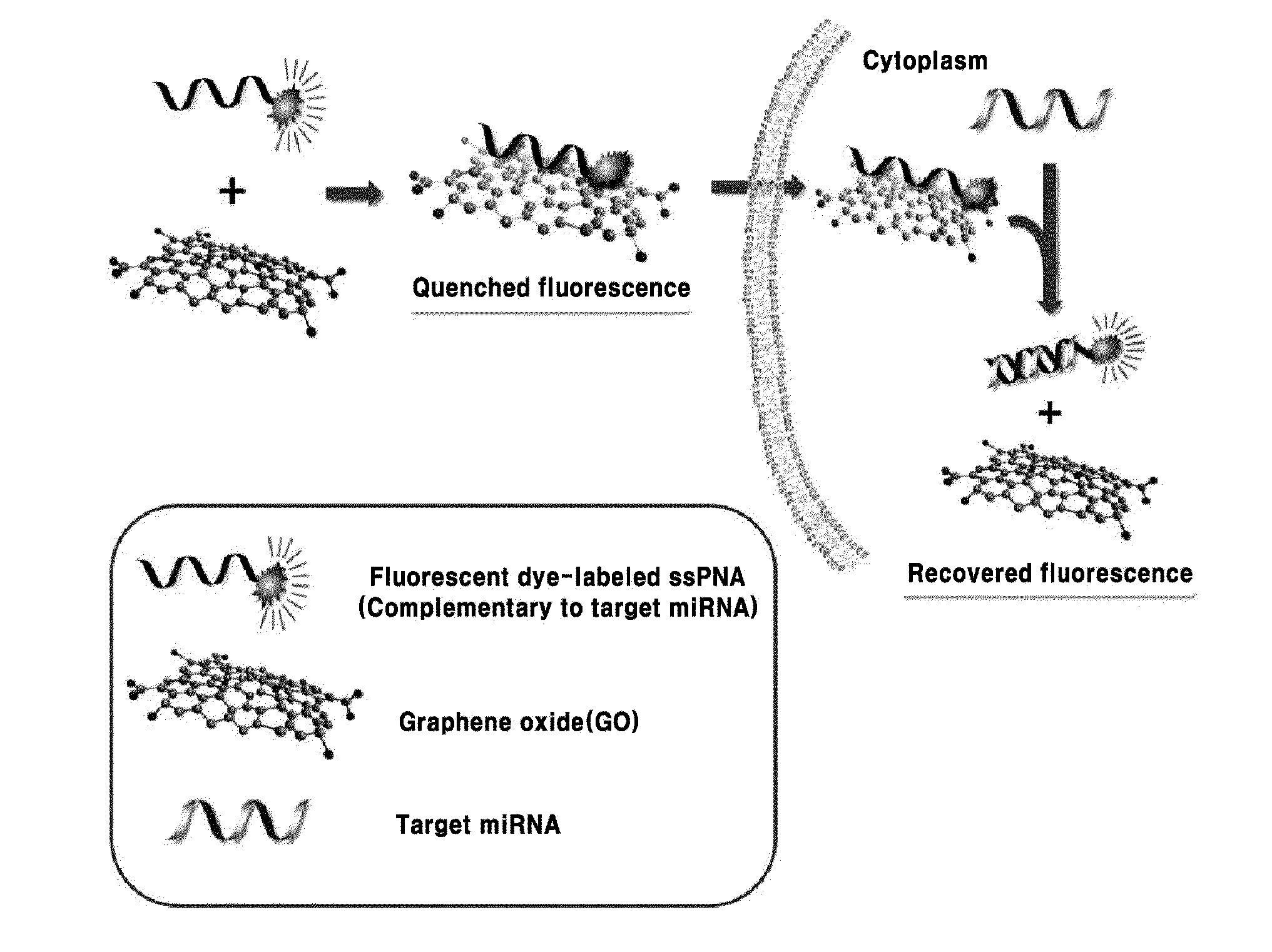
However, these methods have drawbacks in that multiplexed-detection is imp

Method used


Image

  • Composition for detecting nucleic acid and method for detecting nucleic acid using same
  • Composition for detecting nucleic acid and method for detecting nucleic acid using same
  • Composition for detecting nucleic acid and method for detecting nucleic acid using same

Examples


example 1

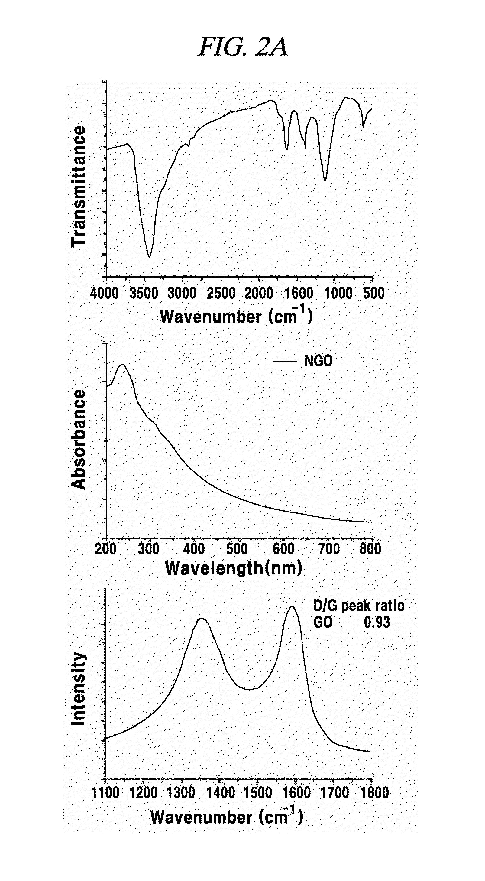
Preparation and Characterization of Graphene Oxide

[0079]In an example, a graphene oxide sheet was prepared according to the modified Hummer's method which is commonly known in the pertinent art. First, 0.5 g of NaNO3 (produced by Junsei Co., (Japan)) and 23 mL of H2SO4 (produced by Samchun Chemical Co., (Seoul, Korea) were mixed while being intensely agitated within a water tub filled with ice. Then, 3 g of KMnO4 (produced by Sigma-Aldrich Co., (Missouri, USA) was slowly added to the mixture. After the KMnO4 is added, the solution was moved into an ice bath at a temperature of 35° C. and agitated therein for 1 hour. Then, 40 mL of distilled water was added, and the temperature of the water tub was raised to 90° C. for 30 minutes. Then, 100 mL of distilled water was added again. Thereafter, by adding 3 mL of 30% H2O2 (produced by Junsei Co., (Japan)) in drops, the color of the solution was changed from dark brown to yellow. Then, the synthesized graphene oxide solution was filtered t...

example 2

Adsorption of a Single-Stranded PNA Probe to Graphene Oxide and Observation of Resultant Fluorescence Quenching

[0081]Sequences of miRNAs and complementary sequences of PNA probes used in the present example and all of the following examples are specified in Table 1 as bellows.

TABLE 1Base Sequence ofTargetBase SequenceFluorescence-Organ-miRNAof miRNAlabeled ProbeismmiRNA-UAGCUUAUCAGACUGAUGFAM-OO-Human21UUGATCAACATCAGTCTGATAAGCTAmiRNA-UCCCUGAGACCCUAACUUROX-OO-Human125bGUGATCACAAGTTAGGGTCTCAGGGAmiRNA-UUAAUGCUAAUCGUGAUACy5-OO-Human155GGGGUCTATCACGATTAGCATTAmiRNA-UUUGGAUUGAAGGGAGCUFAM-OO-Plant159CUATAGAGCTCCCTTCAATCCAAAmiRNA-UAGCACCAUCUGAAAUCGFAM-OO-Human29aGUUATAACCGATTTCAGATGGTGCTA

[0082]In the above table, the left sequences of nucleic acids are base sequences of miRNAs, whereas the right sequences of nucleic acids are base sequences of PNA probes that are complementary to the respective base sequences of the miRNAs. The miRNA-159 was used as a negative control group. The miRNA used in...

example 3

Evaluation of Oligonucleic Acid Containing a Fluorescent Material, for Detecting miRNA as a Target Material

[0086]Numerous biomolecules exist in a cell. Therefore, in case of graphene oxide having high adsorbability for nonspecific biomolecules, non-specific desorption of a probe may be occur even under the absence of a nucleic acid as a target material, thus emitting a fluorescence signal. In view of this, in the present example, miRNA detection efficiencies of a PNA probe or DNA probe having a base sequence complementary to that of a target miRNA were compared to investigate whether the PNA probe is more suitable than the DNA probe.

[0087]Cells of MDA-MB-231 expressing the miRNA-21 as one of breast cancer cell lines selected as a model disease cell line were collected, and a cytolysate was acquired by repeating freezing an defrosting of the cells. A DNA or PNA probe complementary to the miRNA-21 was used while labeling the DNA or PNA probe with an organic fluorescent pigment FAM.

[00...


PUM

PropertyMeasurementUnit
Sizeaaaaaaaaaa
Sizeaaaaaaaaaa
Lengthaaaaaaaaaa

Abstract

The present invention relates a composition, including an RNA probe which contains a fluorescence material absorbed in graphene oxide, for detecting a nucleic acid, and to a method for detecting a nucleic acid using the composition. By means of the composition and the method, the presence and expression pattern of a target nucleic acid in a sample or a cell can be observed in real time, and a plurality of target nucleic acids can be detected in multitude.

Description

TECHNICAL FIELD[0001]The embodiments described herein pertain generally to a composition, which includes a PNA probe containing a fluorescent material and adsorbed on graphene oxide, for detecting a nucleic acid, and also pertain to a method of detecting a nucleic acid using the composition.BACKGROUND[0002]Graphene has a plane monolayer structure including a 2-dimensional lattice made of carbon atoms. Graphene is a basic structural element of various allotropes of graphite having different dimensional structures. That is, the graphene may be a basic structure of fullerene (O-dimensional structure), carbon nanotube (1-dimensional structure) or graphite stacked in a 3-dimensional structure. In many of recent researches, peculiar properties of graphene, such as a zero band gap, derived from its hexagonal crystalline structure, two interpenetrating triangular subordinate lattice structures and its one-atom-size thickness are attracting attention. Further, graphene also has a peculiar el...

Claims


Application Information

Patent Timeline
IPC IPC(8): C12Q1/68
CPCC12Q1/6841C12Q2600/178C12Q1/6823C12Q1/6876C12Q2523/308C12Q2527/125C12Q2561/113
Inventor MIN, DAL-HEELEE, JIEONRYOO, SOO-RYOON
Owner SEOUL NAT UNIV R&DB FOUND
Why Eureka
Try Eureka