High sensitivity biosensor using pixel analysis of CMOS image sensor

a biosensor and image sensor technology, applied in the field of cmos image sensors, can solve the problems of inability to analyze a biomarker having a concentration of pg/ml unit or less in the human body, almost impossible to analyze cytokine by using biochip technologies, and most existing analysis equipment analyzing biochips, such as fluorescent scanners, can achieve the effects of increasing measurement sensitivity, low cost, and small siz

Active Publication Date: 2015-04-30
GWANGJU INST OF SCI & TECH
View PDF6 Cites 10 Cited by
  • Summary
  • Abstract
  • Description
  • Claims
  • Application Information

AI Technical Summary

Benefits of technology

[0012]According to the present invention, measurement sensitivity may be increased by using a CMOS image sensor itself as a substrate for fixing a bioreceptor, and measuring light intensity in a luminescent reaction in a state in which the substrate is adjacent to the sensor itself to analyze variables of each pixel.
[0013]In addition, since trace amounts of the biomarker which is difficult to be measured in a protein chip, and the like, of the related art, may be analyzed and screened by using the analysis method according to the present invention, the analysis method of the present invention may be applied to develop new medicines and new biomarkers.
[0014]Further, instead of a high cost analysis equipment used for analysis of the protein chip in the related art, a high sensitivity biosensor having small size and low manufacturing cost may be implemented to be applied as an economical biochip analysis system.

Problems solved by technology

However, a detection sensitivity of the biochip researched up to now is several pg / ml to several tens of pg / ml, and thus, it is not possible to analyze a biomarker having a concentration of a pg / ml unit or less in a human body.
As an example, in the case of cytokine intermediating signal transmission between cells among important biomarkers present in the human body, most of cytokine is present at a concentration of a pg / ml unit or less, such that it is almost impossible to analyze the cytokine by using biochip technologies.
In addition, since most of the existing analysis equipments analyzing the biochips, such as a fluorescent scanner, a SPR sensor, and the like, are slightly expensive, economic feasibility of the measuring equipments is an important problem to be solved.

Method used


Image

  • High sensitivity biosensor using pixel analysis of CMOS image sensor
  • High sensitivity biosensor using pixel analysis of CMOS image sensor
  • High sensitivity biosensor using pixel analysis of CMOS image sensor

Examples


example 1

Measurement of Streptavidin-Horseradish Peroxidase (STA-HRP)

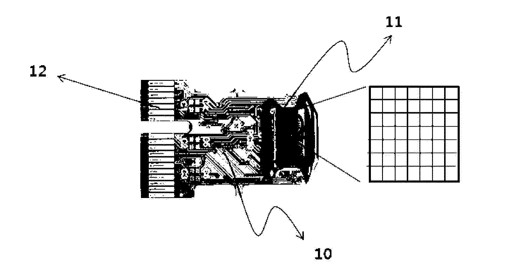
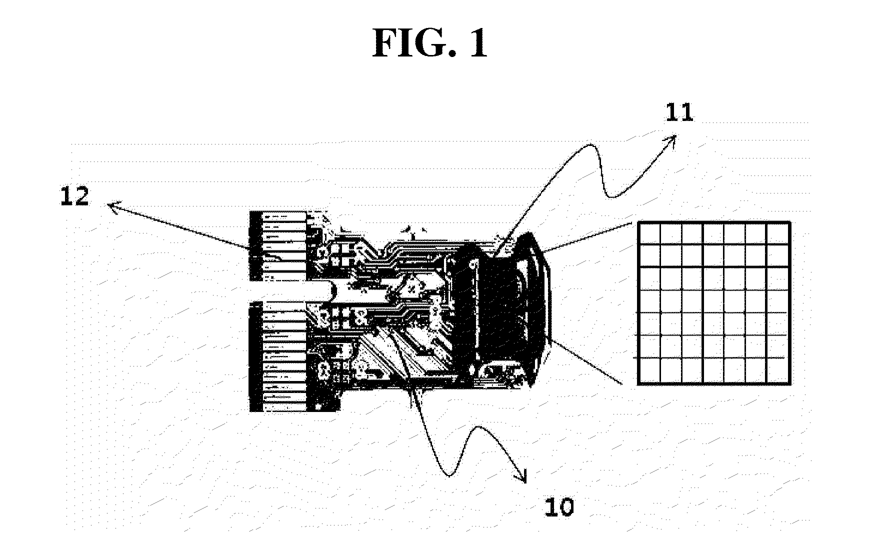
[0037]46000 pixels having each width of about 4 μm and each length of about 4 μm, were formed on the surface of the measuring unit 11 of the CMOS image sensor of FIG. 1. A solution (concentration of 0, 1, 100, 10000 fg / ml) obtained by dissolving streptavidin-horseradish peroxidase (STA-HRP) manufactured by Calbiochem, USA in 10 mM PB buffer (pH 7.0) was reacted onto the surface of the measuring unit modified by a biotin group at room temperature for 30 minutes, to fix enzymes onto the surface by an adsorption method. Then, the surface was washed with 10 mM PB buffer, distilled water, and 0.1M carbonate buffer (pH 9.0) in sequence.

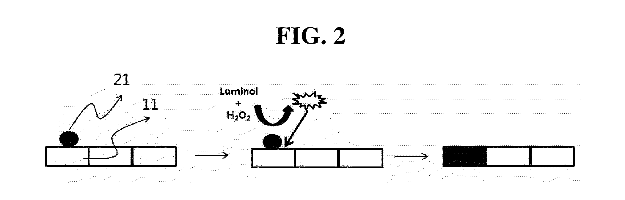
[0038]Next, the measuring unit was treated with 2 mM luminol dissolved in 0.1M carbonate buffer (pH 9.0) and 2 mM hydrogen peroxide solution.

[0039]The process of forming the chemiluminescent signal according to the treatment of the luminol and the hydrogen peroxide solution was shown in FIG. 2.

[004...

example 2

Measurement of IL-5

[0044]The measuring unit 11 of the CMOS image sensor of FIG. 1 was ultrasonic-washed in 0.1N NaOH solution for 15 minutes, then washed with ethanol three times, and APTMS ((3-Aminopropyl)trimetoxysilane) having 1% concentration dissolved in ethanol was reacted for about 2 hours. Then, the reaction product was washed with ethanol about two times, then ultrasonic washed in ethanol for five minutes again, and dried with high purity nitrogen gas. In addition, in order to prepare a surface reacting with an antibody, 2 vol % of glutaraldehyde dissolved in 10 mM PB buffer was reacted for about 4 hours to finally modify the surface of the measuring unit with an aldehyde group.

[0045]Then, a 0.1 mg / ml IL-5 capture antibody was reacted onto the modified surface for about 1 hour, followed by reaction with 1% BSA solution for about 2 hours in order to remove non-specific reaction. In addition, IL-5 antigen dissolved in PBS buffer and biotinylated 5 μg / ml IL-5 detection antibod...


PUM

PropertyMeasurementUnit
lengthaaaaaaaaaa
widthaaaaaaaaaa
concentrationaaaaaaaaaa

Abstract

Provided are a method of analyzing a target substance to be measured, the method including: dividing a surface of a measuring unit of a CMOS image sensor into a plurality of pixels, directly fixing a bioreceptor onto the surface of the measuring unit of the CMOS image sensor, and measuring chemiluminescent signals depending on concentrations of the target substance to be measured, and a CMOS image sensor applied to the same.

Description

TECHNICAL FIELD[0001]The present invention relates to a CMOS image sensor maximizing detection sensitivity in the same reaction signal by measuring and analyzing chemiluminescent signals depending on reaction concentrations of a target substance to be measured as a pixel unit of the CMOS image sensor, on a substrate of the CMOS image sensor having a bioreceptor fixed thereonto, and more specifically, to a CMOS image sensor capable of maximizing the detection sensitivity by directly analyzing the signal of each pixel arrangement in a measurement region and measuring the number of pixels having a cut-off value or more.BACKGROUND ART[0002]A biochip is a biosensor technology in which several tens to several tens of thousands different analysis substances are fixed onto a solid surface, and a specific signal is analyzed by using an analysis equipment to be utilized in a wide range of fields such as a biolabeling substance screening, a disease diagnosis, an environment monitoring, and the...

Claims


Application Information

Patent Timeline
Patent Type & Authority Applications(United States)
IPC IPC(8): G01N21/76H04N5/374G01N21/64
CPCG01N21/76H04N5/374G01N21/6454G01N33/52G01N33/53H04N25/00H04N25/76
Inventor KIM, MIN-GONJOUNG, HYOU-ARMHONG, DONG-GU
Owner GWANGJU INST OF SCI & TECH
Why Eureka
Try Eureka