Organ Mounted Electronics

a technology of electronic components and organs, applied in the field of organ mounted electronics, can solve the problems of prolonging ep clinical procedures, affecting the real-time mapping of transient abnormal rhythms, and sudden cardiac arrest, so as to reduce the risk of damage, reduce the size or volume, and prevent or reduce the current of leakage.

Active Publication Date: 2015-05-21
WASHINGTON UNIV IN SAINT LOUIS +1
View PDF6 Cites 71 Cited by
  • Summary
  • Abstract
  • Description
  • Claims
  • Application Information

AI Technical Summary

Benefits of technology

[0013]In an embodiment, the device at least partially surrounds, and optionally entirely surrounds, the internal biological tissue. In an embodiment, for example, at least a portion of the sensors, the actuators or both of the device are in physical contact, optical contact, thermal contact and / or fluid contact with the internal biological tissue. In a specific embodiment, for example, the flexible and stretchable substrate and the electronic device or device component provide a net flexural rigidity of the device less than or equal to 1×10⁻⁴ Nm, optionally for some applications less than or equal to 0.5×10⁻⁴ Nm and optionally for some applications less than or equal to 0.1×10⁻⁴ Nm. These and other embodiments advantageously provide for the ability to conform to and interface with biological tissues having complex surface geometries and for the ability to interface with biological tissues that undergo complex motion and / or a change in shape as a function of time.
[0014]Devices and methods of the invention are particularly useful for sensing, characterizing and actuating a range of biological tissues due to the unique ability of the devices to conform to and maintain conformality with complex surface shapes characteristic of many types of organs, muscle, vasculature and / or bone. Various aspects of the disclosed devices provide for the ability to directly interface with a biological tissue, such as electronically, optically or thermally interface, and permit real-time investigation of biological tissues in vivo or ex vivo. In addition, the flexibility and stretchability of the devices provide the ability to interface with biological tissues that change shape and / or size as a function of time, thereby permitting spatial and temporal characterization and actuation of biological tissues as they move, change shape, grow and / or reduce in size or volume. For example, devices of the invention provide the ability to interface with muscle tissue as the muscle moves due to voluntary and / or involuntary muscle contractions, such as experienced by the heart during cardiac cycling. This aspect of the present systems and methods enables advanced systems-level diagnostic and therapeutic functionality for characterization and treatment of organ function.
[0015]For example, in various embodiments, the internal biological tissue is selected from the group consisting of: an organ, a blood vessel, muscle, a bone, any combinations thereof, and any portions thereof. In an embodiment, the biological tissue in conformal contact with the device is epicardium tissue. In some embodiments, for example, the internal biological tissue has a time-varying outer surface shape. For example, in a specific embodiment, the internal biological tissue comprises a heart having an outer surface corresponding to heart epicardium. In embodiments, the device in conformal contact with the internal biological tissue is immersed in a fluid. For example, the tissue is optionally immersed in a body fluid, a saline solution or water. In some embodiments, the device includes one or more barrier layers for preventing fluid, such as blood or water-based physiological ionic solution, from the environment from penetrating to an interior layer of the device. Such a configuration optionally also prevents or reduces leakage current from / to the sensors, actuators and / or electrical interconnects to tissue at the device interface and, thus, reduces or minimizes the risk of damage thereto due to current flow, electronic biasing, etc. Optionally, minimizing or reducing a leakage current maintains the sensing, actuating and / or interconnecting components in working electronic condition. In a specific embodiment, the internal biological tissue comprises an ex vivo explant. In an exemplary embodiment, the internal biological tissue comprises tissue within a living animal.

Problems solved by technology

Sudden cardiac arrest is a leading cause of death in developed countries.
The iterative nature of this approach prolongs EP clinical procedures, thereby impeding real time mapping of transient abnormal rhythms.
Despite explosive growth and innovation in the broader electronics industry, a key limitation of many state of the art EP devices is that they retain the simple electronics-tissue interface developed decades ago.
Unfortunately the planar shapes and rigid, brittle mechanical properties associated with conventional ICs severely limit their non-destructive, intimate integration with the curvilinear, soft surfaces of cardiac tissue undergoing periodic deformation due to cardiac contractions.

Method used


Image

  • Organ Mounted Electronics
  • Organ Mounted Electronics
  • Organ Mounted Electronics

Examples


example 1

3D Multifunctional Integumentary Membranes for Spatiotemporal Measurement / Stimulation Across the Entire Epicardium

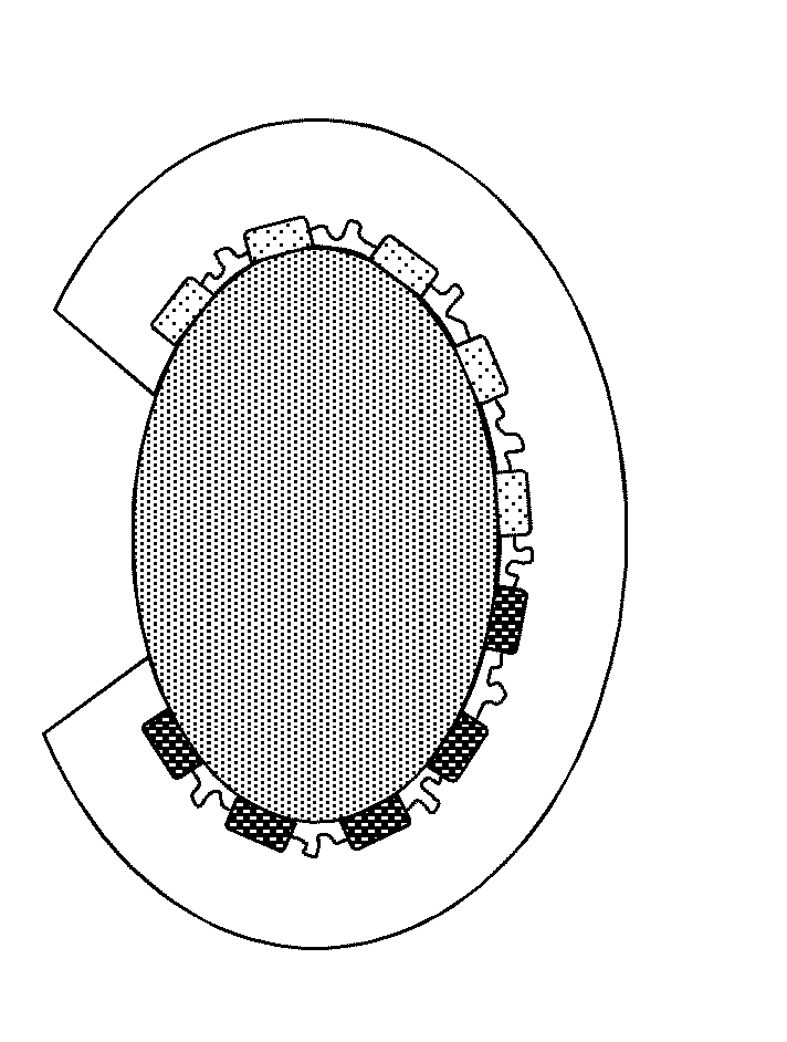
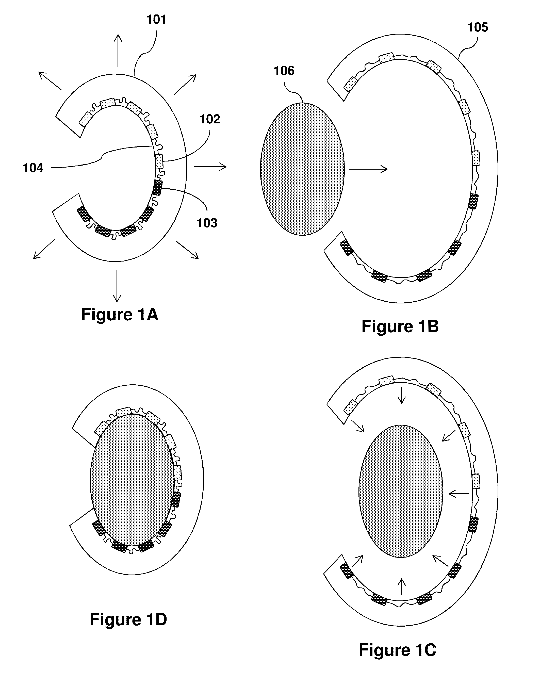
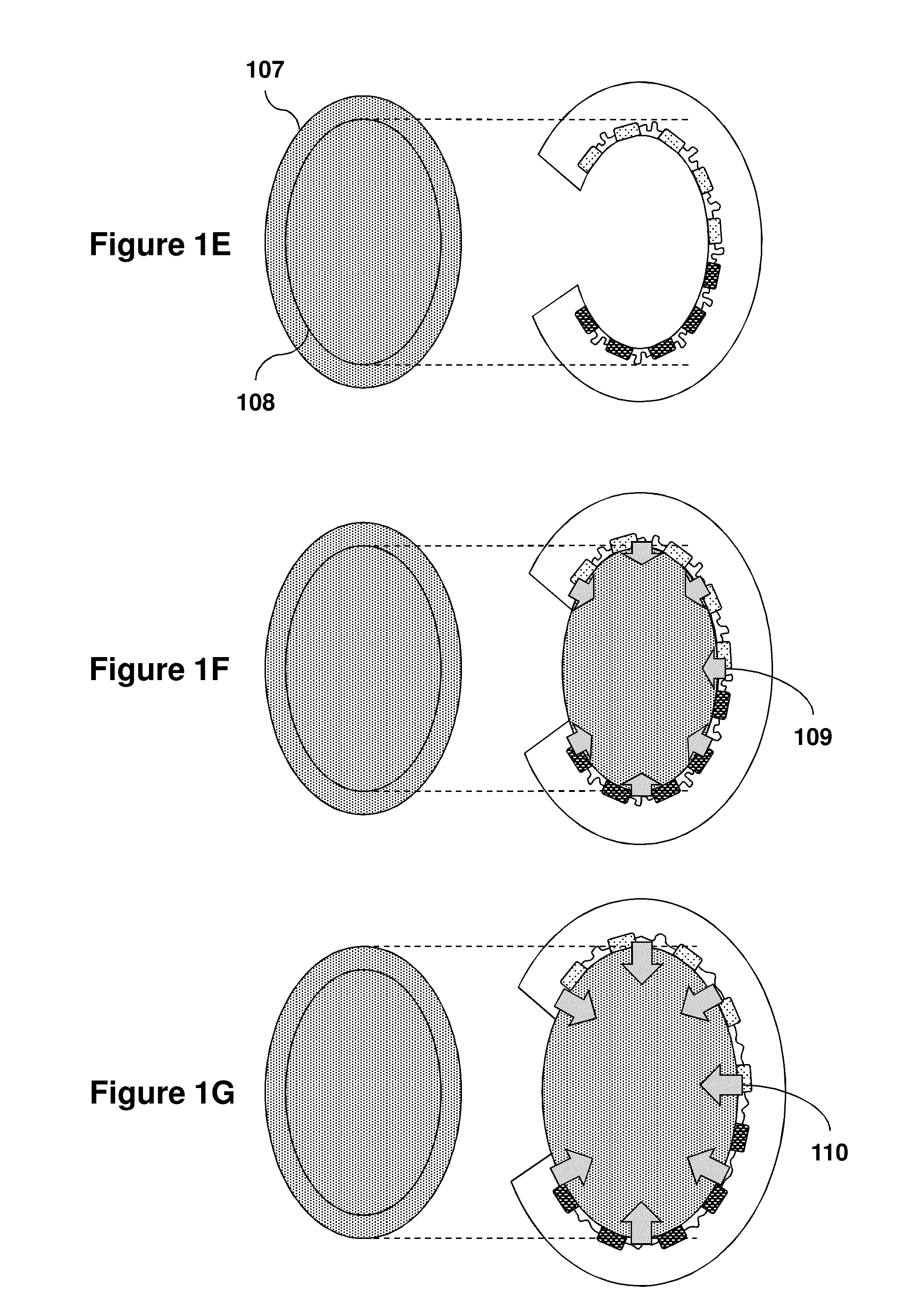
[0142]Means for high-density, large area, conformal multiparametric physiological mapping and stimulation are critically important in both basic and clinical cardiology. Recently developed conformal electronic systems enable important capabilities, but their embodiments as 2D sheets prevent integration over the full 3D epicardial surface and reliable contact for chronic use without sutures or adhesives. This example presents a qualitatively different approach that exploits 3D elastic membranes shaped precisely to match the epicardium of the heart via the techniques of 3D printing, as a platform for deformable arrays of multifunctional sensors, electronic and optoelectronic components. Such integumentary devices completely envelop the heart, in a form-fitting manner, as a sort of artificial pericardium. Inherent elasticity in the soft membranes provides a mechanically sta...

example 2

Devices and Methods for Low-Energy Defibrillation

[0488]The biomedical devices and methods of the present invention support a broad range of therapeutic and diagnostic applications including multiparametric cardiac mapping and stimulation. This example provides experimental results demonstrating the efficacy of the present devices for use in low-energy defibrillation and thermal sensing via establishing a conformal interface with large areas of cardiac tissue.

[0489]FIGS. 23 and 24 provide optical images of a 3D-multifunctional integumentary device of the invention. FIG. 23 shows the 3D-multifunctional integumentary device integrated with a rabbit heart demonstrating the ability to establish conformal contact with large areas of the heart surface. In this figure, the black arrows indicate the defibrillation electrodes having a fractal-based design and temperature sensor arrays. The defibrillation electrodes are for delivering epicardial electrotherapy and the temperature sensors provi...

example 3

Devices and Methods for Detection of Extracellular Potassium and Hydrogen Ion Concentrations

[0497]The biomedical devices and methods of the present invention support a broad range of therapeutic and diagnostic applications including multiparametric cardiac mapping and stimulation. This example provides experimental results demonstrating real-time detection of extracellular potassium and hydrogen ion concentrations in myocardial ischemia using a 3D-MIM device comprising intimately integrated ion selective electrodes.

[0498]Extracellular potassium (i.e. K+) and hydrogen ion (i.e., pH) have been recognized as major determinants of changes in cardiac electrical activity and the occurrence of arrhythmias in myocardial ischemia. Thus, flexible and stretchable ion selective potentiometric sensors have been developed to monitor these ions quantitatively in situ, as well as to provide intimate bio-integration on a cardiovascular 3D-MIM device. The sensors were fabricated by photolithography a...


PUM


Abstract

Provided are devices and methods capable of interfacing with biological tissues, such as organs like the heart, in real-time and using techniques which provide the ability to monitor and control complex physical, chemical, biochemical and thermal properties of the tissues as a function of time. The described devices and methods utilize micro scale sensors and actuators to spatially monitor and control a variety of physical, chemical and biological tissue parameters, such as temperature, pH, spatial position, force, pressure, electrophysiology and to spatially provide a variety of stimuli, such as heat, light, voltage and current.

Description

CROSS-REFERENCE TO RELATED APPLICATIONS[0001]This application claims the benefit of U.S. Provisional Pat. App. 61 / 885,994, filed Oct. 2, 2013, U.S. Provisional Pat. App. 61 / 937,187, filed Feb. 7, 2014, and U.S. Provisional Pat. App. 62 / 003,786, filed on May 28, 2014, each of which is hereby incorporated by reference in its entirety.STATEMENT REGARDING FEDERALLY SPONSORED RESEARCH OR DEVELOPMENT[0002]This invention was made with governmental support under Award Nos. R01 HL115415, R01 HL114395 and R21 HL112278 awarded by the National Institutes of Health and Award No. DGE-1144245 awarded by the National Science Foundation. The government has certain rights in the invention.BACKGROUND[0003]Sudden cardiac arrest is a leading cause of death in developed countries. Many patients at risk for arrhythmic death have advanced structural heart disease, and preexisting non-lethal ventricular arrhythmias. In these and other cases, cardiac electrophysiologic (EP) characterization is useful to aid ...

Claims


Application Information

Patent Timeline
Patent Type & Authority Applications(United States)
IPC IPC(8): A61B5/00A61B5/03A61B5/11A61H23/02A61B5/145A61B5/0205A61B5/042A61N2/00A61B5/1473A61B5/296
CPCA61B5/686A61H2201/10A61B5/036A61B5/1116A61B5/1473A61B5/14539A61B5/0084A61B5/02055A61B5/14546A61B5/0422A61B5/4836A61H23/0245A61B5/6885A61B5/0538A61B5/01A61N2/002A61B5/6869A61B5/015A61B5/1107A61B2562/028A61B2562/046A61B2562/164A61N2/006A61N5/0603A61N5/0622A61F7/007A61B18/14A61B2018/00351A61N1/375A61N1/0563A61N1/0597A61N1/3621A61B2562/0215A61N1/37512A61B5/0036A61B5/287A61B5/6843
Inventor ROGERS, JOHN A.EFIMOV, IGORGUTBROD, SARAHXU, LIZHIBONIFAS, ANDREWWEBB, RICHARD CHADKOH, AHYEON
Owner WASHINGTON UNIV IN SAINT LOUIS
Why Eureka
Try Eureka