
Noninvasive Detection of Human Brain Conditions and Anomalies

a technology of human brain and anomalies, applied in the field of noninvasive detection of human brain conditions and anomalies, can solve the problems of affecting the normal functioning of the brain, so as to achieve the effect of reducing the risk of stroke, preventing the onset of stroke, and improving the neurological defici

Active Publication Date: 2016-03-24
JAN MEDICAL
View PDF3 Cites 16 Cited by
  • Summary
  • Abstract
  • Description
  • Claims
  • Application Information

AI Technical Summary

Benefits of technology

The invention is a system and method for detecting vascular conditions in the brain using sensors attached to the skull. These sensors detect small movements of the skull caused by blood flow pulsations. The signals from the sensors are processed using a computer model to create a signature unique to each patient. This signature is then used to diagnose and localize vascular abnormalities, such as strokes, aneurysms, and vasospasms, in a non-invasive way. The invention can also be used to monitor and screen high-risk patients for potential stroke symptoms.

Problems solved by technology

Additionally, a patient may experience transient ischemic attacks, which are in turn a high risk factor for the future development of a more severe episode.
Thrombi or emboli can result from atherosclerosis or other disorders, for example, arteritis and lead to physical obstruction of arterial blood supply to the brain.
Stroke-associated symptom(s) of intra cerebral hemorrhage are abrupt, with the onset of headache and steadily increasing neurological deficits.
Prior to rupture, aneurysms may be asymptomatic, or occasionally associated with headaches.
However, headache typically becomes acute and severe upon rupture and may be accompanied by varying degrees of neurological deficit, vomiting, dizziness, and altered pulse and respiratory rates, phobophobia and severe headache and or neck stiffness.
Current diagnostic methods for stroke include costly and time-consuming procedures such as non-contrast computed tomography (CT) scans, electrocardiogram, magnetic resonance imaging (MRI) and angiography.
Determining the immediate cause of stroke is difficult.
Advanced imaging systems are not widely available at present and are limited to major hospital centers.
It is, however, an invasive procedure and is also limited by cost and availability.
Moreover, delays in confirmation of stoke diagnosis and identification of stroke type limit the number of patients that may benefit from early intervention therapy.
In addition, continuous monitoring of stroke patients is not possible with CT or MRI scanners, thus only one snapshot in time is available for diagnosis and treatment.
Once an aneurysm ruptures, however, the patient declines rapidly due to major brain injury, and over 50% of aneurysm rupture patients die acutely.
None of these devices provides reliable and clinically relevant information clinicians can rely upon to guide treatment, and none has made an impact on treatment of stroke.
Devices and methods to date do not optimize the detection of useful signals from the brain or other parts of the body, have not localized the detected vascular conditions or tissue conditions and do not present the collected data in a clinically useful way.

Method used


Image

  • Noninvasive Detection of Human Brain Conditions and Anomalies
  • Noninvasive Detection of Human Brain Conditions and Anomalies
  • Noninvasive Detection of Human Brain Conditions and Anomalies

Examples


Embodiment Construction

[0061]General Outline of Process

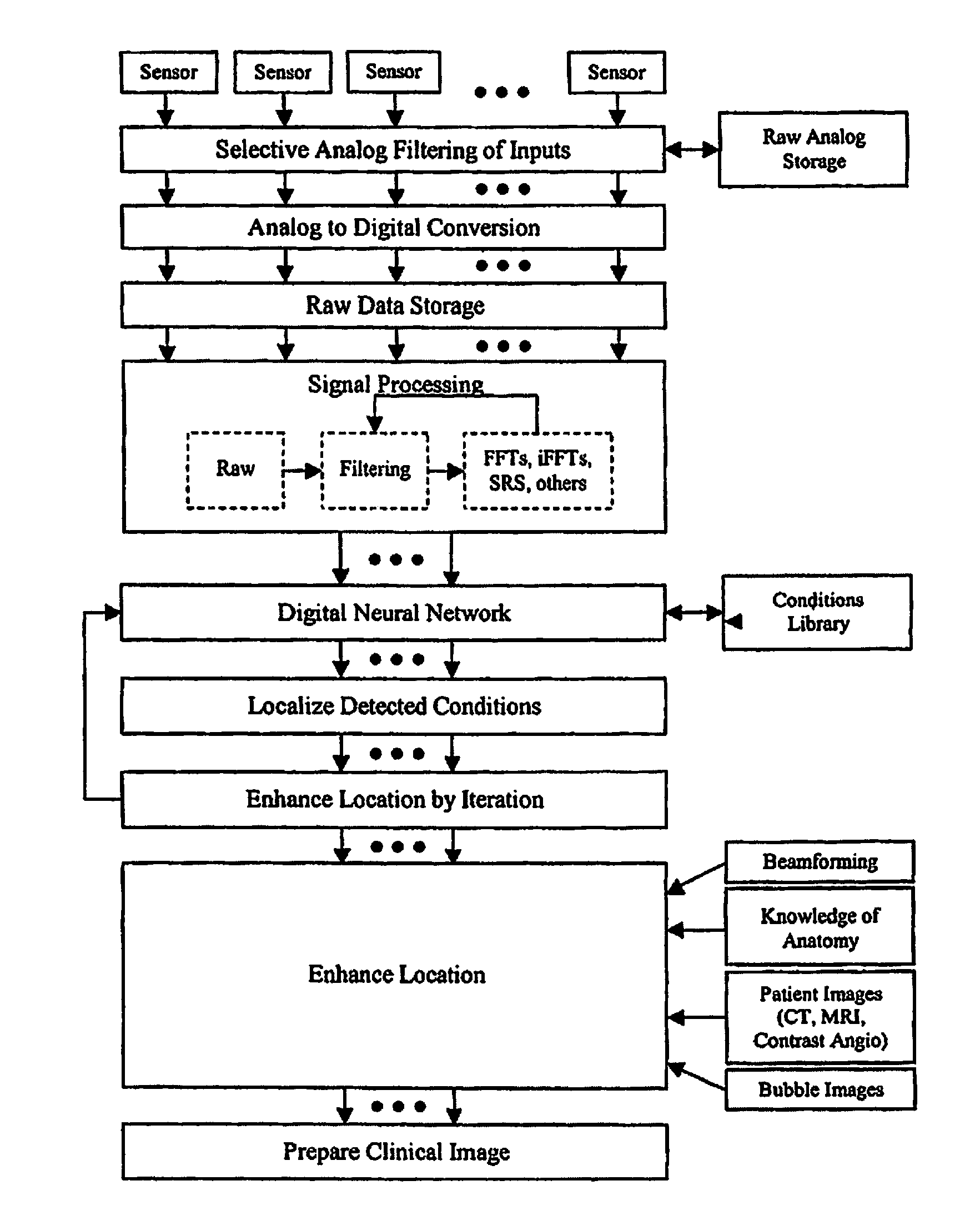
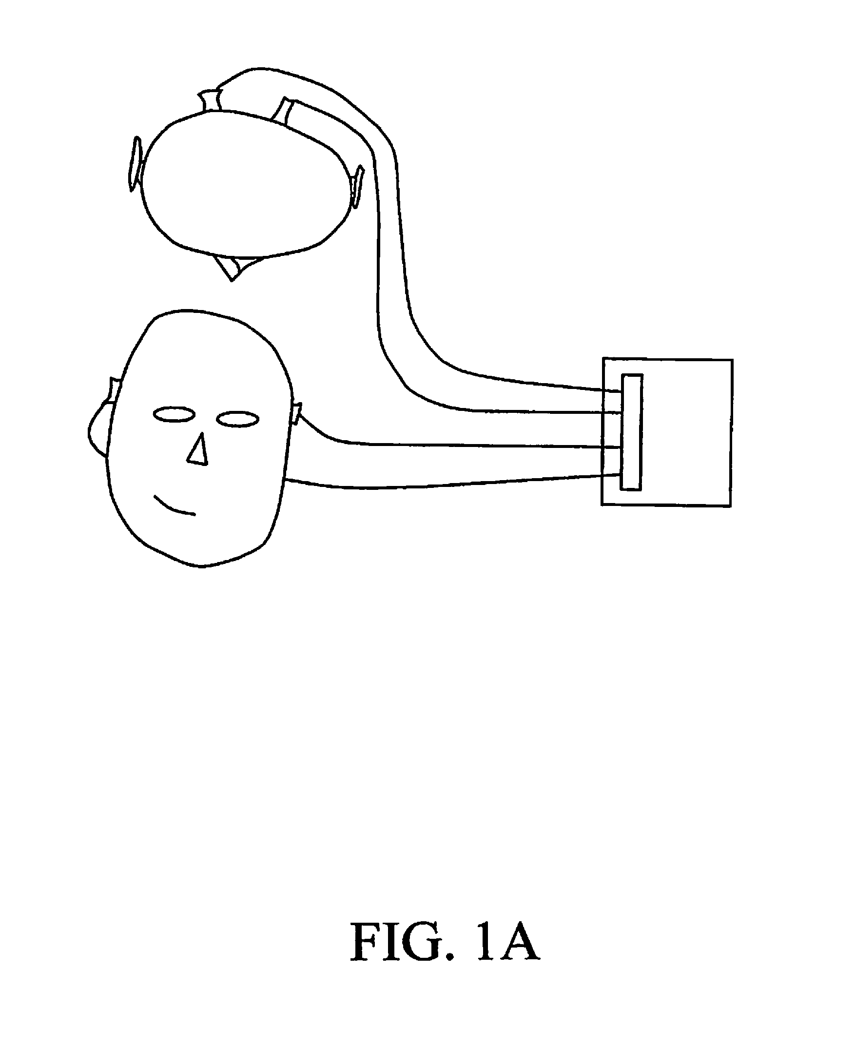
[0062]Periodic pulses from the heart produce waves of expanding blood vessels within the body. The tissue surrounding the blood vessels is displaced during the pulse and contracts again after the pulse. This displacement propagates outward from the blood vessel. In the case of the brain, this displacement reaches the skull and displaces the bone in response the displacement of the blood vessel wall. Extremely sensitive accelerometers record this displacement. The character of the signal recorded by the accelerometers is dependent on the nature of the displacement caused by the blood pulse. At a restriction in the vessel the displacement before the restriction is larger than it would be without the restriction and the displacement beyond the restriction is less. The spatial distribution of displacement produces a different signature than that recorded by an unrestricted vessel. Likewise an aneurysm allows a circulation of blood within the bulb of the a...


PUM


Abstract

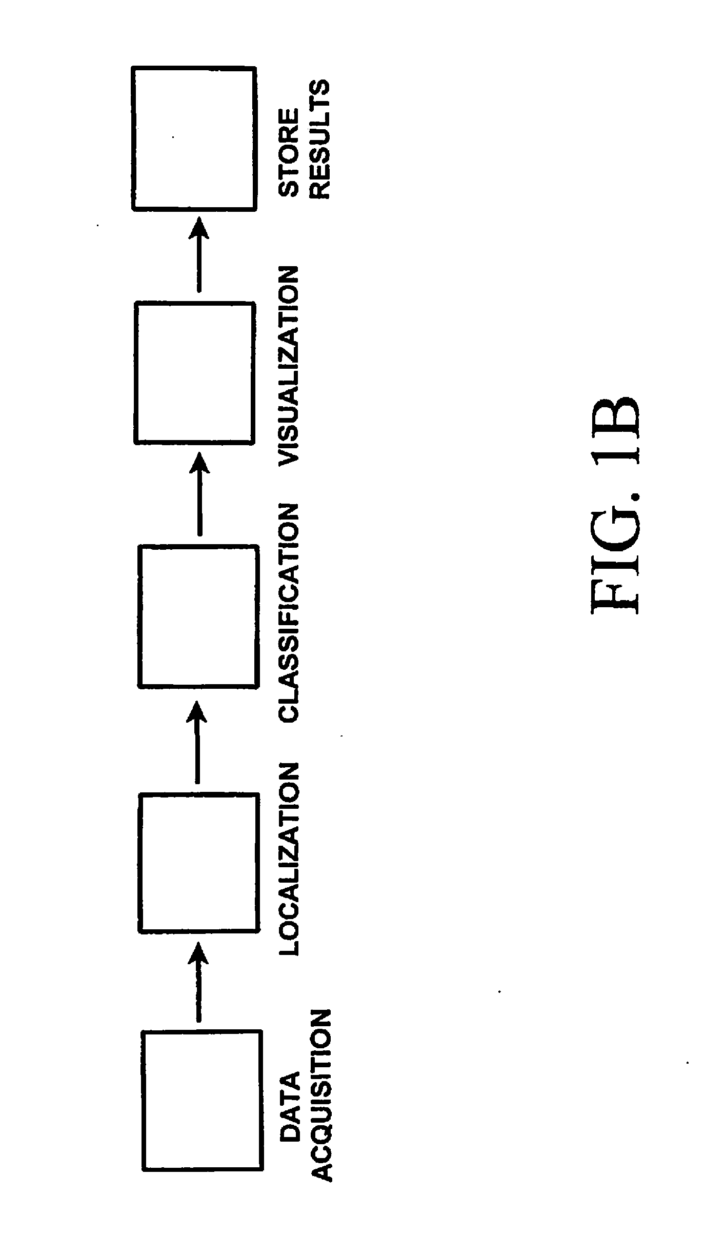
Vascular conditions are detected non-invasively in the human body using a collection of information from small local regions of the vasculature, or from a specific signature or “BrainPulse” that can be derived from a patient's heartbeat-induced cranium movements. An array of accelerometers or other sensors are engaged against the head of a patient and skull movements, preferably under 100 Hz, are recorded. Vibration signatures of vessel structures such as branches, aneurysms, stenosis, etc. using random, periodic, band limited or transient analysis provides a library for further processing. The signature library is used to localize the origin of the recognized vascular feature, and the localized feature is presented to the physician in a clinically relevant manner.

Description

[0001]This application is a continuation-in-part of application Ser. No. 11 / 894,052, filed Aug. 17, 2007, now ______, which was a regular filing from provisional application No. 60 / 838,624, filed Aug. 17, 2006.BACKGROUND OF THE INVENTION[0002]The invention concerns detection of conditions of human vasculature or non-vascular brain tissue non-invasively, from outside the body, and in particular, locating aneurysms, vasospasm, partial or complete stenoses, ruptures, peripheral bleeding and other abnormal conditions pertaining to the cerebral vasculature and other cerebral tissue, profiling blood or other fluid flow through or around the cerebral vasculature and detecting and diagnosing abnormal cerebral conditions. The invention also encompasses mapping based on gathered data to provide a spatially resolved characterization of the major vasculature within the head. The system and method of the invention are also useful to detect vascular conditions in other parts of the body, especial...

Claims


Application Information

Patent Timeline
IPC IPC(8): A61B5/00A61B5/0205A61B5/11
CPCA61B5/4064A61B5/0205A61B5/024A61B5/7257A61B5/7282A61B5/11A61B5/7267A61B7/04A61B5/02007A61B5/4076A61B5/6814A61B2562/0219A61B5/7246G16H50/50G16H50/70G16H50/20G16H30/20G16H70/60G06F2218/16G16H70/00
Inventor LOVOI, PAUL, A.SCHUMACHER, RAYMACVEAN, CHARLIE
Owner JAN MEDICAL
Learn More