Microfluidic System and Method for Real-Time Measurement of Antibody-Antigen Binding and Analyte Detection

a microfluidic system and antibody technology, applied in fluorescence/phosphorescence, laboratory glassware, instruments, etc., can solve the problems of inability to monitor the continuous analyte immunoassay and pharmacokinetic characterization of biomolecules in real-time, laborious and time-intensive procedures, and methods that are impractical for real-time monitoring

Inactive Publication Date: 2017-03-16
THE GENERAL HOSPITAL CORP +1
View PDF3 Cites 30 Cited by
  • Summary
  • Abstract
  • Description
  • Claims
  • Application Information

AI Technical Summary

Benefits of technology

[0029]Another aspect of the invention is a method of determining the binding affinity of an antibody for an antigen, the method including: providing a microfluidic device for continuous flow optical detection of an analyte in a sample, a liquid suspension of microspheres that are conjugated to the antigen, a liquid comprising the antibody, and a liquid comprising an antibody binding agent conjugated to a label; flowing the liquid comprising the antibody into the first inlet at a first flow rate; flowing the liquid comprising the labeled antibody binding agent into the second inlet at a second flow rate, whereby mixing of the antibody and the labeled antibody binding agent in the first mixing channel enables binding of the antibody to the labeled antibody binding agent in a diffusion-independent manner, resulting in formation labeled antibody complexes; flowing the liquid suspension of conjugated microspheres into the third inlet at a third flow rate, whereby mixing of the conjugated microspheres and the labeled antibody complexes in the second mixing channel enables binding of the antigen to the antibody in a diffusion-independent manner, resulting in formation of labeled antibody-coated microspheres; detecting an amount of microsphere-bound label at one or more different points in the second mixing channel; and determining the binding affinity of the antibody for the antigen by comparing the amount of microsphere-bound label at one or more different points in the second mixing channel.

Problems solved by technology

A major challenge in the detection of soluble molecules such as cytokines, protein antigens and antibodies is the ability to monitor time-varying or dynamic concentrations in real-time.
Currently there are no available online monitoring approaches for continuous analyte immunoassays and pharmacokinetic characterization of biomolecules in real-time.
These diagnostic methods are performed on samples obtained at pre-defined times and are therefore laborious and time-intensive procedures.
Additionally, these methods are impractical for real-time monitoring since they cannot be performed rapidly enough to assess dynamic fluctuation of analyte concentration in vivo.
This limits their utility in clinical settings where it is of critical importance to generate real-time profile of analytes such as cytokines or administered drugs in vivo (Crowther, 2001; Mannerstedt et al., 2010; Mao et al., 2009; Wild, 2001).
Nevertheless, most of these methods still require incubation and are unable to measure the dynamic changes in the analyte concentration in real time (Hu and Gao, 2007; Singhal et al., 2010).
Although diffusion distances in microchannels are significantly reduced in comparison to conventional microtiter well plate formats, analytes are still transport-limited in micro channels at low sample concentrations (Parsa et al., 2008).

Method used


Image

  • Microfluidic System and Method for Real-Time Measurement of Antibody-Antigen Binding and Analyte Detection
  • Microfluidic System and Method for Real-Time Measurement of Antibody-Antigen Binding and Analyte Detection
  • Microfluidic System and Method for Real-Time Measurement of Antibody-Antigen Binding and Analyte Detection

Examples


example 1

Materials and Methods

[0138]Microfluidic Device Fabrication.

[0139]The polydimethylsiloxane (PDMS) microfluidic device was fabricated using well-established soft lithography method. Negative photo resist SU-8 2100 (MicroChem, Newton, Mass.) was spin-coated on Silicon wafers to a thickness of 150 μm, and patterned by exposure to UV light through a transparency photomask (CAD / Art Services, USA). PDMS (Sylgard 184, Dow Corning, MI) was mixed with the crosslinker (Sylgard 184 curing agent) in a ratio of 10:1, poured onto the photoresist patterns, degassed thoroughly and cured for 12 hours at 65° C. Next, the PDMS was peeled off the wafer and placed in oxygen-plasma chamber in order to bond with the glass slide. The device consisted of three inlets and two mixing channels. Tygon Micro Bore PVC Tubing 0.010″ ID, 0.030″ OD, 0.010″ Wall (Small Parts Inc., FL, USA) was connected to the channels and to 1 mL syringes. Syringe pumps (Harvard Apparatus, USA) were used to maintain a flow rate of 5 ...

example 2

Flow Dynamics in a Microfluidic Device

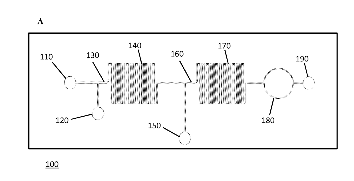
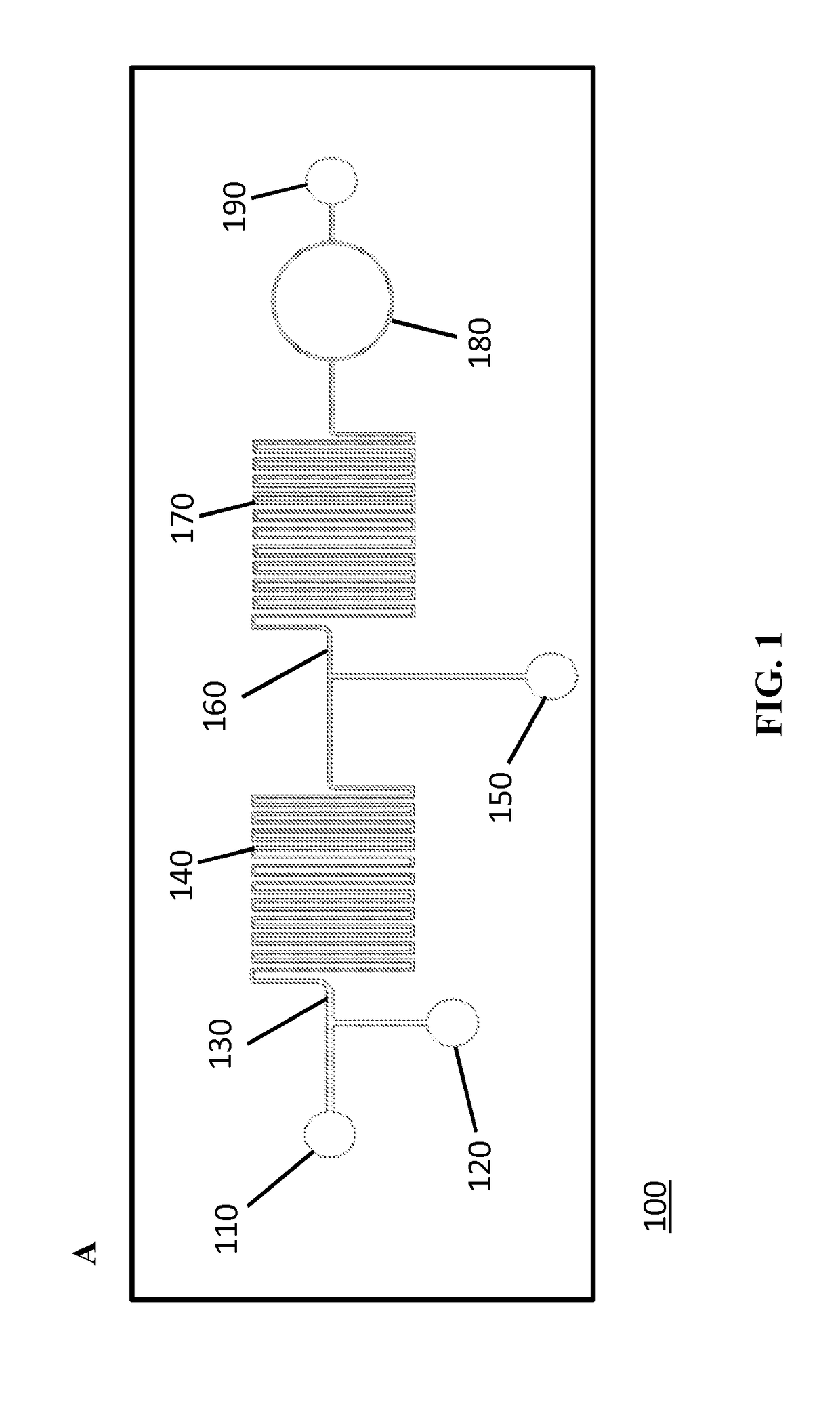
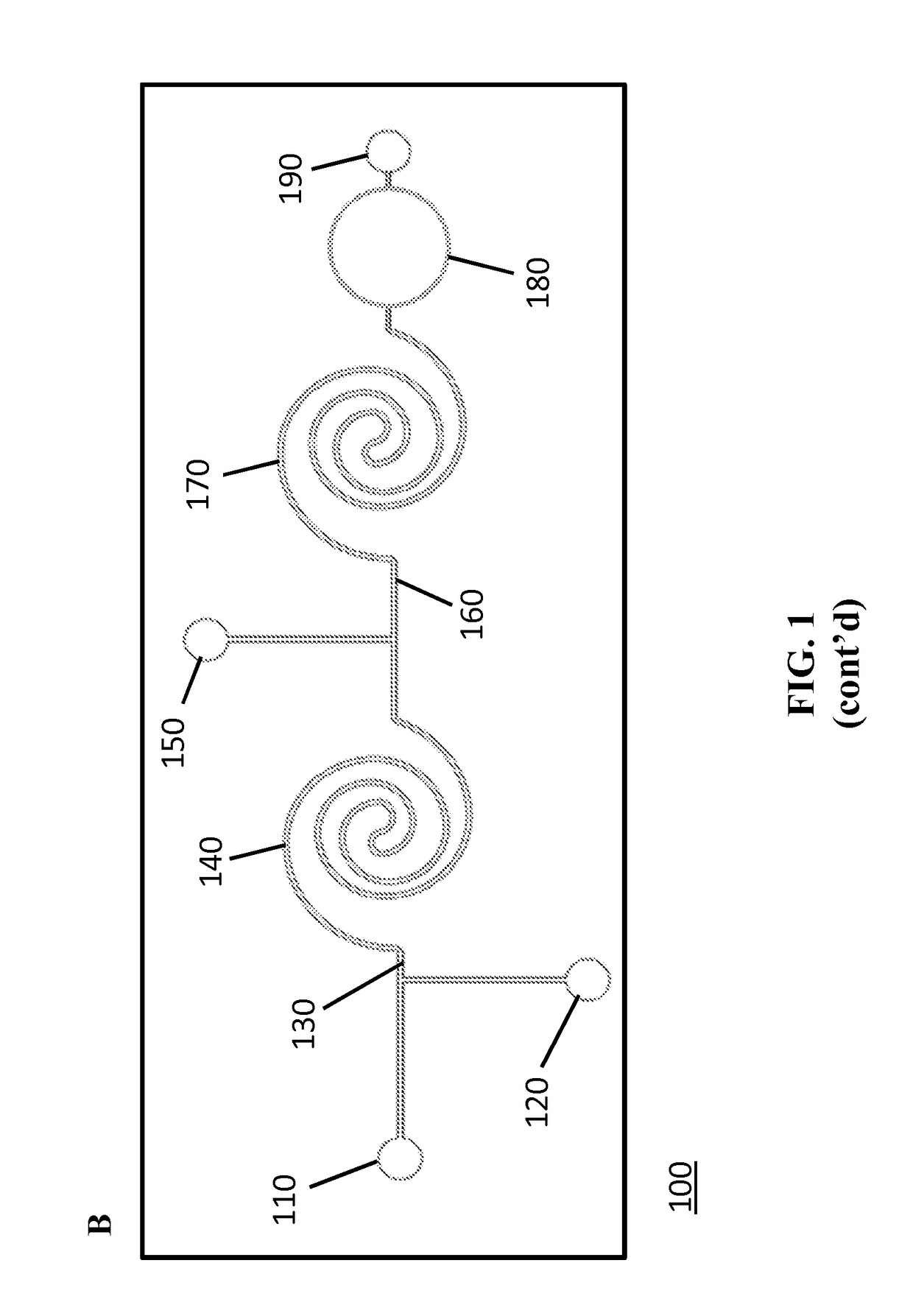
[0185]FIGS. 1A-1C schematically illustrates the developed flow-through LOC device. The device consisted of three inlets and two mixing regions. The inlets were connected with syringe pumps that were operated individually to obtain desired flow rates for the detection of microspheres downstream. First, a solution containing the analyte molecules was introduced into inlet 1 (110) and mixed with a suspension of functionalized microspheres that were introduced via inlet 2 (120) to capture the target analyte in the first mixing channel (140) (FIGS. 1A-1C). The specific interaction that occurs between the conjugated microspheres and the target analyte in the first mixing channel leads to the analyte recognition and capture. A detection (reporter) antibody against the analyte, conjugated with a specific fluorophore, was then introduced to the flow stream via inlet 3 (150) just before the second mixing channel (170). The sandwich complex formation, comp...

example 3

Anti-TNF-α Antibody Immunoassay

[0187]To demonstrate the real-time detection capabilities of the LOC device, efforts were focused on detecting anti-TNF-α antibody. FIG. 12A describes the microsphere-based assay that was introduced into microfluidic format for anti-TNF-α detection. Avidinilated microspheres were conjugated off-chip to biotinylated human TNF-α protein via avidin-biotin bridge (Konry et al., 2009; Diamdandis et al. 1991) as described in Example 1. Next, the generated anti-TNF-α microsphere-based sensors were introduced into the microfluidic device via inlet 2 while the analyte, mouse monoclonal anti-human TNF-α antibody, was introduced via inlet 1. The interaction of the two components resulted in the capture of anti-TNF-α antibodies by microsphere-based sensors in the first mixing channel of the device. Next, the detection antibody, FITC-labeled anti-mouse IgG, was introduced into inlet 3. The fluorescent signal on the microsphere sensor, generated by the conjugation o...


PUM

PropertyMeasurementUnit
areaaaaaaaaaaa
cross-sectional areasaaaaaaaaaa
fluidic path lengthaaaaaaaaaa

Abstract

Microfluidic devices for use with reagents bound to microspheres for determination of the concentration of an analyte in a liquid sample are provided. The devices include two sequential mixing channels that promote rapid binding of microsphere-bound reagents with reagents in solution and a means for detecting labeled microsphere-bound reaction products. Also provided are methods for using the devices with microsphere-bound reagents to determine the concentration of an analyte in a liquid sample and to measure the binding affinity of antibody for an antigen.

Description

BACKGROUND[0001]Rapid, sensitive and quantitative detection methods of disease markers are necessary for timely and effective diagnosis and therapy (Martinez et al., 2008). A major challenge in the detection of soluble molecules such as cytokines, protein antigens and antibodies is the ability to monitor time-varying or dynamic concentrations in real-time. Currently there are no available online monitoring approaches for continuous analyte immunoassays and pharmacokinetic characterization of biomolecules in real-time. At present, state-of-the-art analyte detection techniques for biomolecules include immunoassays such as enzyme-linked immunosorbent assays (ELISA), which are based on specific recognition of clinical antigens by the respective antibodies (Reichert, 2001). These diagnostic methods are performed on samples obtained at pre-defined times and are therefore laborious and time-intensive procedures. Additionally, these methods are impractical for real-time monitoring since the...

Claims


Application Information

Patent Timeline
Patent Type & Authority Applications(United States)
IPC IPC(8): G01N33/543B01F5/06B01F13/00B01L3/00G01N21/64
CPCG01N33/54313B01L3/502715B01L3/502776B01L3/502761G01N33/54366G01N21/6428G01N2201/061B01F5/0647B01L2300/0654B01L2200/0605B01L2200/0647B01L2300/0883G01N2021/6439B01F13/0059B01L3/5027B01L2300/0816B01L2300/0867B01L2300/087B01L2400/0487G01N33/5304B01F25/4331B01F33/30
Inventor KONRY, TANIAYARMUSH, MARTIN L.
Owner THE GENERAL HOSPITAL CORP
Why Eureka
Try Eureka