
Hand-held optical scanner for real-time imaging of body composition and metabolism

a real-time imaging and optical scanner technology, applied in the field of optical scanners and computational models for scanning and imaging of human body composition, can solve the problems of inability to separate scattering from absorption in a single measurement, and inability to accurately calculate the concentration of absorbers in tissue, etc., to achieve rapid information about tissue structure and composition, and low cost

Active Publication Date: 2017-07-27
RGT UNIV OF CALIFORNIA
View PDF2 Cites 19 Cited by
  • Summary
  • Abstract
  • Description
  • Claims
  • Application Information

AI Technical Summary

Benefits of technology

The patent describes a new system that can measure the health of tissues in the body. The system can scan the body or be placed in specific locations. It can measure pulsations in abdomen, muscles, and brain tissues. The system is very fast and can provide high-resolution data. It can also be used with other methods like frequency-domain and time-domain methods, pressure sensing devices, and other imaging devices. Overall, this system can help with the diagnosis and treatment of various medical conditions.

Problems solved by technology

This technique provides fast measurements and simple circuit designs, but is unable to separate scattering from absorption in a single measurement.
Moreover, these techniques assume constant scattering and neglect possible changes in scattering occurring during a continuous measurement.
This assumption can introduce significant errors when accurately calculating absorber concentrations in the tissue.
Despite its ability to obtain both scattering and absorption information, time domain imaging has a few limitations that prevent the translation of this technology to a portable real-time clinical friendly system.
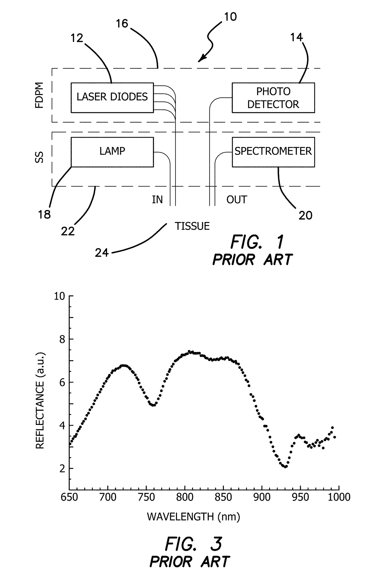
TD's optoelectronic high cost and complex circuitry reduces spectral bandwidth; thereby in applications such as breast cancer, information about water and fat content are inaccessible.
Achieving this goal, covering a large spectral bandwidth, using time-resolved techniques requires tunable sources or a large collection of laser diodes resulting in a bulky slow expensive system with complex maintenance requires tunable sources or a large collection of laser diodes resulting in a bulky slow expensive system with complex maintenance.
Although this platform is powerful and has rich information content, however it has a few limitations such as speed, cost and size.

Method used


Image

  • Hand-held optical scanner for real-time imaging of body composition and metabolism
  • Hand-held optical scanner for real-time imaging of body composition and metabolism
  • Hand-held optical scanner for real-time imaging of body composition and metabolism

Examples


first embodiment

[0104]In a first embodiment, the apparatus 10 is used to extract the heart rate from the fingertip, which is also a common target for pulse-oximetry instruments. The raw data from the left index finger, where two laser diodes (780 nm and 820 nm) were used in FDPM module 16 and / or CW module 18 and data recorded at sample rate of 250 Hz is shown in FIG. 13. The wavelengths of the laser diodes were selected below and above the isosbestic point (810 nm) where both deoxygenated hemoglobin and oxygenated hemoglobin have the same absorption coefficients. As our control, we used a commercial system that was placed on the index finger of the right hand. We calculated the heart rate from raw optical and oxygenated hemoglobin concentrations signals with two different approaches. First, we ran a peak searching algorithm to find the corresponding peak in the photoplethysmogram (PPG) signal and divide the number of peaks by measurement duration to obtain an average per second, and then multiplied...

second embodiment

[0123]In the second embodiment, we used our system to recover a respiration rate which is another important vital signal in clinical settings. Subjects were instructed to synchronize their breath to a 0.25 Hz signal using a video clip. Optical data (four wavelengths) was recorded from wrist and triceps muscle tissue with 2 cm source-detector separation at 80 Hz rate. Combination of EMD-FFT was again utilized to extract desired information. We were able to characterize the vasculature response to the stimuli by extract the induced frequency from both time domain and frequency domain signals of oxygenated and deoxygenated hemoglobin concentrations. In the wrist tissue, time domain result (EMD) showed four peaks during sixteen seconds which corresponds to a 0.25 Hz signal in frequency domain. The respiration rate recovered from oxy-Hb by FFT method was 0.235 Hz while the deoxy-Hb showed a 0.230 Hz peak (2.2% difference). In the arm tissue, measurement duration was increased from sixtee...

third embodiment

[0124]In a third embodiment, we used the continuous-wave system for characterizing muscle vasculature reactivity to changes in blood flow. Cardiovascular disease impairs the vessels' ability to change their diameter and architecture in response to stimuli. Cuff occlusion is a common method for assessing vasculature reactivity and changing blood flow. We chose the left arm's brachial artery for the occlusion site and forearm muscle for optical monitoring. It has been established to analyze the rate of tissue ischemia and recovery to assess vascular reactivity. In addition to this parameter, we also took advantage of system high speed data acquisition to look at dynamic changes in oxygenated hemoglobin and recover heart rate during the measurements. We compared our pulse rate results to ones from commercial pulse oximeter system since we used it to monitor the index finger. They are in agreement with less than 8% differences. This shows the system ability to monitor hemodynamics chang...


PUM


Abstract

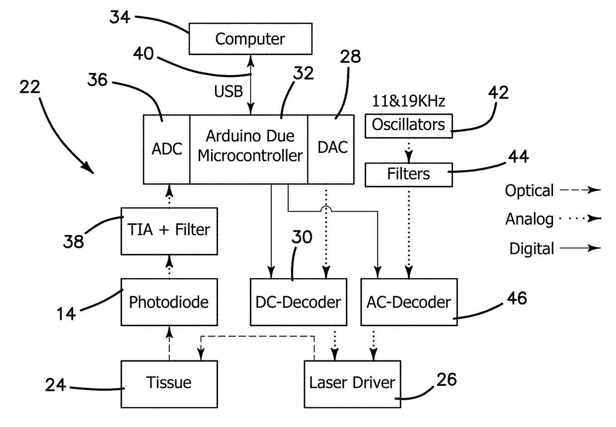
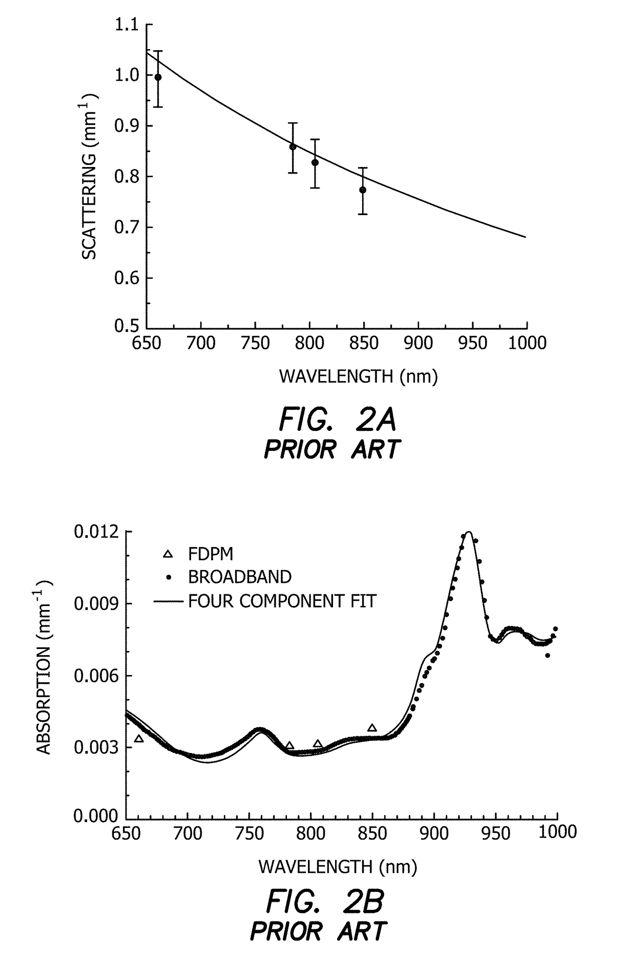
A low cost portable high speed quantitative system for diffuse optical spectroscopic imaging of human tissue. The hybrid system (CWFD) can measure absolute optical properties from 660 nm to 980 nm and recover all tissue chromophore concentrations. The standalone FD module can be utilized to measure scattering at every measurement and recover deoxygenated and oxygenated hemoglobin concentrations. The CW module can operate concurrently with the FD module to also measure water and lipid. The high temporal resolution and large signal-to-noise ratio of the CWFD system may be used to explore tissue oximetry, vascular occlusion, and paced breathing models to measure and analyze tissue hemodynamics response to changes in blood flow. Continuous monitoring of vasculature response to various modified blood perfusion conditions can provide information about local tissue metabolism and physiological state (dysfunction).

Description

RELATED APPLICATIONS[0001]This application is related to provisional patent application, entitled, A HAND-HELD OPTICAL SCANNER FOR REAL-TIME IMAGING OF BODY COMPOSITION AND METABOLISM, Ser. No. 62 / 287,803, filed on Jan. 27, 2016, under 35 USC 119, which is incorporated herein by reference.GOVERNMENT SUPPORT[0002]This invention was made with government support under P41EB015890, R01CA142989, funded by National Institute of Health (NIH). The government has certain rights in the invention.BACKGROUND[0003]Field of the Technology[0004]The invention relates to the field of medical devices and methods, namelly an optical instrument and a computational model for scanning and imaging of human body composition including tissue water, lipid, oxygenated hemoglobin and deoxygenated hemoglobin content.[0005]Description of the Prior Art[0006]Diffuse optical spectroscopic imaging (DOSI) methods provide a low-cost, non-invasive approach for obtaining critical information regarding the structure and ...

Claims


Application Information

Patent Timeline
Patent Type & Authority Applications(United States)
IPC IPC(8): A61B5/1455A61B5/00A61B5/08A61B5/145A61B5/024
CPCA61B5/14551A61B5/14546A61B5/7278A61B5/0816A61B5/02416
Inventor ZARANDI, SOROUSH MOHAMMAD MIRZAEITROMBERG, BRUCE J.O'SULLIVAN, THOMAS D.YAZDI, SIAVASH SEDIGHZADEHCERUSSI, ALBERT
Owner RGT UNIV OF CALIFORNIA
Learn More