Artificial blood vessel and preparation method thereof

a technology of artificial blood vessels and preparation methods, which is applied in the direction of blood vessels, tubular organ implants, prostheses, etc., can solve the problems of inability to meet the requirements of transplantation and replacement of artificial blood vessels in aspects such as anticoagulant activity, suture resistance and growth repair capability, and the limited source of donors

Active Publication Date: 2017-11-16
LIU CHANG
View PDF15 Cites 9 Cited by
  • Summary
  • Abstract
  • Description
  • Claims
  • Application Information

AI Technical Summary

Benefits of technology

[0016]c. removing the second hollow cylindrical mold on the basis of the step 2); sleeving a third hollow cylindrical mold with a larger diameter outside the endothelial cell layer, wherein a gap is reserved in the middle; infusing a natural polymer solution containing smooth muscle cells or mixtures of the stem cells and the smooth muscle cells or mixtures of the stem cells and smooth muscle cell growth factors into the gap between the endothelial cell layer and the third hollow cylindrical mold by adopting the method in the step 2), and enabling the natural polymers in the solution to crosslink or polymerize by utilizing the cross-linking agent or the polymerizing agent, to form a smooth muscle cell layer;
[0017]d. removing the third hollow cylindrical mold on the basis of the step 3); sleeving a fourth hollow cylindrical mold with a larger diameter outside the smooth muscle cell layer, wherein a gap is reserved in the middle; infusing a natural polymer solution containing fibroblasts or mixtures of the stem cells and the fibroblasts or mixtures of the stem cells and fibroblast growth factors into the gap between the smooth muscle cell layer and the fourth hollow cylindrical mold by adopting the method in the step 2) or 3), and enabling the natural polymers in the solution to crosslink or polymerize by utilizing the cross-linking agent or the polymerizing agent, to form a fibroblast layer;
[0025]Compared with the prior art, the present invention has the following advantages and outstanding effects that: (1) accurate spatial positioning of different cells and polymer materials is realized by utilizing integrated technologies such as intra-mold pouring, plasma spraying, electrospraying, electrospining or 3D printing and the like; structures and functions of human blood vessels can be simulated; and construction of the artificial blood vessel on a three-dimensional scale is realized; (2) cell and tissue structures in the artificial blood vessel are trained or cultured by a controllable three-dimensional stress field; the endothelial cells, the smooth muscle cells and the fibroblasts are arranged according to a certain direction to form different tissue areas; mechanical strength of the artificial blood vessel is improved, so that the artificial blood vessel has excellent anticoagulation, suture-resistant and regenerative functions while meeting demands of cell proliferation growth and metabolism; a change from the artificial blood vessel to the natural blood vessel is completed; and a purpose of repairing and regenerating human artery and vein vessels is achieved; and (3) the artificial blood vessel and the preparation method thereof are also applicable to other tubular tissues and organs, such as esophagus, trachea, fallopian tube, bile duct, urethra and bladder.

Problems solved by technology

Accompanying problems are preparation processes of artificial blood vessels and selection of the grafts.
At present, clinical vasotransplantation is mainly in an autotransplantation manner or an allograft manner, and a donor source is greatly limited.
Some blood vessel analogues can be prepared by utilizing tissue engineering or 3D printing technologies, but such products have severe defects in performance [Nerem R M, et al.
The products above reflect some characteristics of natural blood vessels from different points of view, but cannot meet transplantation and replacement requirements of the artificial blood vessel in aspects such as anticoagulant activity, suture resistance and growth repair capability.
The existing engineering technologies, such as intra-mold pouring and three-dimensional printing technologies, cannot be used for preparing implantable artificial blood vessel repair materials which can be directly connected with human artery and vein vessels.

Method used


Image

  • Artificial blood vessel and preparation method thereof
  • Artificial blood vessel and preparation method thereof
  • Artificial blood vessel and preparation method thereof

Examples


embodiment 1

Small-Diameter Blood Vessel and Preparation Method Thereof

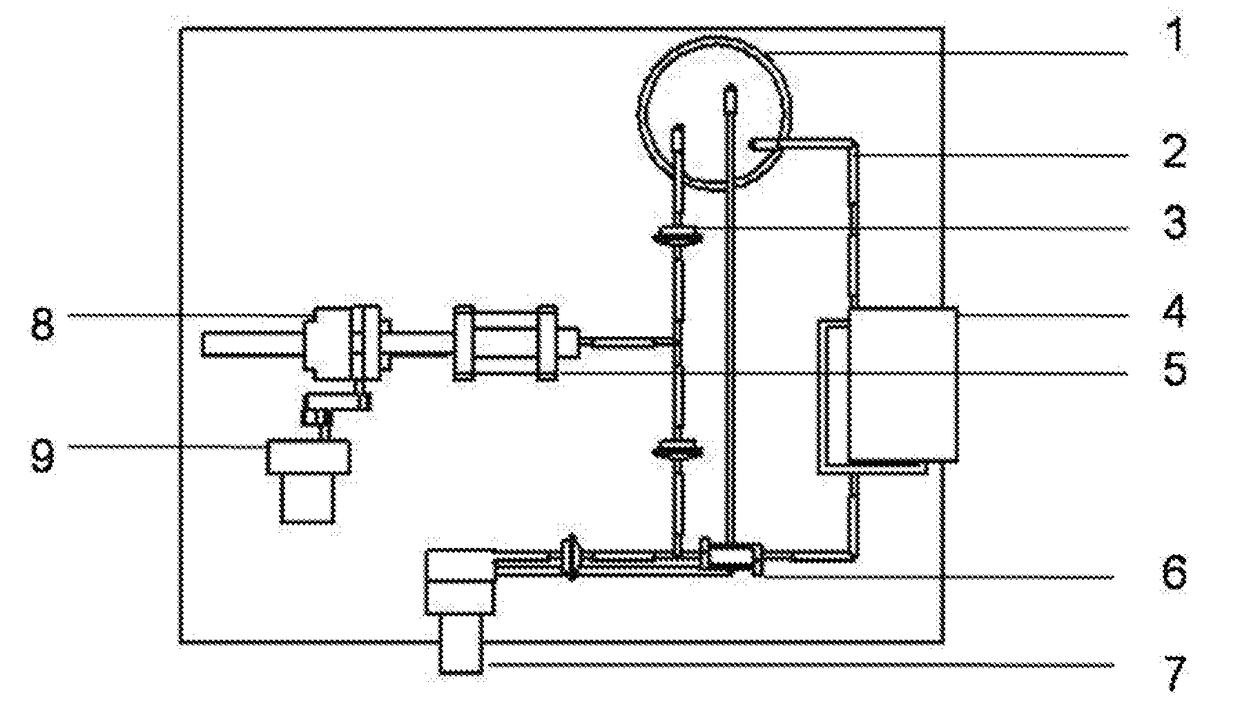
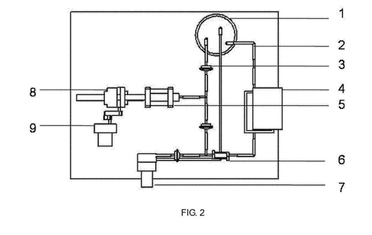
[0047]The preparation method comprises the following steps:[0048]a. respectively preparing a sodium alga acid solution with a mass percent of 0.1% and a gelatin solution with a mass percent of 20%; uniformly mixing the sodium alga acid solution and the gelatin solution according to a volume ratio of 1:1, wherein 1% of an anticoagulation factor heparin and 10% of a cell cryopreservation factor dimethyl sulfoxide are added; and respectively mixing endothelial cells, smooth muscle cells and fibroblasts with the mixed solution, wherein a density of the cells in the sodium alga acid and gelatin mixed solution is 1×10² cells per mL;[0049]b. loading various cell-containing solutions in the step a into a nozzle assembly of 3D printing equipment; printing a hollow cylinder of which an inner cavity has a diameter of 0.1 mm and which sequentially comprises an endothelial cell layer, a smooth muscle cell layer and a fibroblast layer from...

embodiment 2

Large-Diameter Blood Vessel and Preparation Method Thereof

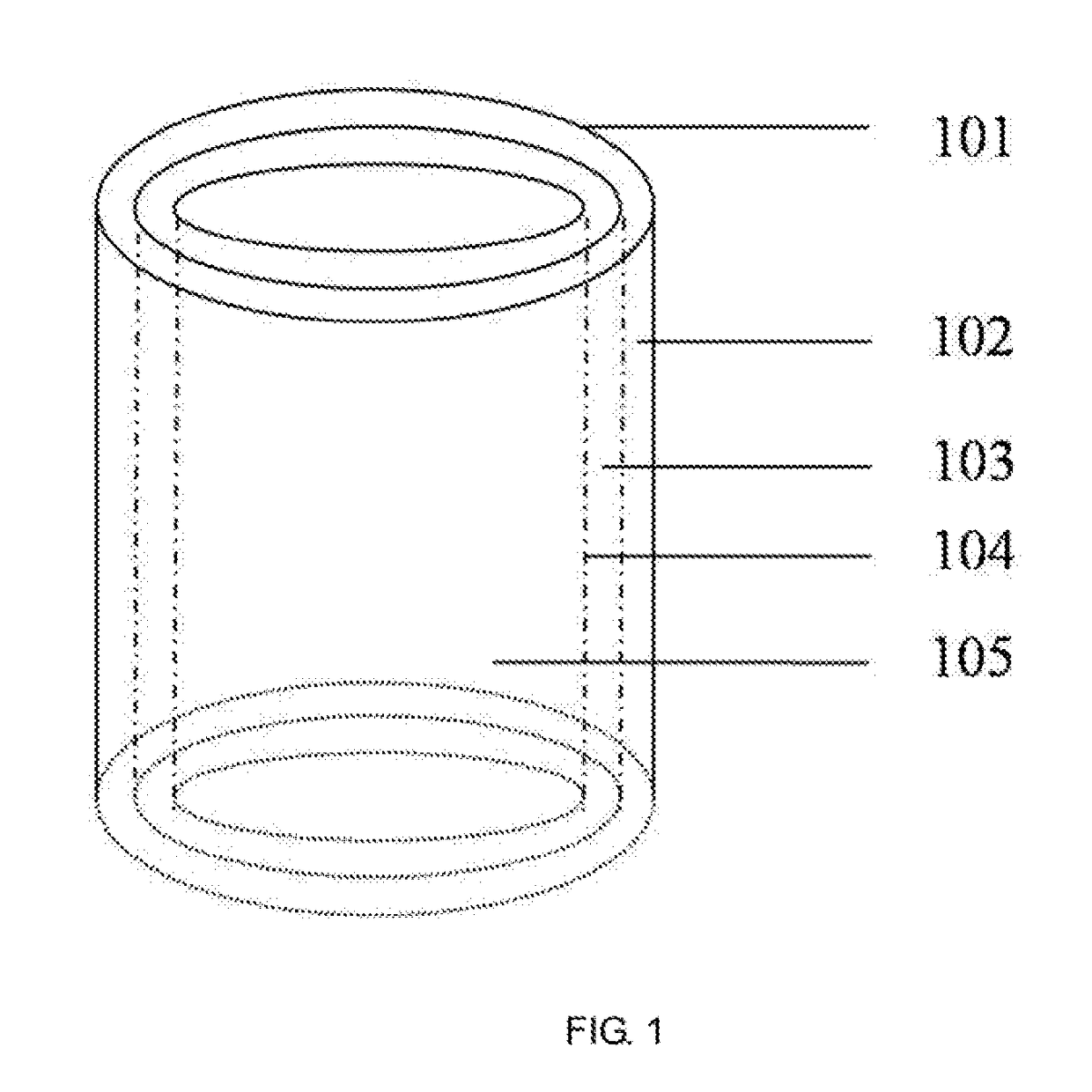
[0052]The preparation method comprises the following steps:[0053]a. preparing a series of hollow cylindrical molds with diameters more than 6 mm; respectively preparing fibrinogen solutions containing bone marrow stem cells, endothelial cells and fibroblasts, wherein the fibrinogen solution has a mass percent of 5% and density of the cells in the fibrinogen solution is 1×10⁷ cells per mL; and respectively adding a smooth muscle cell growth factor with a mass percent of 1%, 1% of anticoagulation factor heparin and 10% of cell cryopreservation factor dimethyl sulfoxide into the bone marrow stem cell solution;[0054]b. sleeving a second hollow cylindrical mold with a larger diameter outside a first hollow cylindrical mold with a smaller diameter, wherein a gap is reserved between the two molds; injecting the fibrinogen solution containing the endothelial cells and heparin into the gap between the two molds by using a dropper; and...

embodiment 3

Medium-Diameter Blood Vessel and Preparation Method Thereof

[0059]The preparation method comprises the following steps:[0060]a. preparing a series of hollow cylindrical molds with diameters of 2-6 mm; preparing collagen solutions containing adipose-derived stem cells, wherein the collagen solutions have a mass percent of 1% and a density of the adipose-derived stem cells in the collagen solution is 1×10⁵ cells per mL; and respectively adding endothelial cell growth factors, smooth muscle cell growth factors and fibroblast growth factors with a mass percent of 0.01%, 0.01% of anticoagulation factor taxol and 1% of cell cryopreservation factor glycerin into the three solutions containing the adipose-derived stem cells;[0061]b. sleeving a second hollow cylindrical mold with a larger diameter outside a first hollow cylindrical mold with a smaller diameter, wherein a gap is reserved between the two molds; injecting the collagen solution containing the adipose-derived stem cells, the endot...


PUM

PropertyMeasurementUnit
mass percentaaaaaaaaaa
thicknessaaaaaaaaaa
mass percentaaaaaaaaaa

Abstract

The artificial blood vessel comprises a cortex layer, a fibroblast layer, a smooth muscle cell layer, an endothelial cell layer and an inner cavity. According to the artificial blood vessel, the endothelial layer, the smooth muscle cell layer, the fibroblast layer and the cortex layer are orderly arranged in a three-dimensional space by utilizing integrated technologies of plasma spraying, electrospraying, electrospining, intra-mold pouring and 3D printing; anticoagulant activity of the artificial blood vessel is enhanced by adopting an anticoagulation factor; step-by-step induced differentiation of stem cells in the artificial blood vessel is realized by adopting a growth factor controlled release method; and the artificial blood vessel is cultured by a pulsatile reactor, so that the artificial blood vessel structurally and functionally simulates natural animal blood vessels and provides a corresponding substitute for vascular transplantation and repair.

Description

CROSS-REFERENCE TO RELATED APPLICATIONS[0001]This application is a continuation of International Patent Application No. PCT / CN2015 / 079792 with a filing date of May. 26, 2015, designating the United States, now pending, and further claims priority to Chinese Patent Application No. 201510097472.8 with a filing date of Mar. 5, 2015. The content of the aforementioned applications, including any intervening amendments thereto, are incorporated herein by reference.TECHNICAL FIELD[0002]The present invention relates to an artificial blood vessel and a preparation method thereof, and belongs to the technical fields of organ manufacture and biological materials.BACKGROUND OF THE PRESENT INVENTION[0003]According to statistics of the World Health Organization, a cardiovascular disease is one of the diseases with the highest global incidence and death rate. Roughly more than 17 million people die from a heart disease in the world every year. The cardiovascular disease has become the biggest caus...

Claims


Application Information

Patent Timeline
Patent Type & Authority Applications(United States)
IPC IPC(8): A61F2/06A61L27/16A61L27/22A61L27/44A61L27/36
CPCA61F2/062A61L27/3604A61L27/16A61L27/222A61L27/44A61F2240/001A61F2/06A61F2210/0076A61L27/18A61L27/20A61L27/225A61L27/24A61L27/3804A61L27/3808A61L27/3826A61L27/3891A61L27/507C08L5/08C08L5/04C08L23/06C08L23/12C08L67/04C08L69/00C08L75/04
Inventor LIU, CHANG
Owner LIU CHANG
Why Eureka
Try Eureka