

Biomarkers Useful in the Treatment of Subjects Having Disease of the Eye
a biomarker and eye disease technology, applied in the field of new methods for detecting oxidative stress in body fluids, can solve the problems of severe oxidative stress in the outer retina, reduced cellular function and eventually apoptosis, and impaired function, and achieves the effects of slowing or stopping the progression of eye disease, increasing or decreasing oxidative stress, and positive correlation between the level of an oxidative damage marker
- Summary
- Abstract
- Description
- Claims
- Application Information
AI Technical Summary
Benefits of technology
Problems solved by technology
Method used
Image

Examples
examples
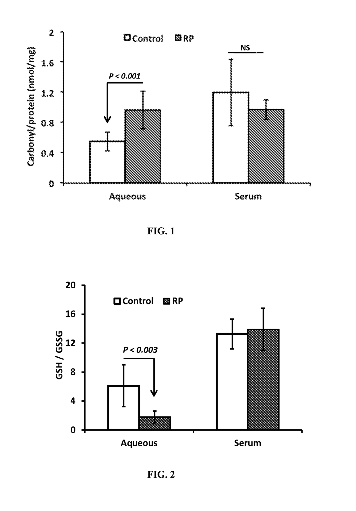
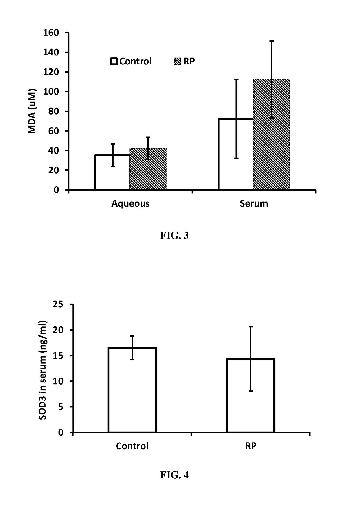
[0073]Subjects. The study was registered on clinicaltrials.gov (NCT01949623), the protocol was approved by the Johns Hopkins University institutional review board, and all participating patients provided informed consent. The diagnosis of RP was made by a retina specialist with expertise in inherited retinal degenerations (HPS) based upon eye examination, electroretinography, visual field testing, and optical coherence tomography. Nine patients with RP were included in this study. An aqueous sample was obtained by administering topical anesthesia and a drop of 5% povidone iodine into the study eye, placing a lid speculum, inserting a 30-gauge needle on a 1 ml syringe into the anterior chamber at the limbus, and gently aspirating aqueous. Samples were frozen and stored at −80° C. until assayed. A blood sample was also obtained and after clotting, the blood was centrifuged. Serum was placed in a small tube and stored at −80° C. until assayed. Eleven control patients who were undergoin...
PUM
| Property | Measurement | Unit |
|---|---|---|
| oxidative stress | aaaaa | aaaaa |
| antioxidant capacity | aaaaa | aaaaa |
| fluorescence polarization | aaaaa | aaaaa |
Abstract
Description
Claims
Application Information