Probes and a methylation in situ hybridization assay

a technology of methylation and in situ hybridization, applied in the field of molecular pathology, can solve the problems of reducing sensitivity, unable to quantify the target at physiological levels, and only 10% techniqu

Inactive Publication Date: 2018-07-05
UNIV GENT
View PDF0 Cites 0 Cited by
  • Summary
  • Abstract
  • Description
  • Claims
  • Application Information

AI Technical Summary

Benefits of technology

[0031]The disclosure also relates to the usage of a probe as defined above to specifically detect small target sequences.

Problems solved by technology

This method combines PCR amplification for sufficient signal amplification and enzymatic restriction to allow probe access; and does thus not allow quantification of the target at physiological levels.
The sensitivity of this technique is only 10% since the enzymatic restriction step that exposes the sense or anti-sense strand is not absolute and is difficult to regulate; further factors contributing to the reduced sensitivity is the low PCR efficiency and DNA loss.
Overall drawbacks of the above-mentioned alternative methods and probes are their complex and expensive protocols, low sensitivity, low quantification possibilities, various signal amplification steps after probe hybridization, and extensive washing steps, resulting in target lost and a high background staining, which hamper their use for routine application.
Thus, this test does not allow detection of the target at physiological levels.
Moreover, because the target is amplified and the amplicons will crowd the nuclei, co-localization of multiple targets (for example, an unmethylated target and a methylated target) will be very difficult to interpret.
However, the efficiency of the probe hybridization is only 10% because enzymatic restriction that should expose the sense or anti-sense strand is not absolute and it is difficult to regulate.
This method thus combines PCR amplification for sufficient signal amplification and enzymatic restriction to allow probe access and, thus, does not allow quantification of the target at physiological levels.
Probe recognition depends on cross-linking of a bipyridine-adenine derivative at the position corresponding to the methylated cytosine in the presence of osmium; therefore, the described method does not allow detection of unmethylated sequences, and so hypomethylation cannot be observed.
The described method can only be used for detection of highly abundant repeats, because these small probes cannot compete with re-hybridization of the complementary strands and will not generate enough signals for microscopic evaluation of single copy genes.

Method used


Image

  • Probes and a methylation in situ hybridization assay
  • Probes and a methylation in situ hybridization assay
  • Probes and a methylation in situ hybridization assay

Examples


example 1

Synthesis of the UniProbe Signal Amplification System (UPSAS) Probe

[0149]Four different UPSAS probes corresponding to the Glutathione S-Transferase Pi 1 (GSTP1), hypermethylated regions in prostate cancer were designed and synthesized by PCR.

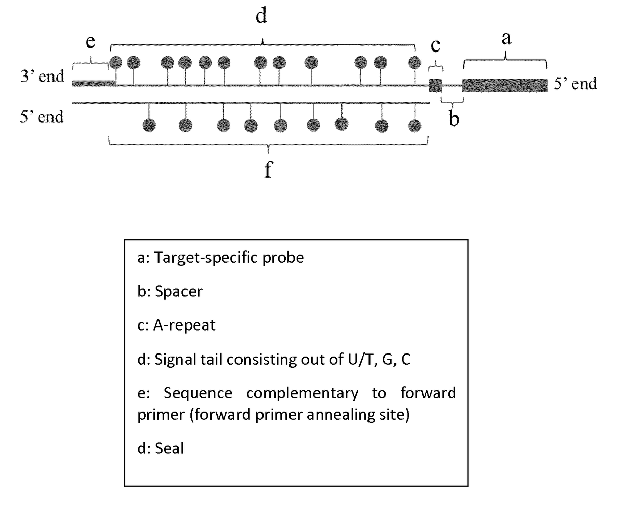
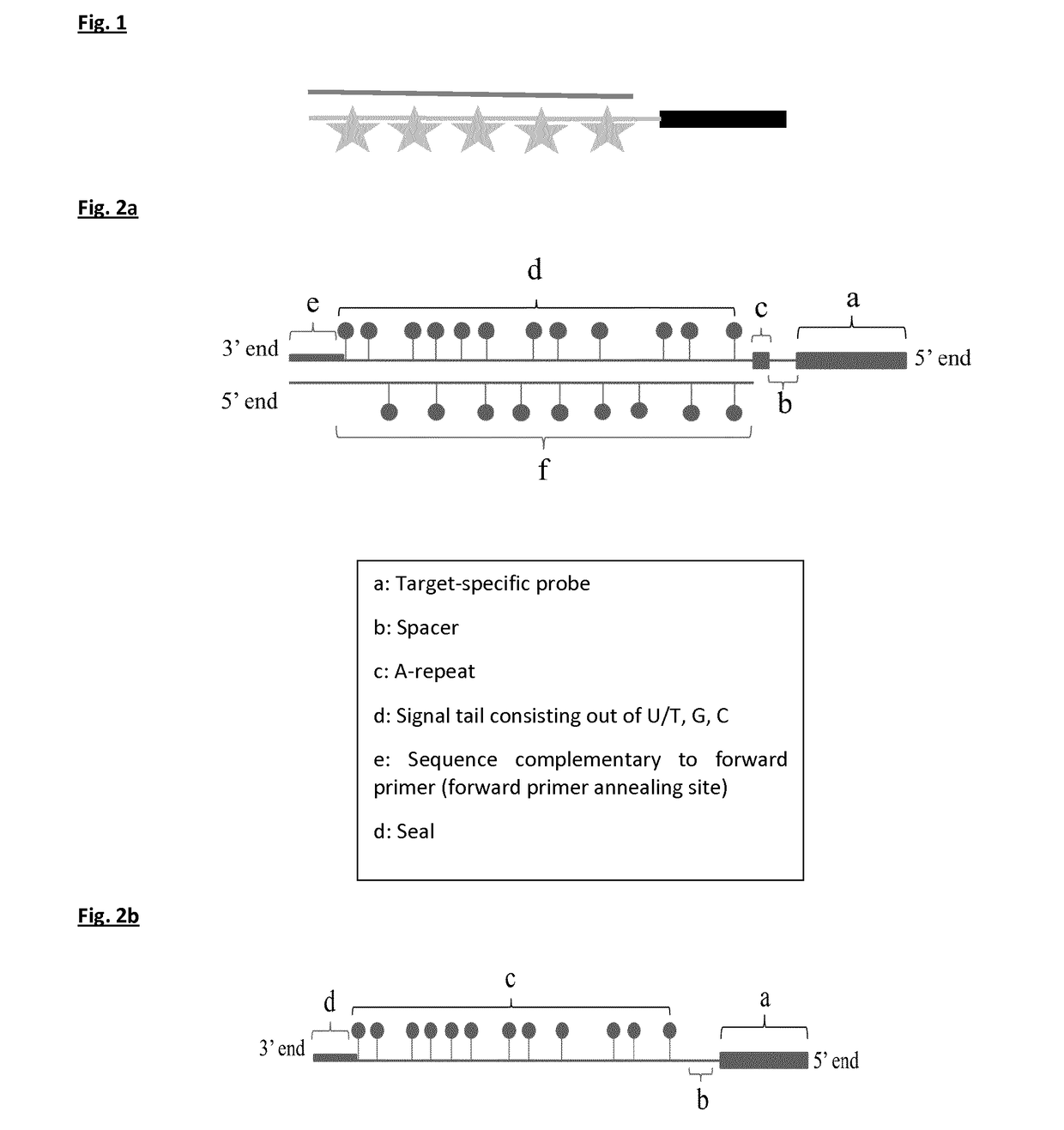
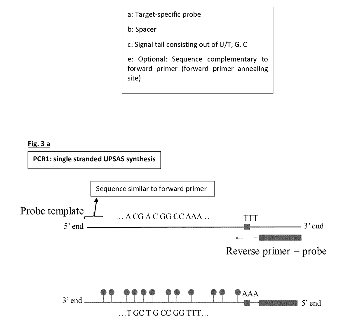
[0150]The four templates used for probe synthesis consist of five major parts (from 5′ end to 3′ end): 1) a sequence that is similar to the forward primer used in probe synthesis and sealing, 2) a part that consists of AGC nucleotides (template for signal tail), 3) a T-stretch of three nucleotides (this is included to stop probe sealing when probe sealing is performed with three nucleotides), 4) a spacer of nine nucleotides, and 5) a sequence that is complementary to the reverse primer and thus to the target-specific probe.

[0151]During the first PCR step, a forward primer (FP) with the same sequence as the sequence found at the 5′ end of the probe template and a reverse primer (RP) that will form the target-specific probe part of UPSAS, were use...

example 2

Probe Labeling is Performed Efficiently

[0154]Probes were run on gel to confirm probe labeling and to estimate the amount of labels per probe. The probes contained at least 250 labels after PCR1 and 500 labels after PCR2.

[0155]Biotin-labeled GSTP1 probes were spotted on a nylon membrane prior to staining with 3,3′-Diaminobenzidine (DAB): spots stained dark brown, indicating that labels were not only incorporated in the probes as could be observed based on the molecular weight size on gel, but also gave strong signals.

example 3

Tissue Morphology is Kept Intact after ISH Pretreatment Steps Combined with Bisulfite Conversion In Situ

[0156]3 μM formalin-fixed and paraffin-embedded (FFPE) cervical tissue sections were cut and stretched on the glass slide and deparaffinized in xylene. The sections were dehydrated two times for 5 minutes in 100% ethanol. Hereupon, the sections were incubated in 0.2 N HCl and washed with ultrapure water. The sections were treated for 28 minutes at 37° C. with porcine pepsin and washed two times for 5 minutes with ultrapure water. The sections were treated for 15 minutes with 0.1% TRITON® and washed for 3 minutes with molecular grade water afterwards. Subsequently, sections were incubated with 150 μl of a bisulfite mix (Zymo) for 4 hours. After a 15-minute desulfonation step, the samples were washed with molecular grade water and stained with hematoxylin and eosin (H&E) to evaluate conservation of the tissue morphology. Tissue morphology was evaluated by an experienced, university ...


PUM

PropertyMeasurementUnit
temperatureaaaaaaaaaa
methylation heterogeneityaaaaaaaaaa

Abstract

The disclosure relates to the field of molecular pathology (for example, cancer diagnosis, prognosis, treatment and/or therapy prediction) through the detection of RNA, mutations, copy number changes and determination of the methylation status of specific sequences of the genome of individual patients in hybridization assays (southern blot, ISH, dot blot) including in situ determination of the methylation status of specific sequences of the genome of individual patients in individual cells. More specifically, this disclosure relates to: a) target-specific probes covalently attached to a labeled tail, b) the synthesis method of said the probe, c) the usage of said the probe such as an in situ hybridization-based method to correlate the methylation status of a promoter region of a gene in a biopsy or cytology specimen of a patient to the morphology and localization in that specimen, and d) kits comprising the target-specific probes. The latter method and products allow detection of (epi) genetic changes in specific cell types of histological or cytological (cancer) specimens or on membranes that will contribute to scientific research and that will help physicians to accurately diagnose diseases and/or start an appropriate treatment.

Description

CROSS-REFERENCE TO RELATED APPLICATIONS[0001]This application is a national phase entry under 35 U.S.C. § 371 of International Patent Application PCT / EP2016 / 066510, filed Jul. 12, 2016, designating the United States of America and published in English as International Patent Publication WO 2017 / 009322 A1 on Jan. 19, 2017, which claims the benefit under Article 8 of the Patent Cooperation Treaty to European Patent Application Serial No. EP15176744.9, filed Jul. 15, 2015.TECHNICAL FIELD[0002]This application relates to the field of molecular pathology (for example, cancer diagnosis, prognosis, treatment and / or therapy prediction) through the detection of RNA, mutations, copy number changes and determination of the methylation status of specific sequences of the genome of individual patients in hybridization assays (southern blot, ISH, dot blot) including in situ determination of the methylation status of specific sequences of the genome of individual patients in individual cells. More...

Claims


Application Information

Patent Timeline
Patent Type & Authority Applications(United States)
IPC IPC(8): C12Q1/6827
CPCC12Q1/6827C12Q2525/161C12Q2537/163C12Q2565/102C12Q2523/125C12Q1/686C12Q1/682C12Q1/6886C12Q2600/154
Inventor LIEVELD, MARUSYAVAN CRIEKINGE, WIMVANDEN BROECK, DAVY
Owner UNIV GENT
Why Eureka
Try Eureka