Microarray Based Multiplex Pathogen Analysis and Uses Thereof

a microarray and multiplex technology, applied in the field of microarray based multiplex pathogen analysis, can solve the problems of inability to accurately detect the presence of pathogens in culture-based testing

Active Publication Date: 2018-09-06
PATHOGENDX INC
View PDF5 Cites 13 Cited by
  • Summary
  • Abstract
  • Description
  • Claims
  • Application Information

AI Technical Summary

Problems solved by technology

These tests require a substantial investment of time, effort, cost as well as labile products.
Current techniques are not ideal for testing large numbers samples.
Culture-based testing is fraught with inaccuracies which include both false positives and false negatives, as well as unreliable quantification of colony forming units (CFUs).
There are issues with the presence of viable but non-culturable microorganisms which do not show up using conventional culture methods.
Certain culture tests are very non-specific in terms of detecting both harmful and harmless species which diminishes the utility of the test to determine if there is a threat present in the sample being tested.
To analyze a pathogen using PCR, DNA is extracted from a material prior to analysis, which is a time-consuming and costly step.
This technique was a partial success but was not as sensitive as culture indicating a possible issue with interference of the PCR by constituents in the specimens.
Although this possible interference may not be significant enough to invalidate the utility of the testing performed, such interference can be significant for highly sensitive detection of pathogens for certain types of tests.
Consequently, Colony PCR did not eliminate the pre-analysis extraction step for use of PCR, especially for highly sensitive detection of pathogens.
However, for blood it is known that the result of such direct PCR is prone to substantial sample to sample variation due to inhibition by blood analytes.
Additionally, attempts to perform direct PCR analysis on plant matter have generally been unsuccessful, due to heavy inhibition of PCR by plant constituents.
However, the technique is costly in the use of chemical consumables and has a long response time.
Optical sensor technologies produce fast real-time detection but such sensor lack identification specificity as they offer a generic detection capability as the pathogen is usually optically similar to its benign background.
However, qPCR is largely limited to the analysis of a single pathogen.
However, processing a large number of PCR reactions for downstream microarray applications is costly and requires highly skilled individuals with complex organizational support.
Because of these challenges, microarray techniques have not led to the development of downstream applications.
While such chemistry may be readily implemented during oligonucleotide synthesis, it raises the cost of the DNA Probe by more than a factor of two, due to the cost associated with the modification chemistry.
However, the need for high energy UV for efficient crosslinking results in substantial side reactions that can damage the nucleic acid probe beyond use.
Previous Attempts at addressing these problems have not met with success.
Attachment of nucleic acid probes to surfaces via their entrapment into a 3-Dimensional gel phase such as that created by polymerizing acrylamide and polysaccharides among others have been problematic due to the dense nature of the gel phases.
While the pore size (about 10 nm) in these gels permit entrapment and / or attachment of the nucleic acid probes within the gel, the solution-phase DNA Target, which is typically many times longer than the nucleic acid probe, is blocked from penetrating the gel matrix thereby limiting use of these gel phase systems due to poor solution-phase access to the Target DNA.
Thus, the prior art is deficient in methods of DNA based pathogen analysis that interrogates a multiplicity of samples, uses fewer chemical and labile products, reduces processing steps and provides faster results while maintaining accuracy, specificity and reliability.

Method used


Image

  • Microarray Based Multiplex Pathogen Analysis and Uses Thereof
  • Microarray Based Multiplex Pathogen Analysis and Uses Thereof
  • Microarray Based Multiplex Pathogen Analysis and Uses Thereof

Examples


example 1

Fabrication of 3-Dimensional Lattice Microarray Systems

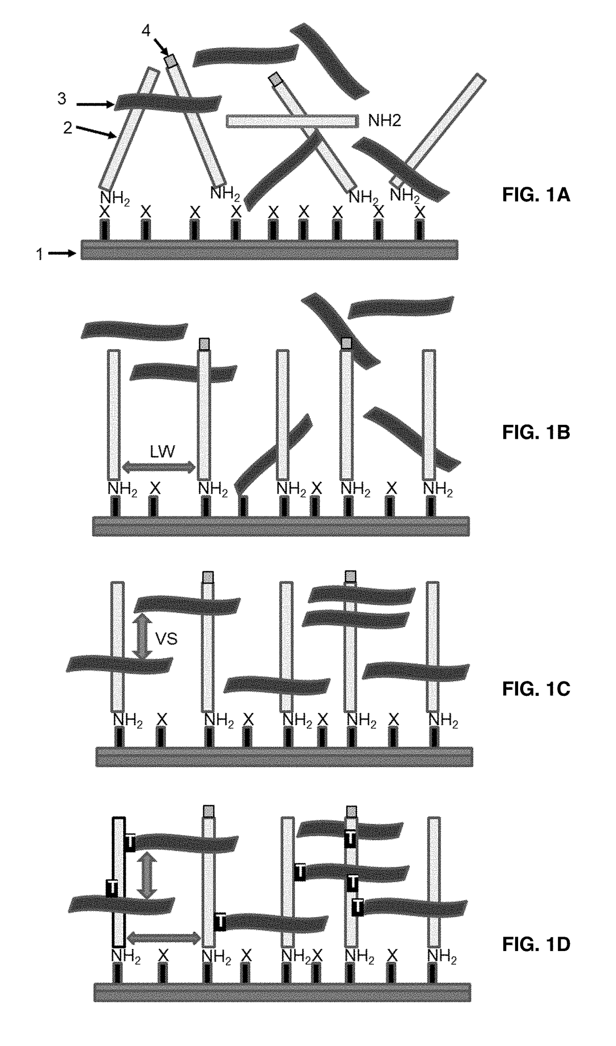
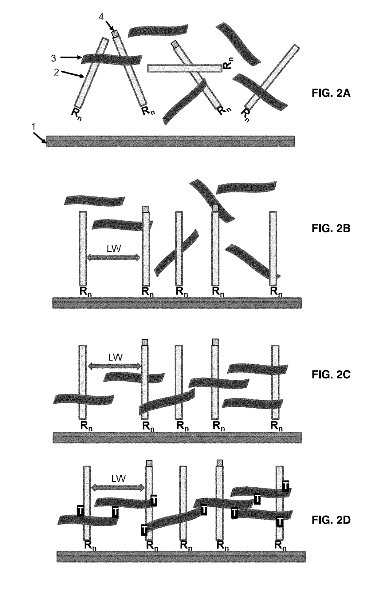
[0078]The present invention teaches a way to link a nucleic acid probe to a solid support surface via the use of a bifunctional polymeric linker. The nucleic acid probe can be a PCR amplicon, synthetic oligonucleotides, isothermal amplification products, plasmids or genomic DNA fragment in a single stranded or double stranded form. The invention can be sub-divided into two classes, based on the nature of the underlying surface to which the nucleic acid probe would be linked.

1. Covalent Microarray System with Activated Solid Support.

[0079]The covalent attachment of any one of these nucleic acid probes does not occur to the underlying surface directly, but is instead mediated through a relatively long, bi-functional polymeric linker that is capable of both chemical reaction with the surface and also capable of efficient UV-initiated crosslinking with the nucleic acid probe. The mechanics of this process is spontaneous 3D self asse...

example 2

Using the 3-Dimensional Lattice Microarray System for DNA Analysis

Sample Processing

[0103]Harvesting Pathogens from plant surface comprises the following steps:

[0104]1. Wash the plant sample or tape pull in 1× phosphate buffered saline (PBS)

[0105]2. Remove plant material / tape

[0106]3. Centrifuge to pellet cells & discard supernatant

[0107]4. Resuspend in PathogenDx® Sample Prep Buffer pre-mixed with Sample Digestion Buffer

[0108]5. Heat at 55° C. for 45 minutes

[0109]6. Vortex to dissipate the pellet

[0110]7. Heat at 95° C. for 15 minutes

[0111]8. Vortex and centrifuge briefly before use in PCR

Amplification by PCR

[0112]The sample used for amplification and hybridization analysis was a Cannabis flower wash from a licensed Cannabis lab. The washed flower material was then pelleted by centrifugation. The pellet was then digested with proteinaseK, then spiked with a known amount of Salmonella DNA before PCR amplification.

TABLE 5PCR Primers and PCRconditions used in amplificationPCR primers (P1...

example 3

[0127]FIG. 4A shows an exemplar of the first PCR step. As is standard, such PCR reactions are initiated by the administration of PCR Primers. Primers define the start and stopping point of the PCR based DNA amplification reaction. In this embodiment, a pair of PCR reactions is utilized to support the needed DNA amplification. In general, such PCR amplification is performed in series: a first pair of PCRs, with the suffix “P1” in FIG. 4A are used to amplify about 1 μL of any unpurified DNA sample, such as a raw Cannabis leaf wash for example. About 1 μL of the product of that first PCR reaction is used as the substrate for a second PCR reaction that is used to affix a fluorescent dye label to the DNA, so that the label may be used to detect the PCR product when it binds by hybridization to the microarray. The primer sequences for the first and second PCRs are shown in Table 6. The role of this two-step reaction is to avert the need to purify the pathogen DNA to be analyzed. The first...


PUM

PropertyMeasurementUnit
boiling pointaaaaaaaaaa
molar ratioaaaaaaaaaa
temperatureaaaaaaaaaa

Abstract

Provided herein is a 3-dimensional lattice microarray system for DNA sequence detection and analysis. The system has a plurality of bifunctional polymer linkers, on one end of which are attached nucleic acid probes where each have a sequence complementary to signature nucleotide sequences in pathogens, plants or animals. The other end of the bifunctional polymer linker is attached to a solid support by non-covalent or covalent means. Each of the nucleic acid probes have terminal thymidine bases at the 5′ and 3′ ends that permit attachment of the probes to the bifunctional polymer linkers. Also provided is a method for fabricating the microarray system by first attaching the bifunctional polymer linkers to the solid support, followed by photochemical coupling of the nucleic probes to the microarray. A customizable microarray kit is provided that contains the solid support, linkers, probes, solvent mixture and instructions to use the kit.

Description

CROSS-REFERENCE TO RELATED APPLICATIONS[0001]This application is a continuation-In-part under 35 U.S.C. § 120 of pending non-provisional application U.S. Ser. No. 15 / 388,561, filed Dec. 22, 2016, which claims benefit of priority under 35 U.S.C. § 119(e) of provisional application U.S. Ser. No. 62 / 271,371, filed Dec. 28, 2015, now abandoned, both of which are hereby incorporated in their entireties.BACKGROUND OF THE INVENTIONField of the Invention[0002]The present disclosure is in the technical field of DNA based pathogen and plant analysis. More particularly, the present disclosure is in the technical field of pathogen analysis for plant, agriculture, food and water material using a multiplex assay and a 3-dimensional lattice microarray technology for immobilizing nucleic acid probes.Description of the Related Art[0003]Current techniques used to identify microbial pathogens rely upon established clinical microbiology monitoring. Pathogen identification is conducted using standard cu...

Claims


Application Information

Patent Timeline
Patent Type & Authority Applications(United States)
IPC IPC(8): C12N15/10
CPCC12N15/1086G01N33/5308C12Q1/6837C12Q2565/513
Inventor HOGAN, MICHAEL EDWARDMAY, MELISSA ROSEEGGERS, FREDERICK HENRY
Owner PATHOGENDX INC
Why Eureka
Try Eureka