Compositions and methods for a three dimensional ex-vivo glomerular cell co-culture biological engineering model

a biological engineering model and glomerulus technology, applied in the field of applicability, methods and systems of a three-dimensional ex-vivo biological engineering model of a glomerulus, can solve the problems of limited ability and problematic culture system types

Inactive Publication Date: 2018-10-18
SIVAKUMARAN ARUNTHATHY
View PDF4 Cites 12 Cited by
  • Summary
  • Abstract
  • Description
  • Claims
  • Application Information

AI Technical Summary

Benefits of technology

[0034]In this system, glomerulus-derived vascular endothelial cells are seeded and cultured within the fiber lumen on the inner wall of the hollow fibers, and podocyte cells are cultured within the extra-capillary space (ECS) on the outside surfaces of the hollow fibers. The cells are associated with the fiber walls, and can be cultured associated with the fiber walls either in the absence or presence of precoated ECM. The cell culture medium that is delivered to the cartridge is not directly sent to the extra-capillary space. The semi-permeable walls of the hollow fibers permit the passage of water and dissolved solutes below a specified molecular weight threshold from the fiber lumen and into the ECS, and prevent the traversal of larger molecular weight proteins and whole cells.

Problems solved by technology

These types of culture systems are problematic because they are greatly limited in their ability to mimic more dynamic in vivo three-dimensional cell environments.

Method used


Image

  • Compositions and methods for a three dimensional ex-vivo glomerular cell co-culture biological engineering model
  • Compositions and methods for a three dimensional ex-vivo glomerular cell co-culture biological engineering model
  • Compositions and methods for a three dimensional ex-vivo glomerular cell co-culture biological engineering model

Examples


example 1

Hollow Fiber Bioreactor Models for Renal (Glomerular) Health and Renal (Glomerular) Disease Model

[0259]This example provides a description of a hollow fiber bioreactor cell co-culture models of a glomerulus used to simulate renal (glomerular) model systems in both health and diseased conditions finding use with the invention.

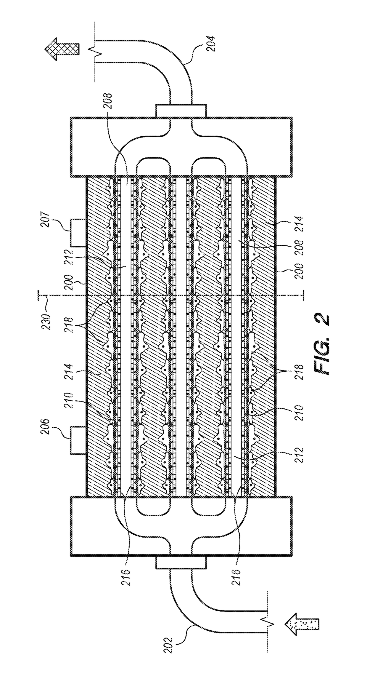
[0260]Cells are co-cultured in a e.g. FiberCell® Systems (Frederick, Md.) PS+ module cartridge (small cartridge, Catalog No. 4300-C2025). The hollow fibers in this cartridge are made from polyvinylidene fluoride (PDVF), are 10 cm in length, have an inner diameter of 700 μm and outer diameter of 1300 μm. The fiber walls have a pore size of 0.1 μm as rated by the manufacturer. The cumulative surface area of all the fibers (20 fibers) in the system is 75 cm2 and the extra-capillary space (ECS) volume is 2.5 mL. The FiberCell® Systems (Frederick, Md.) PS+ fibers have a surface chemistry that facilitates the attachment of extra-cellular matrix proteins, antibodies, c...

example 2

Hollow Fiber Bioreactor (HFBR) Model System No. 2—Cell Culture Establishment

[0266]This example describes the inoculation and co-culture maintenance of a hollow fiber bioreactor of the invention.

[0267]Glomerular endothelial cells are cultured in a suitable culture media with VEGF (5 ng / mL) for one week in order to avoid subsequent detachment of podocyte cells in the glomerular co-culture with podocyte cells in the HFBR. See Satchell et al., 2006. “Conditionally immortalized human glomerular endothelial cells expressing fenestrations in response to VEGF,”Kidney International, 69: 1633-1640.

[0268]The glomerular endothelial cells are seeded onto the inner lumen of the hollow fibers in the bioreactor. After all ports are closed, place the HFBR in the incubator for one hour, rotating the HFBR 180 degrees at the halfway point without any flow so that the endothelial cells can attach (see FiberCell Systems. 2015b. “User Manual). Following introduction of the cells into the luminal space of ...

example 3

Assays for Glucose and Albumin

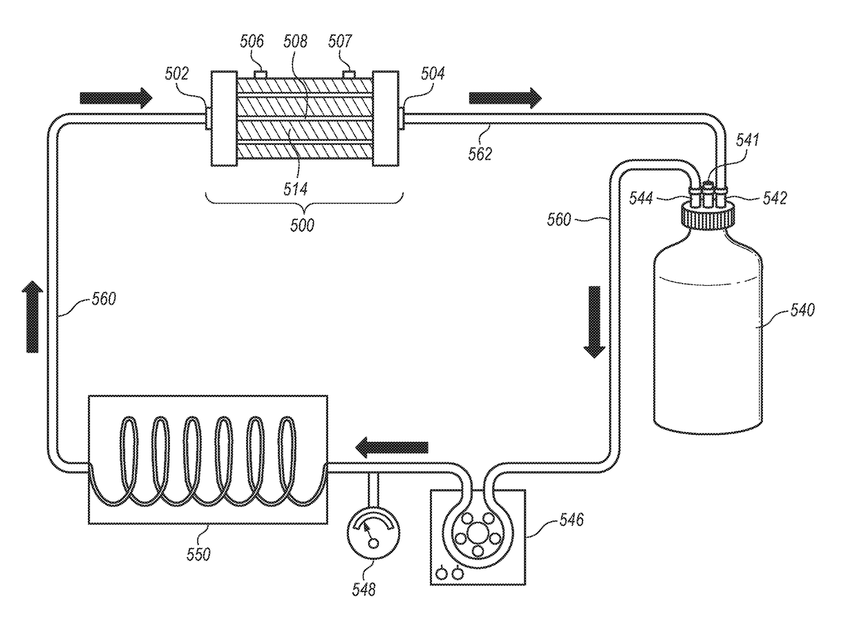
[0274]This example describes testing protocols for glucose in a defined cell culture media during cell culture establishment. In the scenario of a batch mode (retentate recycle) system (FIG. 5), the media samples are obtained at the time of each medium replacement and analyzed, for example, for glucose.

[0275]Glucose Assay.

[0276]A kit provided by Sigma-Aldridge, Inc.® (catalog# GAGO20-1KT) can be used to determine glucose levels, more specifically, the change in glucose level from the previous day. The reagent employed causes, over a 15-minute incubation, the conversion of glucose to glucose-6-phosphate through a phosphorylation reaction, followed by the oxidation of the latter in the presence of NAD (nicotine adenine dinucleotide) to 6-phosphogluconate. The oxidation reaction results in an equal number of moles of NAD being reduced to NADH, the latter of which increases the absorbance of the sample at a 340 nm wavelength as measured by a spectrophotomet...


PUM

PropertyMeasurementUnit
volumetric flow rateaaaaaaaaaa
molecular weight cutaaaaaaaaaa
inner diametersaaaaaaaaaa

Abstract

The invention provides compositions, methods and systems for the ex vivo co-culture of renal cells, more specifically, for the co-culture of glomerulus-derived vascular endothelial cells and podocyte cells using apparatus that mimics the in vivo cellular architecture of the renal corpuscle. The invention described herein finds a variety of uses, for example, as a model system for the study of renal corpuscle function, including the filtration of the blood supply that occurs at the interface of the glomerulus and Bowman's capsule, normal physiology of those cell types, and as a model system for the study of renal disease, including the study of drug effects on the functioning of these cell types.

Description

FIELD OF THE INVENTION[0001]This invention provides apparatus, methods and systems for a three dimensional ex-vivo biological engineering model of a glomerulus. The ex vivo partitioned co-culture of glomerulus-derived vascular endothelial cells and podocyte cells, along with a glomerular basement membrane is used to create a model that mimics the in vivo three-dimensional cellular architecture of the glomerular filtration structure of the glomerulus-Bowman's capsule interface. This model enables the study of the effects of biological engineering variables i.e. shear rate (fluid mechanics), pressure, and mass transport e.g. of drugs, on glomerular model system function and cell physiology in health or disease conditions.BACKGROUND OF THE INVENTION[0002]The kidney is one of a pair of organs in the back of the abdominal cavity, which has a number of significant roles. It acts as a filter, removing water soluble metabolic wastes (i.e. urea, ammonia) and foreign materials from the blood,...

Claims


Application Information

Patent Timeline
Patent Type & Authority Applications(United States)
IPC IPC(8): C12N5/071C12M3/00C12M1/12C12M1/00G01N33/50C12M1/34
CPCC12N5/0686C12N5/0691C12M21/08C12M25/10C12M29/04C12M27/18C12N2513/00C12M41/46C12M23/20G01N33/5044C12N2502/28C12N2500/84G01N33/5082C12M29/10C12M29/26C12N2521/00G01N33/5064G01N33/5088
Inventor SIVAKUMARAN, ARUNTHATHY
Owner SIVAKUMARAN ARUNTHATHY
Why Eureka
Try Eureka