
Eureka
For R&D, Eureka makes reading and utilizing patents & technical documents easy.
Eureka AIR
Designed for self-driven R&D workflows. Generate viable solutions, solve complex R&D challenges, empower your innovation with AI.

Eureka Materials
Designed for material experts only. Revolutionize your material R&D, from search, analyze, to developing new materials.

TechResearch
Generate reliable direction feasibility study reports for your R&D in just a few steps.

TechSeek
Discover and master advanced knowledge NOW. Basics, ideas, possibilities, all at once.

TechMind
As an expert in R&D Theories, TechMind can generates customized viable solutions instantly.

TechRisk
Analyze your overall solution with one click, know your potential R&D risks in advance.

TechMonitor
Get weekly tech updates, stay abreast of the latest tech innovations and key insights.
Method for determining cell clonality
- Summary
- Abstract
- Description
- Claims
- Application Information
AI Technical Summary
Benefits of technology
Problems solved by technology
Method used
Image

Examples
example 1
ection and Subcloning
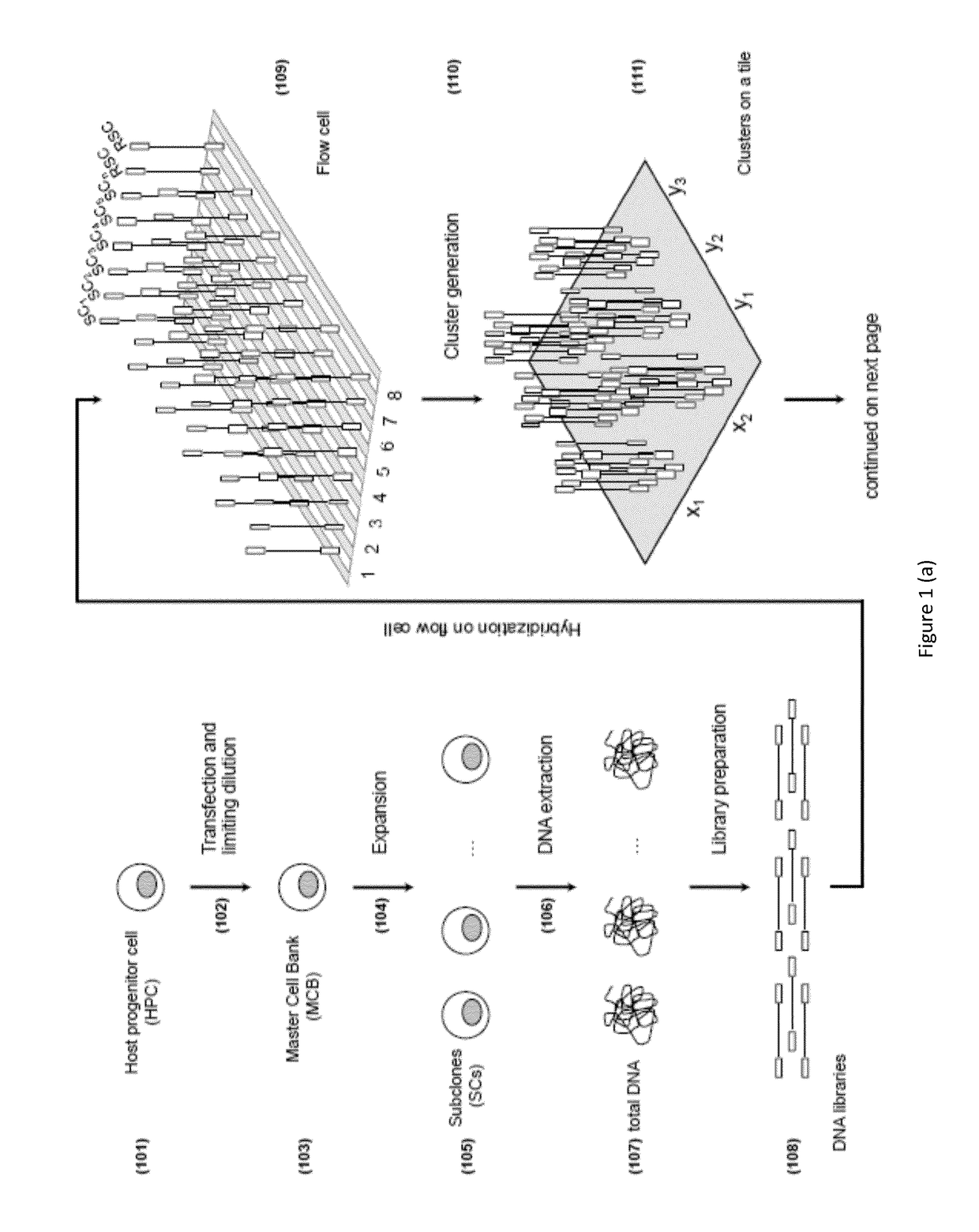
[0163]The MCB, the clonality of which is to be assessed, was generated by transfection of two transgenes carrying light and heavy chain into the genome of a Chinese hamster ovary (CHO) cell line that served as the host progenitor cell (HPC). The transfection was performed by Gala® (Catalent) using the GPEx® technology and a single limiting dilution was performed in order to obtain the monoclonal cell line (See “GPEx®: a flexible method for the rapid generation of stable, high expressing, antibody producing mammalian cell lines”, Gregory T. Beck, Book: Current Trends in Monoclonal Antibody Development and Manufacturing, Publisher: Springer New York, 2010). In order to investigate the clonality of this cell line 25 subclones (SCs) where generated by dilution of the MCB. The limiting dilution was performed as follows. The MCB was thawed at room temperature and then incubated in a T 75 flask with 20 mL of DMEM with 10% FBS for 24 hours at 37° C., 5% CO2. The next da...
example 2
ction
[0165]DNA extraction from the 25 subclones, the MCB and the divergent MCBΔ was performed on an affinity column using the QIAamp Blood DNA Mini kit (QIAGEN) according to the manufacturer's and internal working instructions. Briefly, cell pellets were resuspended in phosphate buffered saline (PBS) according to the sample concentration and split into different aliquots. 200 μL of lysis buffer and 20 μL of proteinase K were added to each sample. Samples were mixed thoroughly by vortexing, and incubated at 56° C. for 10 minutes. Then, 200 μL ethanol (96 to 100%) was added and the mixture was transferred into the DNeasy Mini spin column (QIAGEN) placed in a 2 mL collection tube. Samples were washed and centrifuged for a minute at 13,000 rpm to remove any residual ethanol. Elution was performed with 150 μL of water directly added to the DNeasy membrane (QIAGEN). Eluates of the same clone where combined.
[0166]Each sample was incubated with RNase enzyme (Roche) at 37° C. for 30 minutes ...
example 3
Library Preparation and Sequencing
[0167]Library preparation for the 25 subclones, the MCB and the MCBΔ was performed using the TruSeq DNA kit (Illumina) according to manufacturer's instructions. Briefly, 2.6 μg of each subclone DNA were fragmented by the Covaris S220 instrument to obtain 300 bp dsDNA fragments with 3′ or 5′ overhangs. Overhangs were converted enzymatically into blunt ends. A single adenine (A) nucleotide was added to the 3′ ends of the blunt fragments to prepare fragments for adapter ligation. The ligation of multiple indexing adapters to the ends of the DNA fragments allows the hybridization onto a flow cell. Selective enrichment of those DNA fragments that have adapter molecules on both ends was performed to increase library yield.
[0168]The quality of DNA libraries was analyzed by an Agilent 2100 Bioanalyzer to validate the mean size of the fragments in the DNA libraries. Libraries were further quantified by Fluorometer Qubit® 2.0.
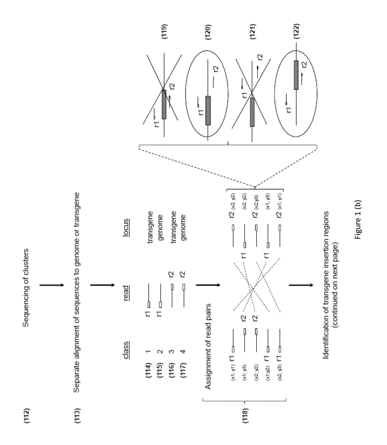
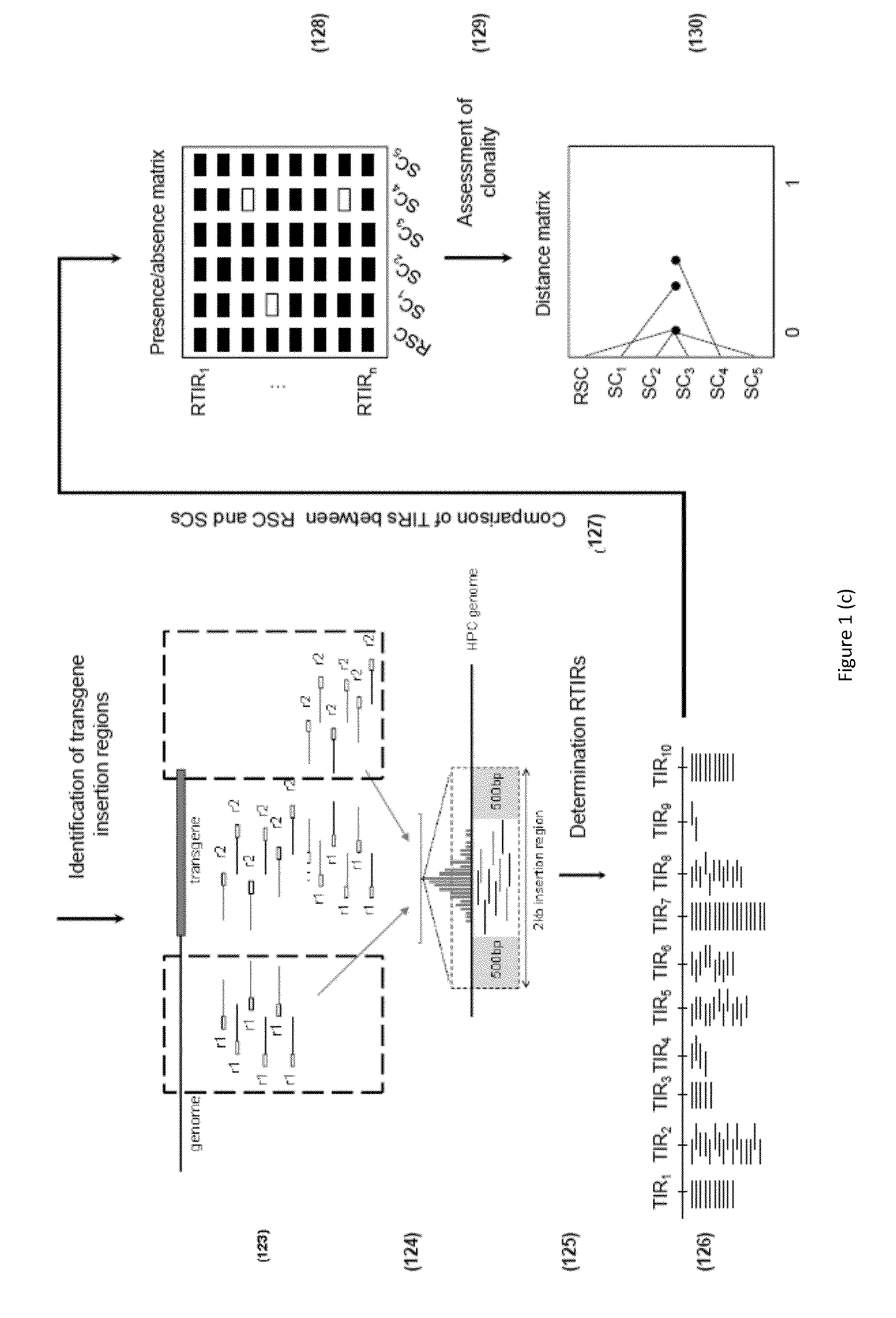
[0169]DNA library clusters were g...
PUM
| Property | Measurement | Unit |
|---|---|---|
| Digital information | aaaaa | aaaaa |
| Dimension | aaaaa | aaaaa |
| Distance | aaaaa | aaaaa |
Abstract
Description
Claims
Application Information
 - R&D Engineer
- R&D Manager
- IP Professional
- Industry Leading Data Capabilities
- Powerful AI technology
- Patent DNA Extraction
Browse by: Latest US Patents, China's latest patents, Technical Efficacy Thesaurus, Application Domain, Technology Topic, Popular Technical Reports.
© 2024 PatSnap. All rights reserved.Legal|Privacy policy|Modern Slavery Act Transparency Statement|Sitemap|About US| Contact US: help@patsnap.com