Improved cytometric assays

a flow cytometric and assay technology, applied in the field of flow cytometric assays, can solve the problems of ineffective treatment of patients, insufficient standard patient management, and inability to accurately identify and characterization of various til populations, and achieve high accuracy and informative separation of cell populations, the effect of high accuracy and accurate identification and characterization

Active Publication Date: 2018-12-13
THE MEDICAL RES INFRASTRUCTURE & HEALTH SERVICES FUND OF THE TEL AVIV MEDICAL CENT
View PDF0 Cites 4 Cited by
  • Summary
  • Abstract
  • Description
  • Claims
  • Application Information

AI Technical Summary

Benefits of technology

[0016]Thus, the cytometric panels, reagents, kits and methods, collectively referred to herein as the assays of the invention, surprisingly provide for highly accurate and informative separation of cell populations using minimal marker combinations.
[0017]According to a first aspect of the invention, there is provided a cytometric kit comprising a viability dye and a plurality of labeled antibodies. In another embodiment, the kit is useful for characterizing multiple cell populations in a human tissue sample (dissociated from solid tiss...

Problems solved by technology

Quantification and characterization of the various TIL populations, including rare populations such as dendritic cell (DC) subtypes, is often challenging.
In particular, accurate immune phenotyping of solid tumors characterized by low TIL counts (such as in brain tumors), has not yet been established in standard patient management.
Consequently, the patients may be administered with ineffective therapies that waste the time of their therapeutic window, and even with potentially harmful therapies.
However, mass cytometry entails the destruction of the examined cells during the process, thus precluding isolation and further examination of the identified cell populations.
The simultaneous staining for a large number of markers, enabling discrimination between multiple cell populations, involves many techni...

Method used


Image

  • Improved cytometric assays
  • Improved cytometric assays
  • Improved cytometric assays

Examples


example 1

Tissue Dissociation to Viable Single Cells

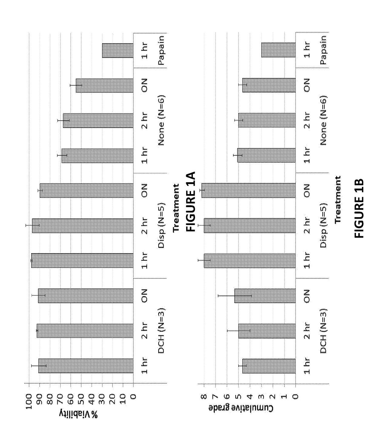
[0209]Freshly isolated brain tissue and brain tumor (BT) tissue, was transferred to the lab in saline or in Ringer lactate (Biological Industries, Beit HaEmek, Israel). The specimens were weighed following the removal of blood clots and necrotic areas. The cleansed tissue was cut into 1-2 mm pieces and resuspended in HBSS(+Ca +Mg) without phenol red (Biological Industries) at 100 mg tissue per ml. The tumor slurry was divided, 4 ml per 50 ml tube, to allow for complete trituration using a 5 ml Pasteur pipette.

[0210]The following enzymes or their combination were tested on the tumor slurry:

1. DNase-I (Sigma St. Louis, Mo., USA): DNase is an endonuclease used to reduce viscosity (‘gooeyness’) resulting from DNA released from dead cells. 2. Collagenase type IV from Clostridium histolyticum (Ch; Sigma): a metalloprotease that cleaves native triple-helical collagen. 3. Papain from papaya latex (Sigma): a relatively nonspecific protease. 4. Hyalur...

example 2

s for Panel Construction

[0225]The following tables depict exemplary antibodies useful in construction of panels.

TABLE 3antibody characteristicsAntibody target andCloneFluorophorenameIsotypeCompanyAlexa Fluor 488-CD11bICRF44Mouse IgG1 kBiolegendPE-CY5.5-CD33WM53Mouse IgG1AbcamAPC eFlour780- HLA-DRLN3Mouse IgG2b keBiosciencePac-Bl-CD14M5E2Mouse IgG2a kBiolegendQD655-CD14TuK4Mouse IgG2a kInvitrogenPE-CD86IT2.2Mouse IgG2b kBiolegendPerCP-Cy5.5-CD163G8Mouse IgG1 kBiolegendPE-Cy7-CDlcL161Mouse IgG1 kBiolegendFITC-CD303AC144Mouse IgG1 kMilteniBiotecPE-CD141AD5-Mouse IgG1 kMilteni14H12BioteceFluor 780-CD3SK7Mouse IgG1 keBioscienceAPC-CD452D1Mouse IgG1 kBiolegendPE-Cy5.5-CD4S3.5Mouse IgG2a kInvitrogenNC650-CD8RPA-T8Mouse IgG1 keBiosciencePE-V-alpha24J6B11Mouse IgG1 keBiosciencePac-Bl-CD3OKT3Mouse IgG2a kBiolegendPac-Bl-CD56MEM-Mouse IgG2a kBiolegend188Pac-bl CD19HIB19Mouse IgG1 kBiolegendAlexa Fluor 647-GFAP1B4Mouse IgG2b kBDPE-A2B5105-Mouse IgMMiltenyiHB29biotecIL-13Ra2 (Primary)N / AGoat IgG...

example 3

g Cytometric Assays for Tumor Infiltrating Leukocytes (TIL)

[0226]Three multicolor (7-8 color) staining panels were developed, identifying all prominent immune subsets within human brain tumors. The constructed lymphocytic, innate and DC staining panels target only cell surface markers, thus enabling live sorting of the identified cells.

[0227]Lymphocytic Panel

[0228]The first lymphocytic panel monitored the following markers: CD3, CD4, CD8, CD56, TcR-Vα24Jα18, CD45, CD11b, and a dump channel removing on the same fluorescent channel, dead cells-using a viability dye (ViViD) and unwarranted cells—CD14+ monocytes / macrophages, CD19+ / 20+ B cells, and CD66b+ granulocytes (see Table 4). As can be seen in FIG. 5A, the panel identified cytotoxic T lymphocytes—CTLs (3+8+4−56−DUMP−), Helper T cells—Th (3+4+8−56−DUMP−), CD56+ NK cells (3−4−8+ / −56+ DUMP−) and NK-T cells (3+4+ / −8+ / −56+Vα24+ / − DUMP−) of 3 subtypes (Type1—iNKT:Vα24Jα18+CD4+ or CD4−CD8-, and Type 2-NKT lacking the Vα24 Jα18 TcR).

[0229...


PUM


Abstract

Provided relates to the field of cytometry, specifically to flow cytometric methods and kits for improved diagnosis, prognosis and monitoring of tumors and other lesions involving immune cell infiltration. Further provided are embodiments of the subject matter which relate to compositions and methods providing high resolution quantitative means for immunophenotyping and immune modeling, and for identification of disease prognostic and therapy predictive biomarkers.

Description

FIELD OF THE INVENTION[0001]The invention relates to the field of cytometry, specifically to flow cytometric assays for improved tumor immunophenotyping, prognosis and immune modeling.BACKGROUND OF THE INVENTION[0002]Immune therapies to cancer, as well as many non-immune cancer therapies (such as photodynamic therapy irradiation, and various chemotherapeutics), induce a complex immune response that may facilitate the systemic rejection of tumor cells. These immune-inducing therapies are emerging as a highly effective means to treat cancers of various tissues. However, the outcome of such therapies is highly dependent on the host's immune status, and particularly on the nature and quantity of resident or infiltrating immune cells (also referred to as tumor-infiltrating leukocytes, or TIL) present within the tumor environment.[0003]For example, different TIL populations may induce either beneficial immune responses capable of attacking the tumor cells, or detrimental tolerogenic respo...

Claims


Application Information

Patent Timeline
IPC IPC(8): G01N33/569G01N33/533
CPCG01N33/56977G01N33/533G01N2333/70514G01N2333/70517G01N2333/70596G01N33/56972G01N2333/705G01N33/53
Inventor VOLOVITZ, ILANLUSTGARTEN, MERAVRAM, ZVISHAPIRA, NETANEL YAKOVBEN HORIN, IDANDIAMANT, GIL
Owner THE MEDICAL RES INFRASTRUCTURE & HEALTH SERVICES FUND OF THE TEL AVIV MEDICAL CENT
Why Eureka
Try Eureka