
Synthetic implant device replicating natural tissue structure and methods of making same

a synthetic material and implant device technology, applied in the field of composite implants, can solve the problems of lack of biological activity, limited ability to elicit beneficial biological responses, and difficulty in repairing and replacing tissues,

Active Publication Date: 2019-01-03
POLY MED
View PDF6 Cites 2 Cited by
  • Summary
  • Abstract
  • Description
  • Claims
  • Application Information

AI Technical Summary

Benefits of technology

This patent is about a new implant design that prevents tissue from sticking to other organs or tissues. The implant uses a special component that reduces tissue attachment. It also has the ability to change its mechanical properties by adding or removing materials, making it suitable for replacing different types of tissues with different physical properties. This can help improve the implant's effectiveness and reduce the risk of complications.

Problems solved by technology

Despite the expanding sophistication of medical devices, repair and replacement of tissues remains a formidable challenge.
However, certain disadvantages exist for meshes comprised of synthetic materials.
These disadvantages, or drawbacks, include a lack of extracellular matrix molecules resulting in a lack of biological activity and minimal ability to elicit beneficial biological responses; the generation of degradation by-products such as lactic and glycolic acid which can have negative effects on the surrounding wound environment and the potential for foreign body reaction that may develop after implantation.
Drawbacks to using biologically-derived mesh materials include varied degradation kinetics, variability in material consistency based on anatomical location and donor species, and the possible transfer of pathogens.
Since then, the use of ADM has expanded in the reconstruction space; however, there have been a wide range of positive and negative results depending on the type of ADM used, the particular surgical technique, and patient population.
In addition to the heterogeneity experienced between ADM brands based on biological source and processing technique, another factor that is prohibitive and discourages the use of ADM is its high cost, which can be as high as US$5,000 per piece.
Prior attempts do not disclose how to mimic the structure or properties of ADM in a synthetic format.
The reference, however, fails to specifically teach or suggest how to fabricate the constructs disclosed herein, and the reference also fails to provide a teaching or motivation for making such constructs.
This barrier is intended to not allow cellular attachment, adhesion, or ingrowth, and moreover not to replicate the natural tissue structure.
Totakura does not disclose the barrier as a means to replicate the topography or structure of natural tissue as found in ADM based products, which are designed to support and promote the adhesion and ingrowth of autologous cells and tissues.
Further, the reference does not teach or suggest the use of elastomeric degradable materials that are critical for making a synthetic construct that mimics certain properties of biologically-derived meshes.
However, the reference does not disclose how to mimic the structure and properties of ADM in a synthetic format nor are the composites fully bioresorbable.
Further, it does not include the use of biomolecules as an additive and minor component within the construct, nor does it disclose the creation of a directional or zonally organized hydrating gradient.

Method used

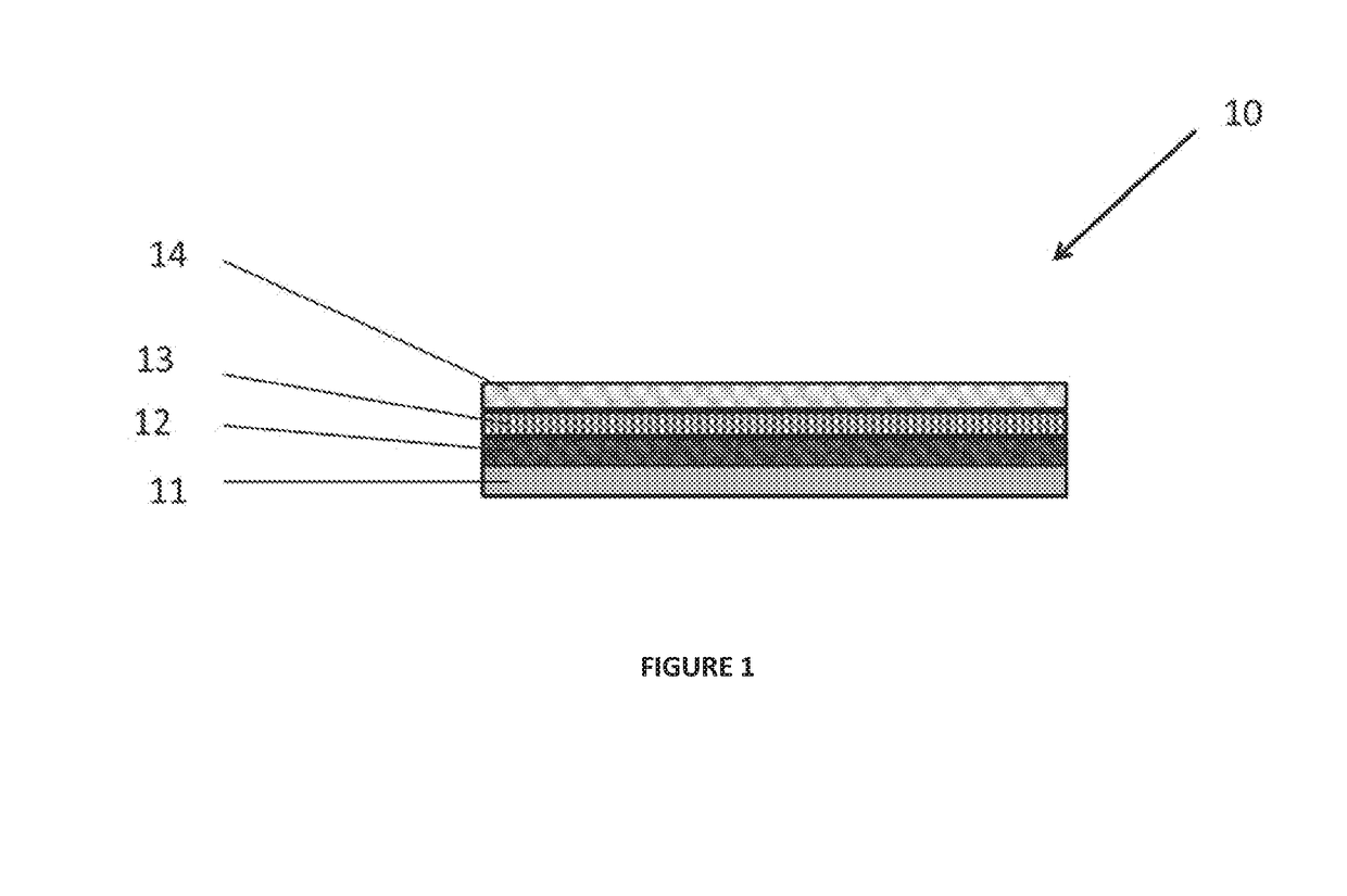
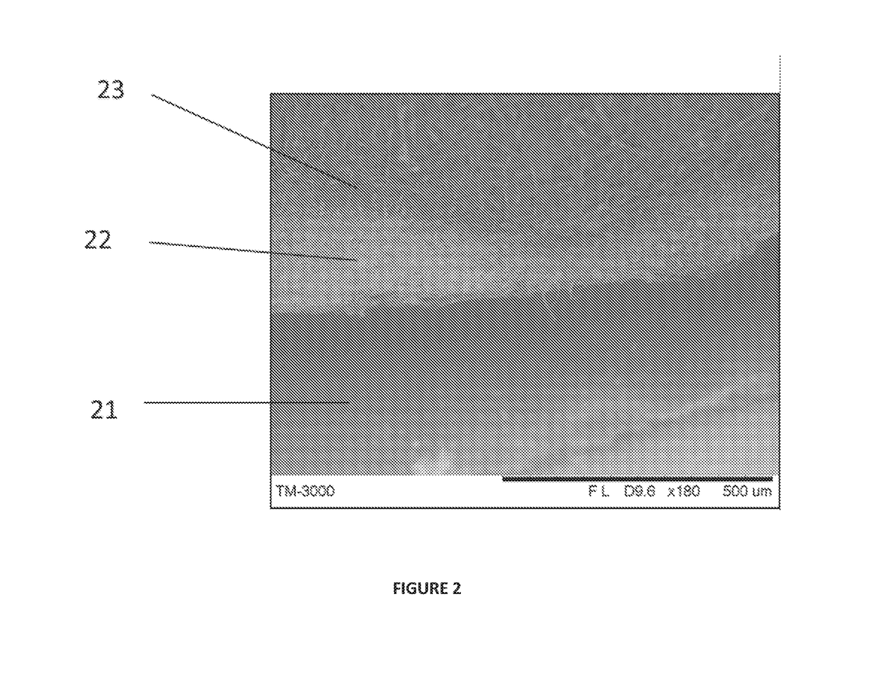

Image

  • Synthetic implant device replicating natural tissue structure and methods of making same
  • Synthetic implant device replicating natural tissue structure and methods of making same
  • Synthetic implant device replicating natural tissue structure and methods of making same

Examples


example 1

[0167]An implant was prepared by dissolving an elastomeric, bioabsorbable polyaxial block copolymer, polymer 1, in chloroform overnight by continuous agitation to form a five (5) wt % solution. Following dissolution of the polymer, D&C Violet #2 was added at 40 ppm and agitated to form a homogenous tinted solution. The dissolved solution was volumetrically measured out and cast into an inert tray at a concentration of 0.5 mL / cm3. The tray was covered with an inert, permeable cover to slow evaporation of the solvent.

[0168]After overnight (12 hr) evaporation, a solid elastomeric polymer film of 0.3-0.6 mm thickness, length of 100 mm, and width of 100 mm was obtained. The resultant film was fixed to a grounded stainless steel collector drum of diameter 12.7 cm and 25 cm in length. Two polymeric solutions were prepared by dissolution in hexafluoroisopropanol (HFIP). Solution 1 comprised of polymer 1 dissolved at a concentration of eight (8) wt %. Solution 2 comprised of linear poly(diox...

example 2

[0173]A composite implant device of the invention was prepared. The implant device displays an impregnated reinforced structure. The implant was prepared by the warp knitting of multifilament (filament count 10, denier 180 g / 9000 m, tenacity of gf / denier) yarn comprising greater than 90% glycolide content into a Tricot pattern producing a porous mesh with a thickness of 0.3 mm, areal density of 75 g / m2, and breaking strength N in both the machine and counter-machine direction. The mesh was coated with a polyaxial elastomeric bioabsorbable polyester polymer (polymer 1) coating. Polymer 1 was dissolved in chloroform overnight by continuous agitation to form a five (5) wt % solution. Following dissolution of the polymer, D&C Violet #2 was added at 40 ppm and agitated to form a homogenous tinted solution. The mesh was cut to 90 mm×90 mm in area and was centrally placed in an inert tray measuring 100 mm×100 mm×5 mm. The dissolved polymer solution was volumetrically measured out and cast ...

example 3

[0175]A composite implant device of the invention was prepared according to the following procedure. The implantable device of Example 2 was produced and further modified to incorporate fibrous elements on the roughened side of the implant device. The resulting implant device was fixed (secured) to a grounded stainless steel collector drum of diameter 12.7 cm and 25 cm in length. Two polymeric solutions were prepared. Solution 1 was made by dissolving polyethylene oxide (molecular weight of 20,000 Da) in deionized water at a concentration of six (6) wt %. Solution 2 was made by dissolving a poly(ether-ester) block copolymer in hexafluorisopropanol (HFIP) at a concentration of eight (8) wt %). The poly(ether-ester) was a triblock copolymer comprised of a central hydrophilic block derived from poly(ethylene glycol) (molecular weight of 12,000 Da) and flanking poly(ether-ester) blocks derived from para-dioxanone.

[0176]The polymeric solutions were volumetrically dispensed out of two sep...


PUM

PropertyMeasurementUnit
Fractionaaaaaaaaaa
Fractionaaaaaaaaaa
Diameteraaaaaaaaaa

Abstract

A composite implant device for use in a medical application, comprising a synthetically-derived mesh that mimics particular critical aspects of a biologically-derived mesh. The composite implant device can be used for the reinforcement and reconstruction of tissues within the body and can be comprised of a majority of synthetic components and minority of naturally-derived components which mimic the structure and function of a naturally-derived mesh.

Description

CROSS-REFERENCE TO RELATED APPLICATION[0001]This application claims the benefit under 35 U.S.C. § 119(e) of U.S. Provisional Patent Application No. 62 / 266,008 filed Dec. 11, 2015, where this provisional application is incorporated herein by reference in its entirety for all purposes.FIELD OF THE INVENTION[0002]The present invention relates to a composite implant device for use in a medical application, comprising a synthetically-derived mesh that mimics certain aspects of a biologically-derived mesh such as composition, mechanical properties, topography, microstructure, and tissue response. The present invention also relates to methods for producing such devices.BACKGROUND[0003]Despite the expanding sophistication of medical devices, repair and replacement of tissues remains a formidable challenge. In reinforcing applications, implant devices are used for internal support and augmentation of injured and / or diseased tissues. For reconstruction of soft tissue, these implant devices ty...

Claims


Application Information

Patent Timeline
IPC IPC(8): A61F2/00A61L27/58
CPCA61F2/0063A61L27/58A61F2210/0004A61F2240/001A61F2250/003A61F2250/0039A61L27/00B32B2250/03B29L2031/7534D10B2509/08D01D5/0084D10B2509/06B29L2031/753D01D5/0076D01D5/0038B29L2031/7532B32B2262/0215B32B5/266B32B27/12B32B2262/0276B32B27/36B32B5/04
Inventor MCCULLEN, SETH DYLANCULBREATH, CLAYTON JOSEPHVAUGHN, MICHAEL AARONTAYLOR, MICHAEL SCOTT
Owner POLY MED
Learn More