
Method for Manufacturing Body Substitutes by Additive Deposition

a technology of additive deposition and body, applied in the field of biotechnology, can solve the problems of toxicological problems, huge therapeutic stakes, and the risk of disturbing the product obtained

Active Publication Date: 2019-01-03
LAB SKIN CREATIONS +4
View PDF0 Cites 7 Cited by
  • Summary
  • Abstract
  • Description
  • Claims
  • Application Information

AI Technical Summary

Benefits of technology

The patent text describes the use of additive deposition methods, such as 3D printing, for fabricating body substitutes and testing the toxicity and efficiency of pharmaceutical or cosmetic active ingredients. These methods involve depositing a powder, paste, or liquid phase in a controlled three-dimensional form on an initial substrate and solidifying it. The technique has been explored in the biotechnology field for several decades and has been used to fabricate complex shaped industrial parts. The use of hydrogels, which are three-dimensional structures based on biological materials, has also been explored in the past for biotechnological applications. The technical effects of the patent text include the fabrication of more realistic body substitutes and the reduction of animal testing for safety purposes.

Problems solved by technology

However, solidification by photochemistry requires significant quantities of photocatalysts, photoinitiators and / or stains (for example: 2% photoinitiator); these additives can create a toxicological problem.
Concerning the photocatalyst, even if it is not toxic in itself, the production of free radicals is always conducive to cell mortality and a risk of disturbing the product obtained.
Although this biomaterial can be used for the culture of cells, it is not a skin substitute.
On the other hand, and still in the field of human skin, the therapeutic stakes are enormous because there are still many unsatisfied needs.
The inventors have observed that traditional methods for fabricating body substitutes, and particularly skin substitutes, take a long time in two respects; firstly they require a long manipulation time (expressed in man-hours) because the fabrication of body substitutes and particularly skin substitutes is a complex process; and secondly the porous substrate (scaffold) necessary for the fabrication of skin substitutes cannot be used as such but must firstly be allowed to age for several months (typically about six months) before it can be used under culture conditions.
It should also be noted that the complex manipulation during fabrication implies a risk of contamination of cultures.
However, the result is not very good because we do not obtain an artificial dermis usable for the culture of a stratified epidermis on its surface, that can be used as a realistic model of a natural skin.
However, the skin substitute thus obtained does not have the stratified structure of a natural epidermis, and it contains many holes.
At the present time, there is no solution to the problem de fabricating a skin substitute with a sufficient structural and functional resemblance to natural skin, using additive techniques.

Method used


Image

  • Method for Manufacturing Body Substitutes by Additive Deposition
  • Method for Manufacturing Body Substitutes by Additive Deposition
  • Method for Manufacturing Body Substitutes by Additive Deposition

Examples


example 1

nd Collection of Cells

[0115]This example illustrates a method for amplification and collection of cells (fibroblasts and keratinocytes) that can then be used in the fabrication of the skin substitute according to the invention.

[0116]Dermal keratinocytes and fibroblasts were isolated from a human preputium.

[0117]The keratinocytes were cultivated on human fibroblasts irradiated using a technique well known to a person skilled in the art, using the culture medium known as “Green's medium” containing DMEM and Ham's F12 (in the ratio 3:1), to which adenine was added (24.3 μg / mL) with the human epidermal growth factor (10 ng / mL), hydrocortisone (0.4 μg / mL), insulin (Humulin®, 5 μg / mL), 2×10⁻⁹ M tri-iodo-L-thyronine (5 μg / mL), 10−10 M isoproterenol, penicillin (100 U / mL), streptomycin (100 μg / mL) and 10% of fetal bovine serum. The keratinocytes collected during passes 2, 3 and 4 were used.

[0118]The fibroblasts were cultivated in an appropriate medium containing DMEM, 20% of new-born calf s...

example 2

n by Additive Technique (Method According to the Invention)

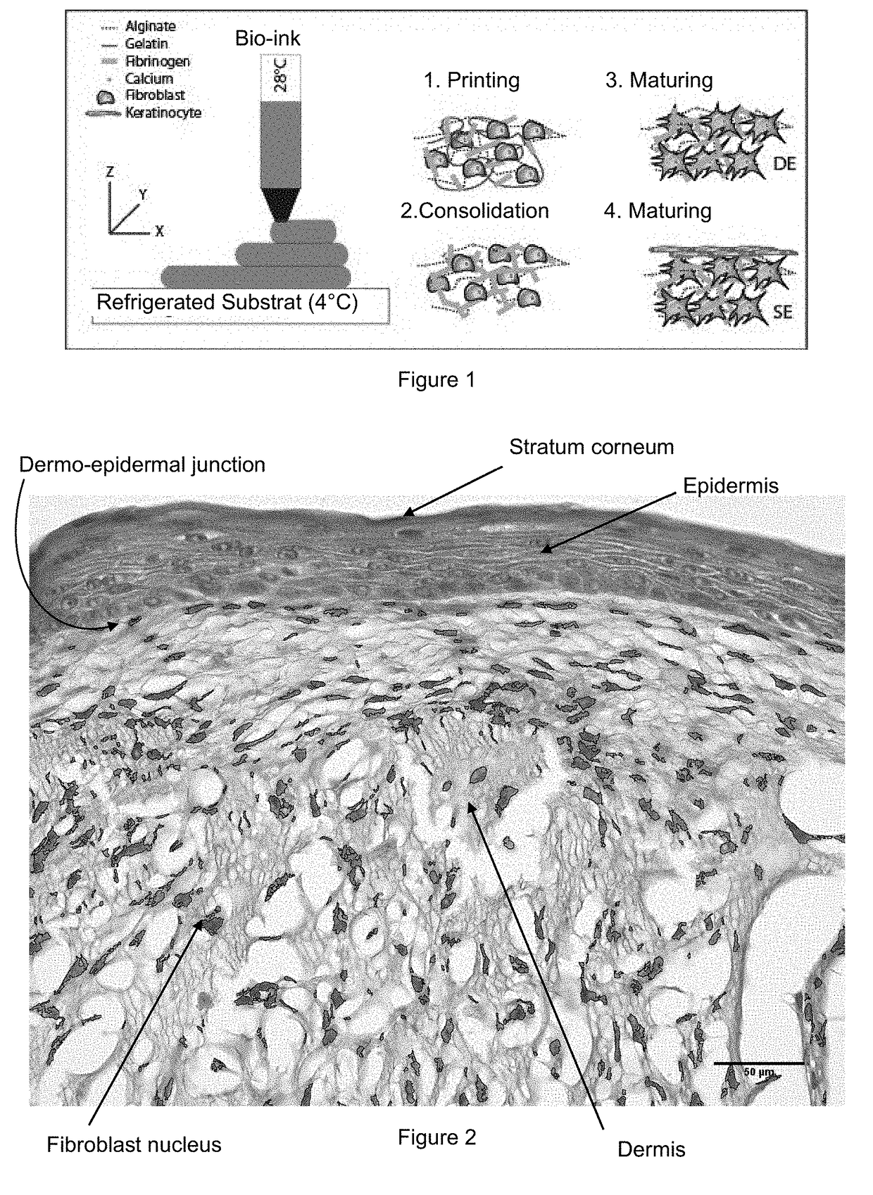
[0119]A first aqueous solution of gelatin was prepared by dissolving a gelatin powder at 20% m / v in a solution of NaCl at 0.9% m / v. A second aqueous solution of alginate was prepared by dissolving alginate powder (Very Low Viscosity) at 4% m / v in a solution of NaCl at 0.9% m / v. A third aqueous solution of fibrinogen was prepared at 8% m / v into which fibroblasts were added in suspension (obtained in example 1) at a cell concentration of 2 million cells / mL.

[0120]These three solutions were then mixed so as to obtain a mixture (called “bio-ink”) that contains 50 vol-% of the first solution (gelatin), 25 vol-% of the second solution (alginate) and 25 vol-% of the third solution (fibroblasts recovered in fibrinogen).

[0121]An aqueous polymerization solution was prepared containing 3% m / v calcium and thrombin at a final concentration of 20 U / mL.

[0122]Said bio-ink has a viscous transition at 29° C. It was heated to a temperature of a...

example 3

of the Skin Substitute Precursor (Method According to the Invention)

[0124]Skin substitute precursors were incubated for 12 days in a fibroblast culture medium containing 1 mM of ascorbic acid 2-phosphate; they were nourished every day. After twelve days, keratinocytes were applied on the surface of skin substitute precursors with a concentration of 2.5×10⁵ cells per cm2.

[0125]Skin substitute precursors were incubated for a first seven-day incubation period immersed in Green's medium as described above, with a concentration of 1 mM of ascorbic acid 2-phosphate and antibiotics; they were nourished every day.

[0126]The skin substitute precursors were then incubated during a second 21-day incubation period, kept at the surface of the liquid in a differentiation medium containing DMEM with hydrocortisone (0.4 μg / mL), insulin (5 μg / mL), ascorbic acid 2-phosphate and antibiotics; the differentiation medium contained 8 mg / mL of bovine serum albumin.

[0127]Skin substitutes were thus obtained. ...


PUM


Abstract

The invention relates to a method for manufacturing a bio-ink by additive deposition, which comprises supplying: a first solution including between 5 and 40 wt. % gelatin; a second solution including between 15 and 35 .wt. % alginate; a third solution including between 1 and 15 wt. % fibrinogen, and optionally living cells in suspension; and creating a mixture including: around 35 to 65 vol. % of the first solution; around 15 to 35 vol. % of the second solution; and around 15 to 35 vol. % of the third solution, said proportions being selected so that they add up to 100%. Said bio-ink allows the additive deposition of objects that can be polymerised by means of a solution including calcium ions and thrombin. Said objects can be incubated and can be used as a substitute for body tissue, for example (with added fibroblasts) as skin substitute.

Description

TECHNICAL FIELD[0001]The invention relates to the field of biotechnology and more particularly body tissue substitutes, especially substitutes for skin tissues. In particular it relates to the fabrication of body substitutes (such as skin) intended to be implanted in the body or the performance of tests on pharmaceutical or cosmetic active constituents to evaluate their toxicity, their efficiency or penetration in the body tissue.STATE OF PRIOR ART[0002]In Europe, chemical products for use in cosmetics can no longer be tested on laboratory animals; therefore continuous attempts are made to improve skin substitutes, to confer characteristics on them more closely resembling the characteristics of a natural dermis, to reduce their production and usage cost. Among the different experimental methods for fabricating biotechnological materials and matrices, additive deposition methods have received considerable attention during the last few years.[0003]Initially designed for fabricating mo...

Claims


Application Information

Patent Timeline
IPC IPC(8): C12N5/071C12N5/077A61L27/38B33Y10/00B33Y70/00
CPCC12N5/0698C12N5/0656C12N5/0629A61L27/3843B33Y10/00B33Y70/00C12N2503/06C12N2502/094C12N2502/1323C12N2533/54C12N2533/74C12N2533/56C12N2533/30C12N2500/38C12N2501/11C12N2501/33C12N2501/39C12N2501/395C12N2501/805C12N2502/091C12N2502/092C12N2502/097C12N2502/1305C12N2533/52B33Y80/00G01N33/50C12N5/0602A61L27/3604B33Y40/10
Inventor MARQUETTE, CHRISTOPHEPOURCHET, LEATHEPOT, AMELIEDOS SANTOS, MORGAN
Owner LAB SKIN CREATIONS
Learn More