Colors for chromogenic ihc and ish staining with multi-dye quinone methide and tyramide conjugates

Active Publication Date: 2019-06-20
VENTANA MEDICAL SYST INC
View PDF0 Cites 4 Cited by
  • Summary
  • Abstract
  • Description
  • Claims
  • Application Information

AI Technical Summary

Benefits of technology

[0074]In addition, Applicants have discovered that the new multi-dye conjugates of the present disclosure provide higher overall signal compared to using mixtures of single-dye conjugates (e.g. TSA or QMSA single-chromogen conjugates). Without wishing to be bound by any particular theory, it is believed that this is the case since a limited number of covalent binding sites exist on tissue, and mixing two or more chromogen conjugates effectively reduces the overall staining concentration compared to utilizing discrete compounds. For example, if overall signal saturates at 2 mM, to generate a novel color by mixing chromogen

Problems solved by technology

An increased background signal interferes with the clinical analysis by obscuring fa

Method used


Image

  • Colors for chromogenic ihc and ish staining with multi-dye quinone methide and tyramide conjugates
  • Colors for chromogenic ihc and ish staining with multi-dye quinone methide and tyramide conjugates
  • Colors for chromogenic ihc and ish staining with multi-dye quinone methide and tyramide conjugates

Examples


example 1 -

Example 1-Quinone Methide-TAMRA-Dabsyl Multi-Dye Conjugate

[0266]The Quinone Methide-TAMRA-Dabsyl Multi-Dye Conjugate comprises two chromophores, namely a TAMRA chromophore and a Dabsyl chromophore. While TAMRA alone produces a magenta color and Dabsyl alone produces a yellow color, the multi-dye conjugate produces a red color.

[0267]The Quinone Methide-TAMRA-Dabsyl Multi-Dye Conjugate was used in an assay to stain the CD8 marker in tonsil tissue. The tissue was first deparaffinized and antigen retrieved (Cell Conditioning 1, CC1, VMSI #950-124) for about 64 minutes. Then rabbit-anti-Ki-67 antibody was applied and incubated (37° C., about 16 minutes). Subsequent washes were followed by incubation with a secondary goat polyclonal anti-rabbit, alkaline phosphatase conjugate (37° C.; about 12 minutes). After incubation with the AP conjugate the slides were washed with saline sodium citrate buffer (SSC, VMSI #950-110). Co-incubation of 200 μL of a 0.5 M tris solution (pH 10.0) and 100 μL ...

example 2

TAMRA-Cy5 Multi-Dye Conjugate

[0272]The Tyramide-TAMRA-Cy5 Multi-Dye conjugate comprises two chromophores, namely a TAMRA chromophore and a Cy5 chromophore. While TAMRA alone produces a magenta color and Cy5 alone produces a cyan color, the multi-dye conjugate produces a violet color.

[0273]The Tyramide-TAMRA-Cy5 Multi-Dye Conjugate was used in an assay to stain the Ki67 marker in tonsil tissue. The tissue was first deparaffinized and antigen retrieved with CC1 for 64 minutes. Endogenous peroxidases were inactivated by incubation with a 1% H2O2 solution for 4 minutes. Rabbit-anti-Ki-67 antibody was applied and incubated (37° C., 16 minutes). Subsequent washes were followed by incubation with a secondary goat polyclonal anti-rabbit, horse radish peroxidase conjugate (37° C.; 8 minutes). After incubation with the HRP conjugate, 200 μL of an about 0.1 to about 1 mM solution of Tyramide-TAMRA-Cy5 Multi-Dye Conjugate and 100 μL 0.1% H2O2 solution were co-incubated for about 32 minutes. The...

example 3

TAMRA-FITC Multi-Dye Conjugate

[0277]The Tyramide-TAMRA-FITC Multi-Dye Conjugate comprises two chromophores, namely a TAMRA chromophore and a FITC chromophore. While TAMRA alone produces a magenta color and FITC alone produces a yellow color, the multi-dye conjugate produces a red color.

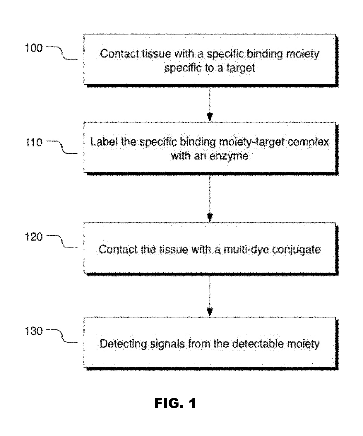
[0278]The Tyramide-TAMRA-FITC Multi-Dye Conjugate was used in an assay to stain the Ki67 marker in tonsil tissue as described in Example 2. A primary antibody specific to Ki67 was first introduced to the tonsil tissue sample to form an antibody-target complex. A secondary antibody was then introduced, namely an anti-Ki67 antibody conjugated to a horseradish peroxidase enzyme. Upon introduction of the Tyramide-TAMRA-FITC Multi-Dye Conjugate, the horseradish peroxidase converted the Tyramide-TAMRA-FITC Multi-Dye Conjugate to a reactive intermediate which was then able to bond to tissue, either proximal to or directly on the labeled Ki67 target. As illustrated in FIG. 6, the Tyramide-TAMRA-FITC Multi-Dye...


PUM


Abstract

Disclosed herein are novel chromogenic conjugates, the conjugates comprising at least two detectable moieties.

Description

CROSS REFERENCE TO RELATED APPLICATIONS[0001]This application is a continuation of PCT / EP2017 / 065795, filed Jun. 27, 2017, which claims the benefit of U.S. Provisional Application No. 62 / 355,398, filed Jun. 28, 2016, the content of each of which is incorporated herein by reference in its entirety.BACKGROUND OF THE DISCLOSUREField of the Invention[0002]The present disclosure relates to the field of chemistry and diagnostics.Description of the Related Art[0003]Immunohistochemistry (IHC) refers to the processes of detecting, localizing, and / or quantifying antigens, such as a protein, in a biological sample using antibodies specific to the particular antigens. IHC provides the substantial advantage of identifying exactly where a particular protein is located within the tissue sample. It is also an effective way to examine the tissues themselves. In situ hybridization (ISH) refers to the process of detecting, localizing, and quantifying nucleic acids. Both IHC and ISH can be performed on...

Claims


Application Information

Patent Timeline
IPC IPC(8): G01N33/533G01N33/58C09B11/24C09B23/08
CPCG01N33/533G01N33/582G01N33/583C09B11/24C09B23/083G01N33/532
Inventor ASHWORTH-SHARPE, JULIAKELLY, BRIAN D.LEFEVER, MARKPOLASKE, NATHAN W.
Owner VENTANA MEDICAL SYST INC
Why Eureka
Try Eureka