Methods and compositions comprising cationic lipids for stimulating type 1 interferon genes

a technology of cationic lipids and interferon genes, which is applied in the direction of antibody medical ingredients, dsdna viruses, immunological disorders, etc., can solve the problems of failure to establish long-lasting protective or therapeutic benefits, and achieve the effect of effective prevention and robust cytotoxic t cell immune respons

Pending Publication Date: 2019-10-24
PDS BIOTECH
View PDF1 Cites 4 Cited by
  • Summary
  • Abstract
  • Description
  • Claims
  • Application Information

AI Technical Summary

Benefits of technology

[0016]Provided herein are novel methods and compositions for vaccines comprising cationic lipids as potent activators of IFN1 (IFN-α/β/γ). As described herein, the present compositions enable the induction of an effective and robust cytotoxic T cell immune response when administered to a host. The compositions comprise cationic lipids, optionally combined with peptides or protein antigens; in certain embodiments, the cationic lipids are in...

Problems solved by technology

However, most adjuvants that are currently approved for human use are poor inducers of type I interfe...

Method used


Image

  • Methods and compositions comprising cationic lipids for stimulating type 1 interferon genes
  • Methods and compositions comprising cationic lipids for stimulating type 1 interferon genes
  • Methods and compositions comprising cationic lipids for stimulating type 1 interferon genes

Examples


example 1

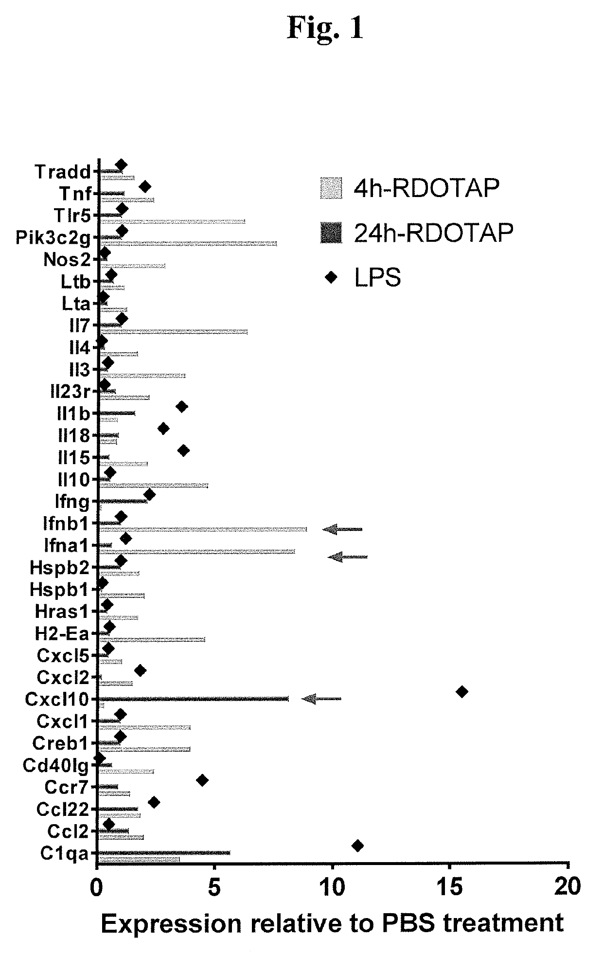
R-DOTAP Induces Type I Interferon Gene Expression in Draining Lymph Nodes

[0095]To assess cationic nanoparticle induced gene expression, C57BL / 6J mice were injected with 100 μl of 12 mM R-DOTAP nanoparticles subcutaneously behind the neck, and inflammatory gene expression in draining lymph nodes analyzed 4 or 24 hours post vaccination. Mice injected with PBS, and LPS (50 μg / mouse) (24 h time point) were used as negative and positive controls respectively. 4 h or 24 hr after injection, axillary and brachial draining lymph nodes from each mouse were pooled and processed by collagenase digestion. Activated dendritic cells (CD11c+) cells in the lymph node cell suspensions were sort-purified and lysed in RLT buffer and processed for relative gene expression analysis using nCounter® mouse inflammation kit and nanostring technologies. R-DOTAP injection was found to significantly alter the expression of several inflammatory genes at both time points. Several genes including IFN-1a and IFN-1β...

example 2

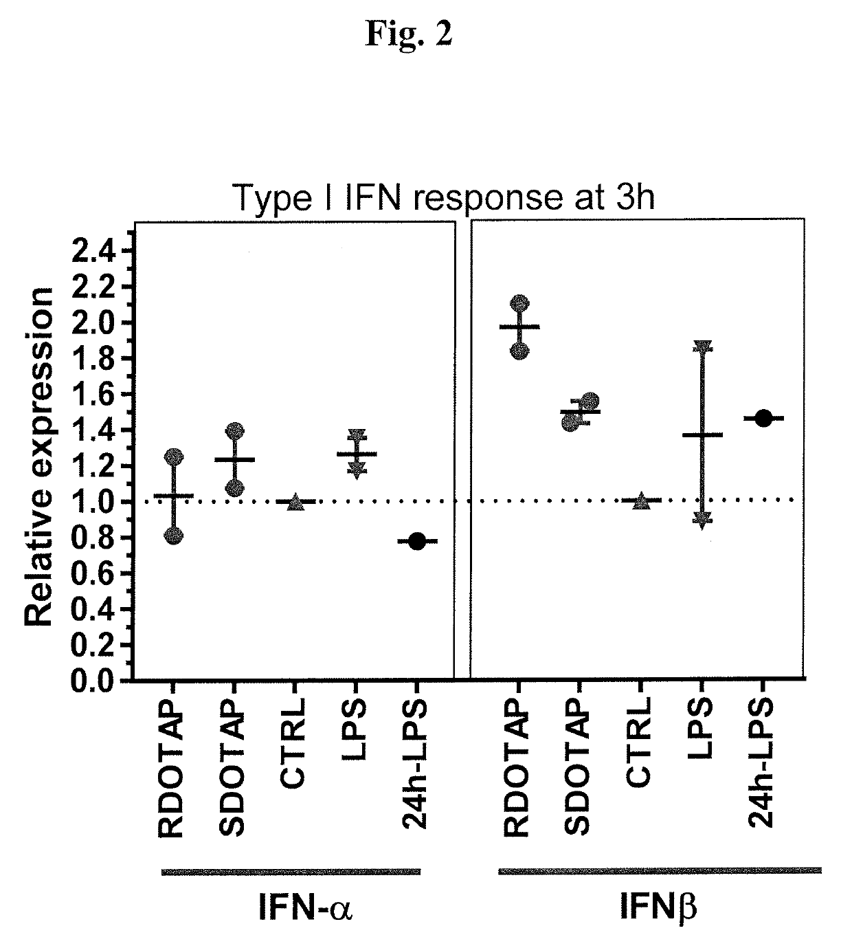
R-DOTAP and S-DOTAP Both Induce Interferon-α and Interferon-β Gene Expression in Draining Lymph Nodes

[0096]To compare type I Interferon gene expression by both R- and S-DOTAP, a similar study as outlined in Example 1 was performed. C57BL / 6J mice were injected with 50 μl of 6 mM RDOTAP nanoparticles subcutaneously in the foot pads. Mice injected with PBS, and LPS (50 μg / mouse) (24 h time point) were used as negative and positive controls respectively. 3 h or 24 hr after injection, popliteal draining lymph nodes from each mouse were pooled and lysed in RLT buffer and processed for relative gene expression analysis using Taqman® gene expression assay and RT-PCR. The studies confirm that both R- and S-DOTAP are strong inducers of the type I interferons (FIG. 2).

example 3

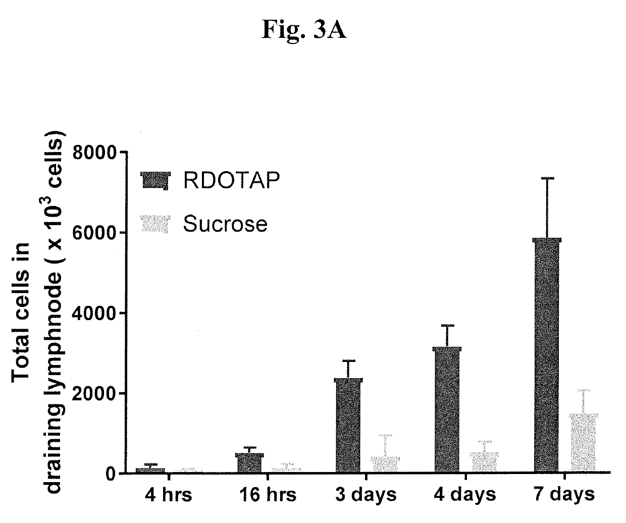
R-DOTAP and S-DOTAP Upregulate Type I Interferon-Associated CD69 Expression on T-cells

[0097]To further understand the effect and ability of cationic lipids to activate the type I interferons, C57BL / 6J mice or IFNAR− / − mice were injected with 50 μl of 6 mM R-DOTAP nanoparticles or 6 mM S-DOTAP or 280 mM sucrose subcutaneously in the foot pads. 24 hr after injection, popliteal draining lymph nodes from each mouse were isolated and single cell suspensions of lymph nodes were stained with fluorochrome conjugated CD3 and CD69. R-DOTAP alone (without antigen) resulted in a visible increase in DLN size and this was due to a steady increase in total cell number over a 7-day period (FIG. 3A). Studies of T-Cell influx into the DLN with pertussis toxin suggest that lipids structurally related to R-DOTAP induce lymph node homing chemokines, most likely a direct result of type I IFN signaling. This increase in total cell number was confirmed to be dependent on type I IFN signaling as it was grea...


PUM

PropertyMeasurementUnit
Immunogenicityaaaaaaaaaa

Abstract

Methods and compositions for modifying type I IFN signaling pathways in a subject comprising the administration a cationic lipid to the subject are provided. The cationic lipids comprise 1,2-dioleoyl-3-trimethylammonium propane (DOTAP), N-1-(2,3-dioleoyloxy)-propyl-N,N,N-trimethyl ammonium chloride (DOTMA), 1,2-dioleoyl-sn-glycero-3-ethylphosphocholine (DOEPC), enantiomers and combinations thereof. The compositions may further comprise one or more antigenic components, wherein such components are autologous or nonautologous.

Description

CROSS-REFERENCE TO RELATED APPLICATIONS[0001]The present application claims priority to and all the benefits of U.S. Provisional Patent Application No. 62 / 594,815, filed on Dec. 5, 2017, which is hereby expressly incorporated herein by reference in its entirety.BACKGROUND OF THE INVENTION1. Field of the Invention[0002]The present invention relates generally to methods and compositions for modifying type I IFN signaling pathways comprising the administration of cationic lipids.2. Description of the Related Art[0003]Type I interferons (IFN-I) in the last decade have been shown to be of critical importance in the mammalian immune response against disease. Type I interferons have been called “viral” interferons due to their direct induction by viral infections, in contrast to “immune” IFN, or IFN-γ, which is synthesized after receptor engagement of T cells and natural killer (NK) cells during immune responses. IFN-I are also well-documented inducers of tumor cell apoptosis and anti-angi...

Claims


Application Information

Patent Timeline
IPC IPC(8): A61K31/21A61K39/12
CPCA61K31/21A61K39/12A61K39/39A61K9/127A61K31/685A61K2039/55555A61K2039/55566A61K2039/572A61K2039/585C12N2710/20034A61P37/04A61P35/00A61K45/06A61K31/231A61K39/0011
Inventor BEDU-ADDO, FRANKCONN, GREGGANDHAPUDI, SIVA K.WARD, MARTINWOODWARD, JEROLD
Owner PDS BIOTECH
Why Eureka
Try Eureka