
Methods and materials for using fibrin supports for retinal pigment epithelium transplantation

a fibrin support and pigment epithelium technology, applied in the direction of prosthesis, drug composition, peptide/protein ingredient, etc., can solve the problems of rpe dysfunction, eventual photoreceptor death, many difficulties in translation to clinical application,

Active Publication Date: 2020-02-27
MAYO FOUND FOR MEDICAL EDUCATION & RES
View PDF0 Cites 1 Cited by
  • Summary
  • Abstract
  • Description
  • Claims
  • Application Information

AI Technical Summary

Benefits of technology

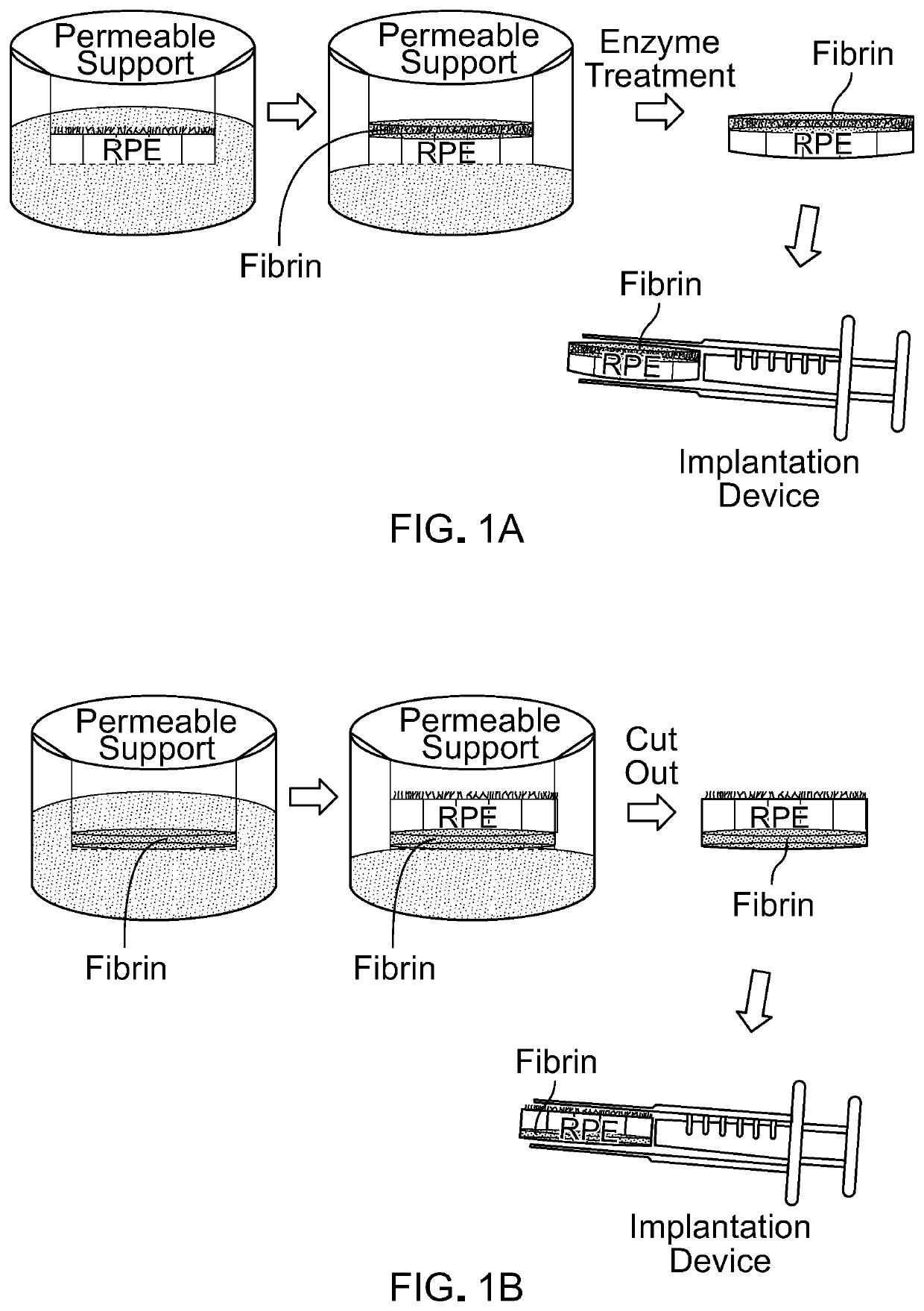
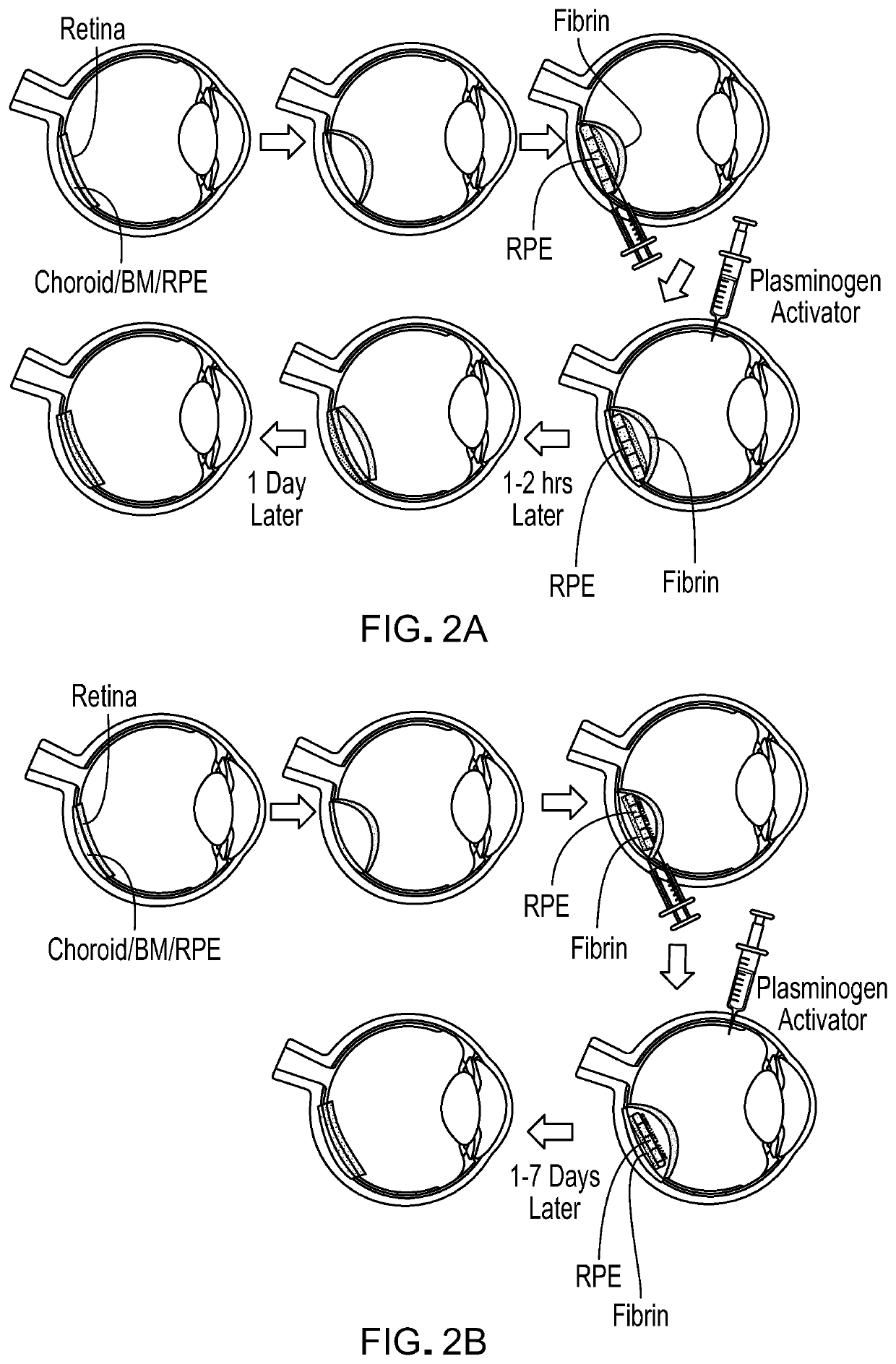
The patent describes a method for using fibrin as a substrate for retinal pigment epithelium (RPE) transplantation. The fibrin is prepared as a thin layer and can be tailored to degrade quickly in the eye. The resulting fibrin-RPE (FRPE) implant can be detached easily and the cells remain viable and phenotypically stable. The fibrin scaffold can be placed on the apical or basal side of the RPE monolayer for better attachment. Multiple fibrin monolayers can be combined to cover large areas, and laser tacking can be used for precise delivery location. The fibrin scaffold can also be degraded under controlled conditions during surgery. Overall, this method provides a safe and effective way to deliver RPE cells for transplantation.

Problems solved by technology

The bestrophinopathies (e.g., Best's disease) arise from a mutation in the Best1 gene, causing RPE dysfunction leading to eventual photoreceptor death.
While in vitro successes of RPE transplantation have been attained, many difficulties have risen in translation towards clinical application.
However, transplantation was characterized by low percentage of RPE attachment and survival.
However, a concern presented was the inability to maintain a flat, wrinkle-free monolayer through the surgical procedure.
Because of this slow degradation, the material can remain between the RPE and choroid after implantation for several months to years, causing concerns of chronic inflammation and fibrosis, low permeability and potentially reduced RPE survival.
Additionally, due to the rigidity of the material, there is concern of damage to the underlying choroid, as seen in previous animal studies (Diniz et al., Invest. Ophthalmol. Vis. Sci., 54:5087-5096 (2013)).

Method used


Image

  • Methods and materials for using fibrin supports for retinal pigment epithelium transplantation
  • Methods and materials for using fibrin supports for retinal pigment epithelium transplantation
  • Methods and materials for using fibrin supports for retinal pigment epithelium transplantation

Examples


example 1

brin Hydrogels for IPSC-RPE Transplantation

Chemicals

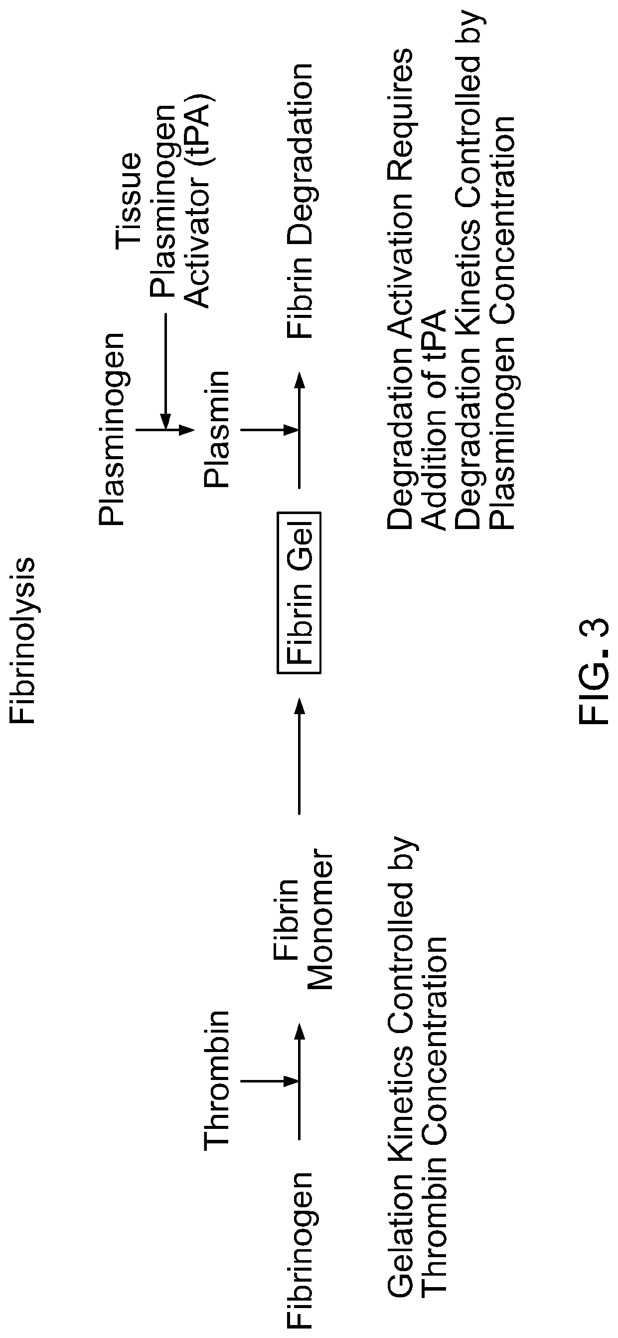
[0087]Fibrinogen was obtained from three sources: as Evicel from Ethicon (60 mg / mL), as Tisseel from Baxter (95 mg / mL), and as research grade material from Sigma-Aldrich (57 mg / mL). Thrombin also was obtained from three sources: part of Evicel from Ethicon, part of Tisseel from Baxter, and research grade material from Sigma-Aldrich. Plasminogen was obtained as research grade material from Sigma-Aldrich. Recombinant tissue plasminogen activator (tPA) was obtained as research grade material from Sigma-Aldrich.

Cells

[0088]IPSC-RPE cells were produced as described elsewhere with modification (Johnson et al., Investig. Ophthalmol. Vis. Sci., 56:4619 (2015)). A membrane support was utilized with apical and basal media, including either transwell or HA membrane. The membrane surface was coated with a collagen gel, per manufacturer's protocol, and, either subsequently or alternatively, coated with a geltrex or matrigel solution, up to 0.1 m...

example 2

f Aprotinin on Fibrin Attachment and Cell Viability

[0113]Studies were conducted to determine the effect of Aprotinin on fibrin gel attachment and maintenance, and on cell viability. iPSC-RPE cells were cultured for two weeks on a fibrin gel with (FIG. 14A) and without (FIG. 14B) geltrex coating, in media containing 50 U / mL Aprotinin. The inclusion of Aprotinin in the media appeared to prevent fibrin gel degradation. In addition, these studies indicated that attachment of the cells to the fibrin gel may occur independent of the presence of geltrex. The cells remained adherent after the gel was released from the plates (FIG. 15A), and there was minimal cell removal after the gel was cut (FIG. 15B).

[0114]To assess the viability of iPSC-RPE cells in fibrin gel after culture with Aprotinin for two weeks, followed by detachment and cutting of the gel, cells were stained with calcein-AM (Live) and ethidium homodimer (Dead). The cells remained viable after detachment and cutting, whether ge...

example 3

for Retinal Pigment Epithelium Monolayer with Apical Fibrin

[0116]1) Gel 2.5 mg / mL collagen onto cellulose ester membrane filter insert.[0117]a. 1.0-5.0 mg / mL collagen.[0118]b. Cellulose ester, polycarbonate, PTFE, TCPS membrane filter.[0119]2) Coat the collagen surface with 1:5 dilution of matrigel.[0120]a. Range: 1:1-1:50 dilution.[0121]b. Matrigel, geltrex or purified laminin.[0122]3) Plate cells at 0.5×10⁶ cells / cm2.[0123]a. 0.1-2.0×10⁶ cells / cm2.[0124]4) Culture for about 2 weeks.[0125]a. 1-6 weeks.[0126]5) Wash cells with PBS.[0127]6) Dry cell surface.[0128]7) Spray 80 μL of mixed 50 mg / mL fibrinogen, 2 U / mL plasminogen, and 100 U / mL thrombin, at total flow rate of 80 uL / sec, 0.8 bar for 1 second at a height of 10 cm.[0129]a. Spray 30-200 μL of mix.[0130]b. 30-70 mg / mL fibrinogen.[0131]c. 0.1-4.0 U / mL plasminogen.[0132]d. 10-600 U / mL thrombin.[0133]e. 30-400 μL / seconds flow rate.[0134]f. 0.6-1.2 bar.[0135]g. 5-15 cm height.[0136]8) Allow fibrinogen to gel 1 hour 37° C.[0137]a. ...


PUM

PropertyMeasurementUnit
Thicknessaaaaaaaaaa
Thicknessaaaaaaaaaa
Massaaaaaaaaaa

Abstract

This document provides methods and materials for performing retinal pigment epithelium transplantation. For example, methods and materials for using fibrin supports for retinal pigment epithelium transplantation are provided.

Description

BACKGROUND1. Technical Field[0001]This document relates to retinal pigment epithelium transplantation. For example, this document relates to methods and materials for using fibrin supports for retinal pigment epithelium transplantation.2. Background Information[0002]Macular degeneration diseases represent a variety of diseases and etiology, but commonly stem from retinal pigment epithelium (RPE) dysfunction. Genetic macular degenerations, including the bestrophinopathies, occur due to protein mutations involved in RPE function. The bestrophinopathies (e.g., Best's disease) arise from a mutation in the Best1 gene, causing RPE dysfunction leading to eventual photoreceptor death. The prevalence has previously been reported as 1 in 16,000-21,500 (Dalvin et al., Ophthalmic Genet., Epub:1-5 (2016)). While the genetically-caused macular degenerations are rare, age-related macular degeneration (AMD) is the leading cause of blindness in the first world. It is estimated to account for 5 milli...

Claims


Application Information

Patent Timeline
IPC IPC(8): A61L27/38C12N5/079A61K35/36A61K35/30A61K38/36A61K38/48A61L27/22A61L27/52A61L27/34A61L27/36
CPCA61K35/30A61L27/3839A61K38/484A61L27/52C12Y304/21007C12N2501/734A61L27/225C12N5/0621C12N2501/999A61L27/3813C12N2533/56C12N2533/90A61K35/36A61L27/3687A61L2430/16A61L27/34A61L2420/02A61L27/3808A61K38/363C12N2506/45A61K38/57C12N5/062A61K31/195A61K31/197A61K2300/00A61L27/54A61P27/02
Inventor MARMORSTEIN, ALAN D.IEZZI, RAYMONDGANDHI, JAREL K.PULIDO, JOSE S.
Owner MAYO FOUND FOR MEDICAL EDUCATION & RES
Learn More