
Indicator Fluids, Systems, and Methods for Assessing Movement of Substances Within, To or From a Cerebrospinal Fluid, Brain or Spinal Cord Compartment of a Cranio-Spinal Cavity of a Human

a technology of indicator fluids and cerebrospinal fluids, which is applied in the direction of diagnostics, sensors, and fluorescence emission detection. it can solve the problems of low accuracy, low accuracy, and low accuracy of measurement results

Active Publication Date: 2020-06-04
BRAINWIDESOLUTIONS AS
View PDF0 Cites 1 Cited by
  • Summary
  • Abstract
  • Description
  • Claims
  • Application Information

AI Technical Summary

Benefits of technology

The patent describes an invention that provides a reference indicator fluid for standardizing magnetic resonance imaging (MRI) acquisitions. The invention also includes an extra-corporal device that allows for determining absolute values of changes in signal fields. This device contains different concentrations of a selectable MRI contrast agent, which can be connected to the MRI coil. The invention has several advantages, including the ability to accurately compare repeated MRI acquisitions within individuals and between individuals, and to express changes in signal fields within different body tissue compartments. The invention also involves using multiple containers with different concentrations of contrast agent to estimate changes in signal fields. Overall, the invention provides a novel and innovative solution for MRI scanner calibration and human imaging applications.

Problems solved by technology

No methods have been established for determination of clearance function from the cranio-spinal cavity.
One reason may be that there are yet no medical treatment strategies to modify removal of substances from the cranio-spinal cavity, making it less relevant at present.
A drawback with MRI is that recorded SUs are highly dependent on the MRI scanner and parameters of the image sequence.
Therefore, absolute values of T1 weighted SU may not be measured quantitatively, making it difficult to directly compare T1 SUs between individuals and at different time points within an individual.
Another approach is T1-mapping, which may provide for absolute values, but is time-consuming and has low image resolution, and is currently applied mostly for research purposes.
However, phantoms are not used while performing scanning of patients, only for scanner calibration.
This compound is metabolized within the cell initially but is unable to progress on to the citric acid cycle, and is also difficult for the cell to excrete.

Method used


Image

  • Indicator Fluids, Systems, and Methods for Assessing Movement of Substances Within, To or From a Cerebrospinal Fluid, Brain or Spinal Cord Compartment of a Cranio-Spinal Cavity of a Human
  • Indicator Fluids, Systems, and Methods for Assessing Movement of Substances Within, To or From a Cerebrospinal Fluid, Brain or Spinal Cord Compartment of a Cranio-Spinal Cavity of a Human
  • Indicator Fluids, Systems, and Methods for Assessing Movement of Substances Within, To or From a Cerebrospinal Fluid, Brain or Spinal Cord Compartment of a Cranio-Spinal Cavity of a Human

Examples


example # 1

Example #1

[0728]Alzheimer's disease and dementia in general. The present invention provides for establishing information about the ability of movement of molecular substances within and from the cranio-spinal cavity in early Alzheimer's disease, or in individuals at risk of Alzheimer's disease, and other variants of dementia. The entorhinal cortex has an important role in cognition and dementia development. Changes within the entorhinal cortex are seen early in dementia. For example, the grid cells, which were described by the 2014 Nobel Prize recipients from NTNU in Norway, are in the entorhinal cortex. The present invention explores paravascular transport within this region as a function of contrast agent within nearby CSF compartment, and as a function of values from the extra-body device. Early changes in paravascular transport within this area may be seen in early dementia development. Hence, impaired paravascular transport within the various layers of the entorhinal cortex may...

example # 2

Example #2

[0729]Brain tumors. The present invention provides for an alternative way of assessing brain tumor invasion. This is particularly relevant for gliomas in areas of the brain not readily detected with current MRI techniques. There are no reliable methods for in vivo characterization of spread of glioma cells. Conventional MRI may present with normal signals though tumor cells are present. Glioma cells typically spread along the outside of the vessels of the brain and within the extracellular space. Diffusion techniques have only revealed alterations in limited areas close to the tumor. Conventional techniques apply intravenous contrast agents, which are restricted to the vascular (intra-vascular) compartment because of the BBB, and only distributes in local, extra-vascular spaces when the BBB is disrupted by tumor. With the present invention (e.g. Aspect 5), we provide a method that may reveal events on the outer side of the vessels. The present invention may reveal spread o...

example # 3

Example #3

[0730]Multiple sclerosis and inflammatory brain disease. The present invention provides for a method of macroscopically imaging the integrity of paravascular fluid spaces in demyelinating disease, which is a perivascular inflammation. This seems to be a main aspect of the pathogenesis of the disease. It is expected that Gd-based contrast agents will facilitate a signal drop in T1 weighted images. Attaching compounds with affinity for inflammation cells to an indicator fluid, such as an MRI contrast agent, may cause SU to increase in areas of inflammatory cell invasion. By this method, assessment of disease load and disease activity may be provided.


PUM


Abstract

The present invention discloses indicator fluids, reference indicator fluid, and usage thereof, and systems and methods for assessing movement of molecular substances within, to or from a cerebrospinal fluid, brain or spinal cord compartment of a human cranio-spinal cavity. Indicator fluid moving from the cerebrospinal fluid compartment enables measurements of levels of indicator fluid in blood or urine and assessment of the cranio-spinal cavity's ability to remove molecular substances. The indicator fluids may be contrast agents used for imaging, such as by computed tomography imaging, and magnetic resonance imaging, or imaging utilizing radioactive substances by positron emission tomography, single-photon emission computed tomography or scintigraphy. Using these imaging modalities, the invention describes indicator fluids, systems and methods enabling assessment of movement of substances within, to or from a cerebrospinal fluid, brain or spinal cord compartment of a cranio-spinal cavity, and from the human cranio-spinal cavity to lymphatic pathways or kidneys.

Description

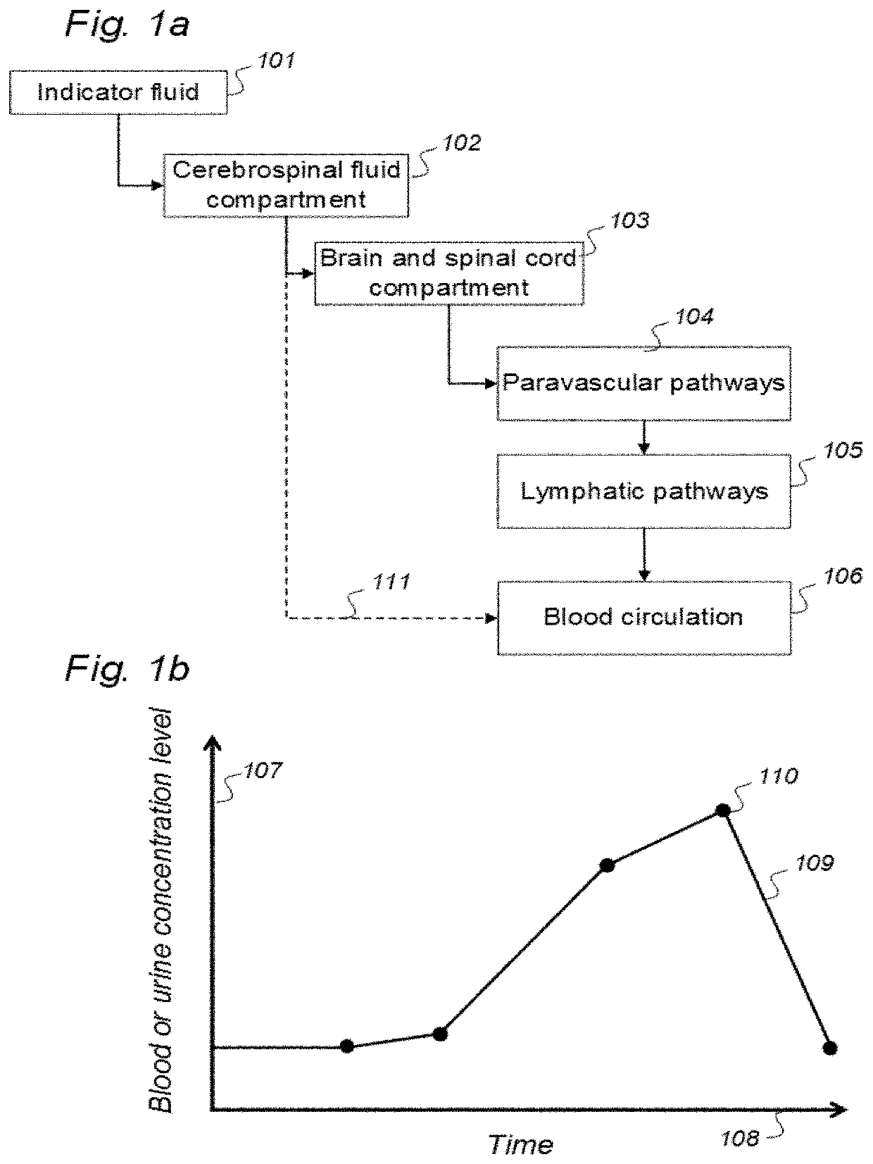
BACKGROUND OF THE INVENTIONField of the Invention[0001]The present invention relates to various aspects of indicator fluids, systems, methods and devices for assessing movement of substances within, to or from a cerebrospinal fluid (CSF), brain or spinal cord compartment of a cranio-spinal cavity of a human.[0002]More specifically, the present invention relates to indicator fluids, reference indicator fluid, and usage thereof, as well as systems and methods for assessing movement of substances within, to or from a cerebrospinal fluid, brain or spinal cord compartment of a cranio-spinal cavity of a human as defined in an introductory part of attached independent claims.[0003]The three main elements of the cranio-spinal cavity are i) CSF compartment, ii) brain and spinal cord tissue compartment, and iii) vascular (blood) compartment. The central nervous system (CNS) consists of the brain and the spinal tissue cord, being confined within a cranio-spinal cavity, and residing within the ...

Claims


Application Information

Patent Timeline
Patent Type & Authority Applications(United States)
IPC IPC(8): A61B5/00A61B6/00A61K49/04A61K49/00A61K51/04
CPCA61B5/055A61K51/048A61K49/0002A61K49/106A61K49/0438A61B6/501A61B2576/026G01R33/5601A61B6/037A61B6/481A61B5/0035A61K49/0004A61B5/4082A61B5/748A61B5/4088A61B5/4076A61B5/14546A61B6/032G16H30/40A61B5/0071A61B5/4064G01R33/5608
Inventor EIDE, PER KRISTIANRINGSTAD, GEIR ANDRE
Owner BRAINWIDESOLUTIONS AS
Learn More