Extracellular Matrix for Treating Pelvic Floor Disorders and Skeletal Muscle Degeneration

a technology of extracellular matrix and pelvic floor, which is applied in the field of extracellular matrix for treating pelvic floor disorders and skeletal muscle degeneration, can solve the problems of poor cell survival and increased connective tissue, and achieve the effects of preventing the development of pfd, promoting differentiation of muscle progenitor cells, and facilitating endogenous cell infiltration

Active Publication Date: 2020-06-25
RGT UNIV OF CALIFORNIA
View PDF0 Cites 0 Cited by
  • Summary
  • Abstract
  • Description
  • Claims
  • Application Information

AI Technical Summary

Benefits of technology

[0013]In embodiments the invention provides an injectable biomaterial scaffold and a minimally invasive delivery system for the treatment of US, EAS, and PFM. Upon injection, the material will set up into a porous and fibrous scaffold that will facilitate endogenous cell infiltration to regenerate and heal the damaged muscles post-vaginal delivery thereby preventing the development of PFD.
[0014]The present invention provides a new use for ECM materials that have already shown to promote differentiation of muscle progenitors in vitro (DeQuach et al., PLoS One, 2010), as well as migration of muscle progenitors, decrease in cell death, and increase in neovascularization and muscle development in vivo in hindlimb ischemia models of muscle damage (DeQuach et al., ECM, 2012; Ungerleider, et al., JACC: BTS, 2016). This approach has significant clinical application as injectable scaffolds can be delivered with minimal invasiveness, thereby enabling their administration in labor and delivery units and outpatient settings, and reducing patient recovery time and perioperative morbidity compared to sur

Problems solved by technology

Despite this, the current standard treatments are compensatory, as they do not directly target sphincteric and supportive muscle dysfunction and do not reverse the existing injury or halt functional

Method used


Image

  • Extracellular Matrix for Treating Pelvic Floor Disorders and Skeletal Muscle Degeneration
  • Extracellular Matrix for Treating Pelvic Floor Disorders and Skeletal Muscle Degeneration
  • Extracellular Matrix for Treating Pelvic Floor Disorders and Skeletal Muscle Degeneration

Examples


example 1

Injectable Skeletal Muscle Matrix

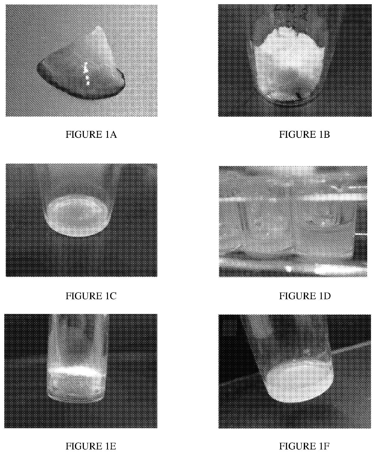
[0104]Skeletal muscle matrix material was derived through decellularization of porcine skeletal muscle tissue (FIG. 1A). Fat and connective tissue was removed, and the skeletal muscle was cut into ˜1 cm3 pieces, rinsed with deioninized water and stirred in 1% (wt / vol) solution of SDS in PBS for 4-5 days. The decellularized muscle was then stirred overnight in deionized water, and agitated rinses under running deionized water were performed to remove residual SDS. In addition to confirmation with lack of nuclei on H&E stained sections (not shown), the DNA content of the material was measured as 26.14±1.67 ng of DNA / mg of dry weight ECM, which confirmed decellularization. The matrix was then lyophilized (FIG. 1B) and milled into a fine particulate. At this stage the material can be hydrated and utilized for in vivo injection or it can be enzymatically digested to form a liquid (FIG. 1C). At this stage, the liquid skeletal muscle matrix can be diluted a...

example 2

Mitogenic Assay

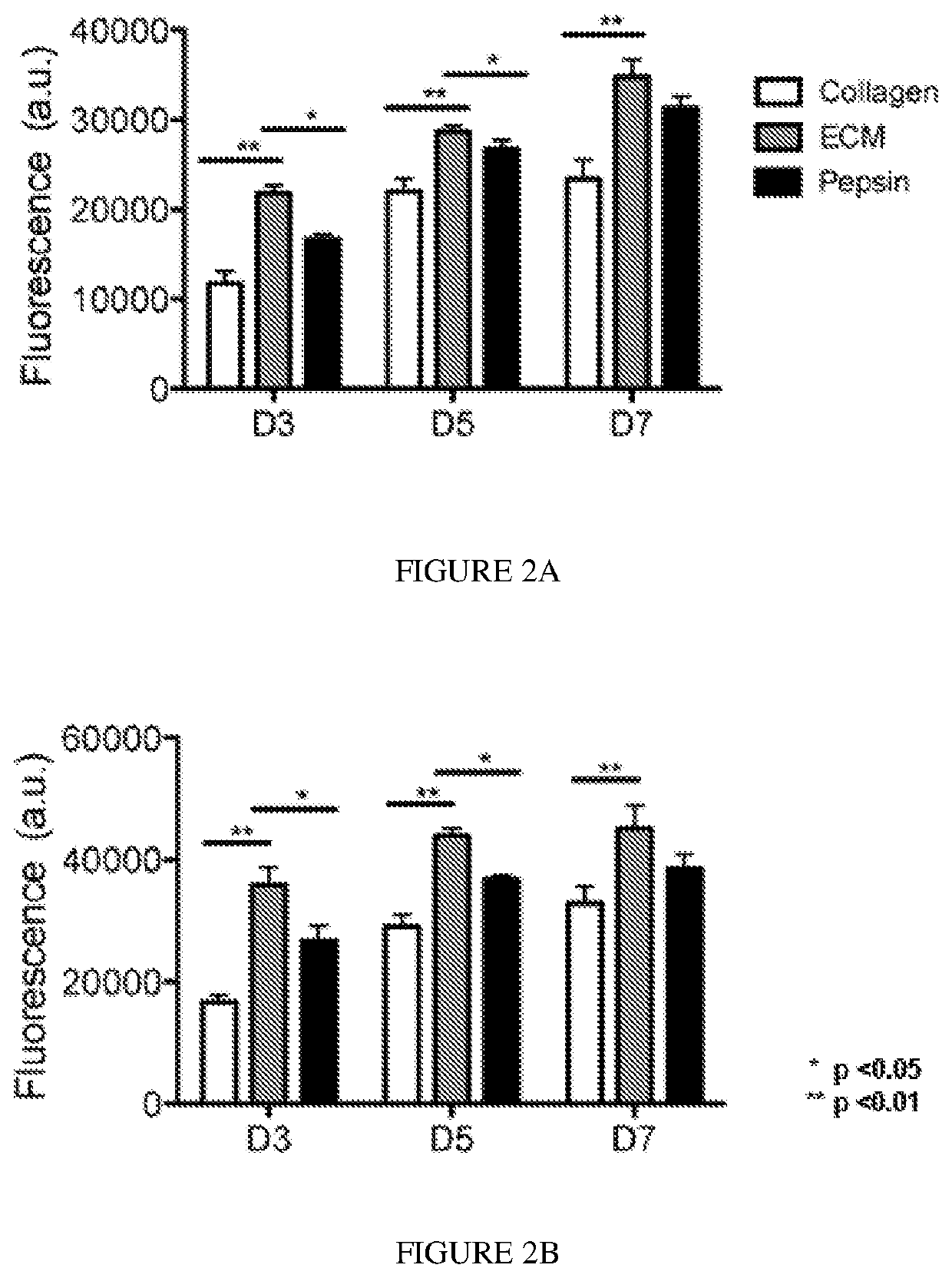
[0105]Degradation products of decellularized ECMs have been previously shown to have mitogenic activity. It was examined whether the degradation products of the skeletal muscle matrix hydrogel had a mitogenic effect on cells in vitro. Proliferation of smooth muscle cells and skeletal myoblasts following exposure to either enzymatically degraded skeletal muscle ECM or collagen was assessed. Pepsin was also included as a control, as pepsin was utilized to digest the matrix material. A Picogreen assay was used to determine double stranded DNA content at days 3, 5, and 7 in culture to quantify cell proliferation. It was found that both smooth muscle cells (FIG. 2A) and myoblasts (FIG. 2B), when cultured in media containing degraded skeletal muscle matrix, had a higher rate of proliferation compared to cells cultured in media containing the same concentration of collagen. The increase in cell number was significantly greater at all time points (p<0.01). At day 3, there was...

example 3

Gelation In Vitro and In Vivo

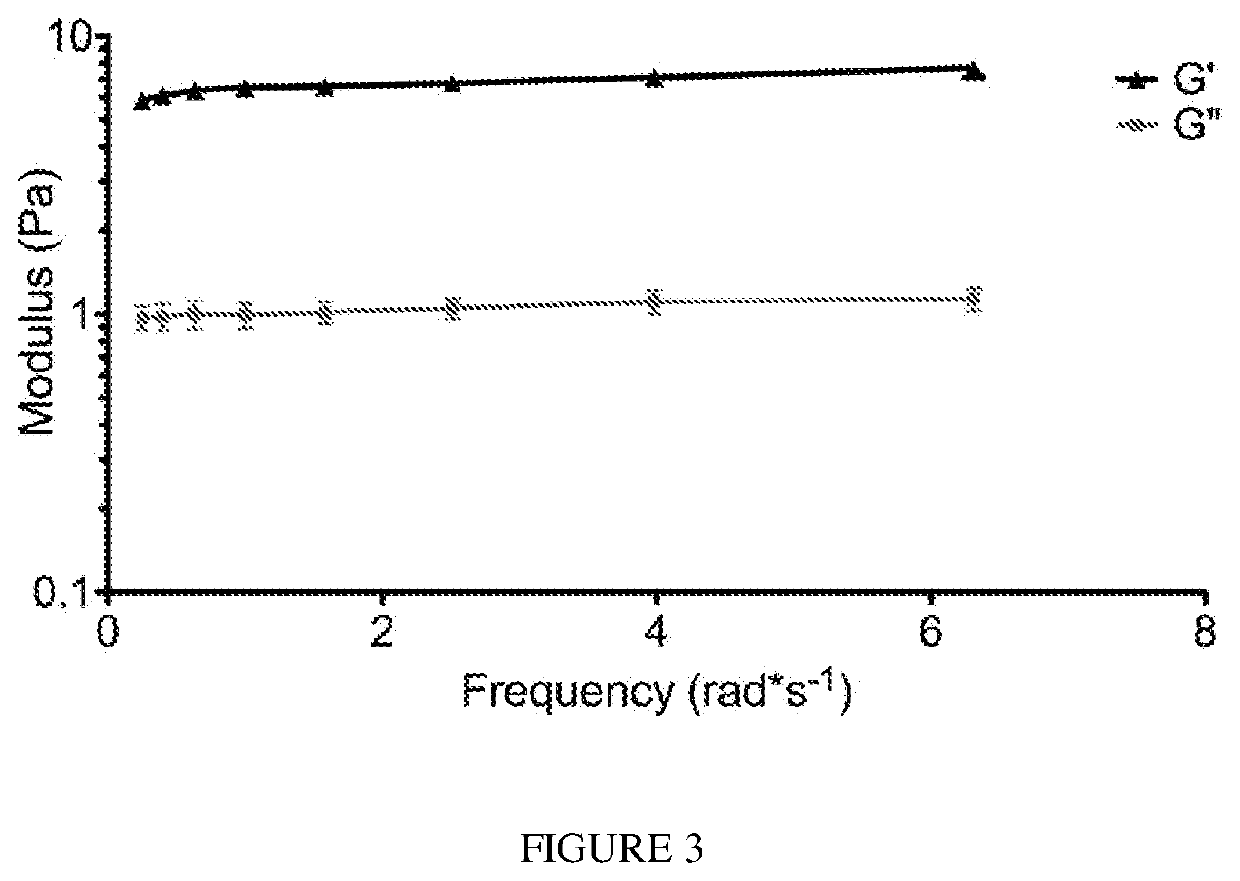
[0106]A gel form of the matrix was initially made in vitro by bringing the material (6 mg / mL) to a physiological pH and incubating the material at 37° C. After gelation, the material was tested for rheological properties where it was determined that the material had a storage modulus (G′) of 6.5±0.5 Pa. A representative trace of rheological data is shown in FIG. 3. The ability of the liquid skeletal muscle matrix to form a gel in situ were then assessed by injecting the material into a healthy rat hindlimb. For all in vivo studies, liquid skeletal muscle matrix, which had been biotinylated and re-lyophilized for storage at −80 C were utilized. Prior to injection, the material was resuspended in sterile water alone. The skeletal muscle matrix was then loaded into a syringe and injected intramuscularly into a rat hindlimb (FIG. 4A). To determine whether the skeletal muscle matrix would assemble and form a scaffold, the injection region was excised after 20...


PUM


Abstract

Described herein are compositions comprising decellularized extracellular matrix derived from skeletal muscle or other suitable tissue, and therapeutic uses thereof. Methods for treating, repairing or regenerating defective, diseased, damage, ischemic, ulcer cells, tissues or organs in a subject preferably a human, with diseases associated with muscular degeneration, using a decellularized extracellular matrix of the invention are provided. Methods of preparing culture surfaces and culturing cells with absorbed decellularized extracellular matrix are provided.

Description

CROSS REFERENCE TO RELATED APPLICATION[0001]This application claims the priority benefit of U.S. Provisional Patent Application No. 62 / 507,850, filed May 18, 2017, the entire contents of which is incorporated herein by reference.STATEMENT OF GOVERNMENT INTEREST[0002]This invention was made with government support under Grant No. R01HL113468 awarded by National Institutes of Health (NIH). The government has certain rights in the invention.BACKGROUND OF THE INVENTION[0003]The extracellular matrix (ECM) of each tissue contains similar components; however, each individual tissue is composed of a unique combination of proteins and proteoglycans (Lutolf and Hubbell, 2005; Uriel et al., 2009). Recent studies have shown that the ECM of various tissues can be isolated through decellularization and utilized as a tissue engineering scaffold (Merritt et al.; Ott et al., 2008; Singelyn et al., 2009; Uygun et al., 2010; Valentin et al., 2010; Young et al., 2011). Other decellularized ECM material...

Claims


Application Information

Patent Timeline
IPC IPC(8): A61L27/36A61L27/58
CPCA61L27/367A61L2400/06A61L2430/30A61L27/58A61L27/3683A61L27/3633A61L27/3604A61L27/50A61K35/34
Inventor CHRISTMAN, KARENWARD, SAMUELALPERIN, MARIANNADURAN, PAMELA
Owner RGT UNIV OF CALIFORNIA
Why Eureka
Try Eureka