
Surgical visualization and medical imaging devices and methods using near infrared fluorescent polymers

a near infrared fluorescent and surgical visualization technology, applied in the field of medical imaging, can solve the problems that surgeons may not be able to fully visualize the target site and discern important aspects about the surgical si

Pending Publication Date: 2020-07-23
UNIVERSITY OF ROCHESTER
View PDF3 Cites 3 Cited by
  • Summary
  • Abstract
  • Description
  • Claims
  • Application Information

AI Technical Summary

Benefits of technology

The present invention is a medical device that emits near infrared light to penetrate biological material such as tissue to a certain depth. This allows surgeons to better visualize different types of tissue during procedures. The device contains a polymer and a near infrared fluorescent dye embedded in the polymer, which produces near infrared fluorescence that can penetrate biological material. The device can be a bowel sizer, vaginal manipulator, uterine manipulator, ureteral catheter, endotracheal tube, or feeding tube, among others. The near infrared fluorescent dye used is indocyanine green. The polymer used is polycaprolactone. The device can be excited with a first frequency of near infrared radiation, and the second frequency of near infrared radiation that has penetrated the tissue to a depth of about ten millimeters can be detected and displayed for viewing. The method includes modulating at least one of intensity, angle of incidence, and duty cycle of the near infrared radiation source. This allows for better visualization of biological material during medical procedures.

Problems solved by technology

While robotically assisted surgeries allow for less invasive and more complex surgical procedures than was possible with traditional surgical approaches, the surgeon may not be able to visualize the target site fully and to discern important aspects about the surgical site, such as the depth and quality of the tissue and the presence and quality of different types of tissue, particularly since robotic surgeries are performed in the absence of any haptic feedback about the tissue.

Method used


Image

  • Surgical visualization and medical imaging devices and methods using near infrared fluorescent polymers
  • Surgical visualization and medical imaging devices and methods using near infrared fluorescent polymers
  • Surgical visualization and medical imaging devices and methods using near infrared fluorescent polymers

Examples


example 1

[0043]To prepare indocyanine green (ICG) for use in an exemplary medical device, ethanol was added to indocyanine green. 10 mil, of ethanol was added to 25 mg of ICG and mixed gently. Steralloy 2380A resin and Steralloy 2380B curative were mixed in the proportion of 50 ml, of 2380B with 200 ml, of 2380A. 2 ml, of the ethanol and ICG solution were added to the resin and curative combination. To provide a resulting surgical tool that has the proper aesthetic qualities, Steralloy PD-7 MP Opaque white color dispersion was added to the mixture. The ICG solution and color dispersion was added to the Steralloy 2380B curative and the resulting mixture was added to the Steralloy 2380A resin. The resulting mixture (approximately 20 parts per million of ICG) was poured into a mold to cast the resulting surgical device. It should recognized that other approaches, such as adding ICG or an ICG solution to a plastic feedstock prior to injection molding, blow molding, extruding, 3D printing, or the...

example 2

[0044]An enhanced NIR polymer was produced according to the present invention by embedding an enhanced NIR powder formed from ICG and milk powder into poly(caprolactone) (PCL; 2-oxypanone), a biocompatible thermoplastic material that is often used in FDA-approved devices such as suture materials, e.g. Monocryl (Ethicon), and then molding the ICG embedded polymer into the desired shape for the medical device. Milk powder was selected over semolina as NIR powder formed from ICG and semolina resulted in a coarse grain and some undesirable properties when used repeatedly under different heating conditions and with repeated water immersion. By contrast, when an ICG and DMSO mixture was used, the resulting polymer was only sufficient fluorescent for about 24 hours as the DMSO dissipated from the material. To achieve the finer grain powder, 50 g of dry evaporated milk was rehydrated with about 100 cc of neat ethanol and ˜500 m of water was then added to 2.5 mg of ICG in 1 ml of water from ...

example 3

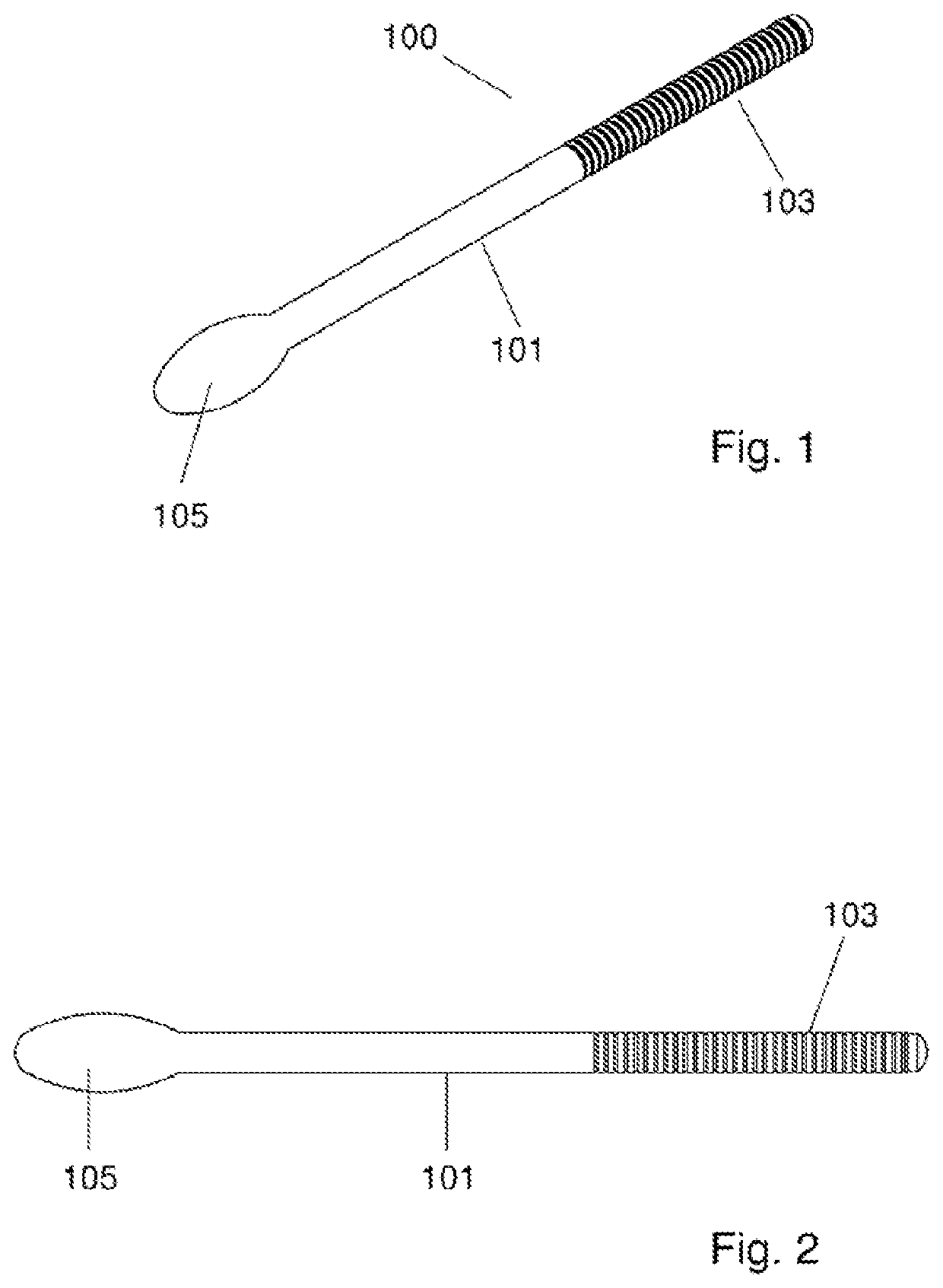
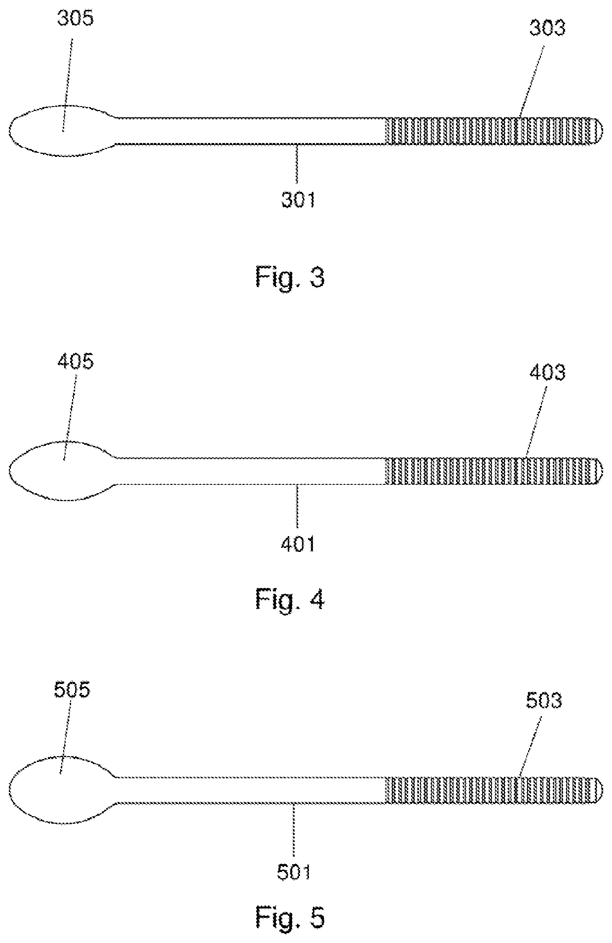
[0046]FIGS. 1-5 illustrate a surgical visualization and medical imaging device in the form of a surgical bowel sizer 100. Bowel sizer 100 comprises a shaft 101, a grip 103 and a head 105. Shaft 101 may be of various lengths dependent on the surgical task required. Head 105 may be, for example, in the range of 25-33 millimeters in length. Bowl sizer 100 is made, at least in part, from a NIR polymer according to the present invention. For example, the entire bowler 100, just head 105, or just a portion of head 105 may be formed from the NIR polymer for near infrared fluorescence when exposed to near infrared radiation.


PUM

PropertyMeasurementUnit
depthaaaaaaaaaa
depth of penetrationaaaaaaaaaa

Abstract

A surgical visualization and medical imaging device and related computer based imaging methods and systems are disclosed. The surgical devices of the present invention use indocyanine green dye combined with a plastic, and are used in enhanced surgical imaging in applications such as robotically assisted surgeries. A near infrared light source, such as an 805 nm laser, may be used to excite the surgical device so that the device emits 835 nm light. Both the excitation and emission wavelengths penetrate tissue and blood, and provide enhanced imaging of surgical procedures. The resulting fluorescence image allows a user to readily determine relative tissue depth, to identify tissue inhomogeneity, to detect masses or tissue irregularities, to pinpoint anatomical holes, and to visualize tears.

Description

CROSS REFERENCE TO RELATED PATENT APPLICATIONS[0001]This application claims priority to U.S. Patent Application Ser. No. 62 / 460,802 filed Feb. 18, 2017 entitled “Surgical Visualization and Medical Imaging Devices and Methods Using Near Infrared Fluorescent Polymers” by Doyle, Reeder and Wood, the entire disclosure of which is incorporated herein by reference as permissible by national or regional laws.TECHNICAL FIELD[0002]This invention relates generally to medical imaging and, more specifically, to devices and methods using near infrared fluorescent polymers for enhanced imaging.BACKGROUND ART[0003]Various surgical techniques require the use of surgical assist devices such as manipulators, sizers, backers, and the like. These surgical assist devices help with the identification of anatomical structures and operative tissues by actions such as physically moving, stretching, and relocating tissue at the surgical site. For example, surgical assist devices provide form and structure to...

Claims


Application Information

Patent Timeline
Patent Type & Authority Applications(United States)
IPC IPC(8): A61B90/30A61B5/107A61B17/42A61B1/04A61B5/00A61B34/30
CPCA61B90/30A61B2090/373A61B1/043A61B5/0086A61B5/1076A61B17/4241A61B1/06A61B2090/304A61B34/30A61B1/267A61B2090/062A61K49/0034A61B2017/00831A61B5/0071A61B2017/00818A61B90/37
Inventor DOYLE, PAULA JAYEREEDER, JAY EVANWOOD, RONALD WESLEY
Owner UNIVERSITY OF ROCHESTER
Learn More