Method for spectral study of a biological fluid

a biological fluid and spectral study technology, applied in biological testing, color/spectral properties measurement, material testing goods, etc., can solve the problems of participating in significant organ damage, exerting toxic effects, local vasoconstriction,

Active Publication Date: 2020-10-29
INST NAT DE LA SANTE & DE LA RECHERCHE MEDICALE (INSERM) +3
View PDF3 Cites 0 Cited by
  • Summary
  • Abstract
  • Description
  • Claims
  • Application Information

AI Technical Summary

Benefits of technology

[0024]It is also proposed to use the spectral data obtained by the previous method to determine the content of one or more proteins present in a biological fluid chosen from HbO2, MetHb, bilirubin, serum albumin/haem, haemopexin/haem, carboxylated haemoglobin (HbCO), a biomarker of haem catabolism by h...

Problems solved by technology

Such a biochemical reaction depends on the haem oxygenase activity which, although overexpressed by the haem, may be insufficient to catabolize an excess of the haem generating reactive oxygen species (ROS), or even alteration of the cell membrane and ultimately vasoconstriction and local inflammation.
The haem, as well as the haemoglobin, may then exert toxic effects by extravasation through the wall of the vessels, accumulation on the surface of the endothelium by reaction with the NO produced by the NO synthase causing local vasoconstriction and participating in significant organ damage (kidney, liver, spleen, heart, brain, lungs).
While ...

Method used


Image

  • Method for spectral study of a biological fluid
  • Method for spectral study of a biological fluid
  • Method for spectral study of a biological fluid

Examples


example 1 (fig.1)

Example 1 (FIG. 1)

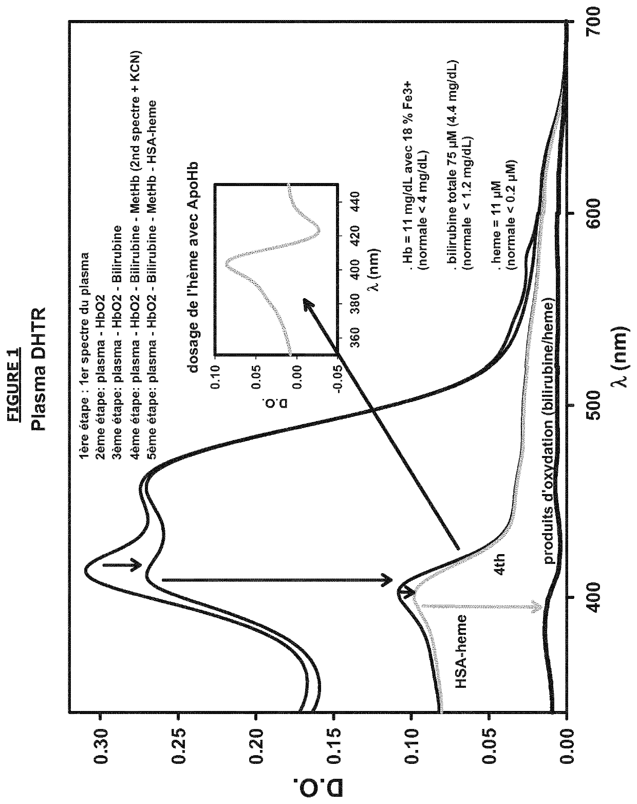
[0293]UV / visible absorption spectrum of a DHTR patient. The different species linked to the haem were measured by a mathematical approach based on experimental spectral measurements coupled to the spectra of the pure forms of each species considered.

[0294]Materials and methods: plasma diluted ⅙th (optical path 4 mm) in a PBS 50 mM potassium phosphate 50 mM NaCl pH 7.4. Addition of 0.2 mM KCN from a 50 mM stock solution. Or 2.5 μl in 600 μl total of diluted plasma (dilution factor 1.004).

[0295]first absorption spectrum: measured spectrum of plasma

[0296]second intermediate calculated spectrum: the HbO2 component was removed by subtraction of a completely oxygenated haemolysate spectrum in air (see FIG. 5a) based on the second derivative between 500 and 600 nm (typically, the peak at 577 nm presents an intense negative band for the second derivative). In fact, the narrow peaks in this absorption domain only come from the HbO2 spectrum while the other major spectral co...

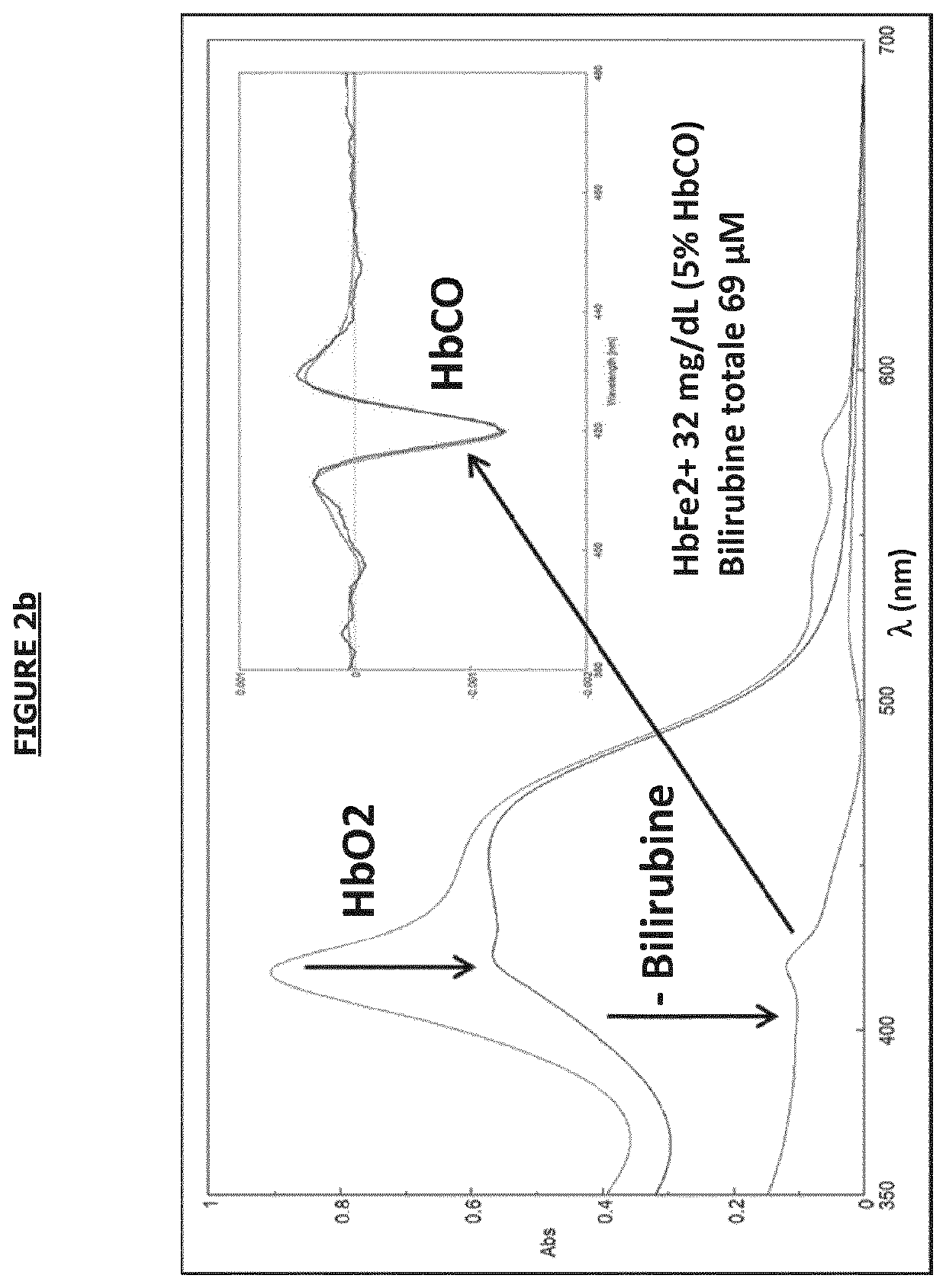
example 2 (figs.2a , b , c , d)

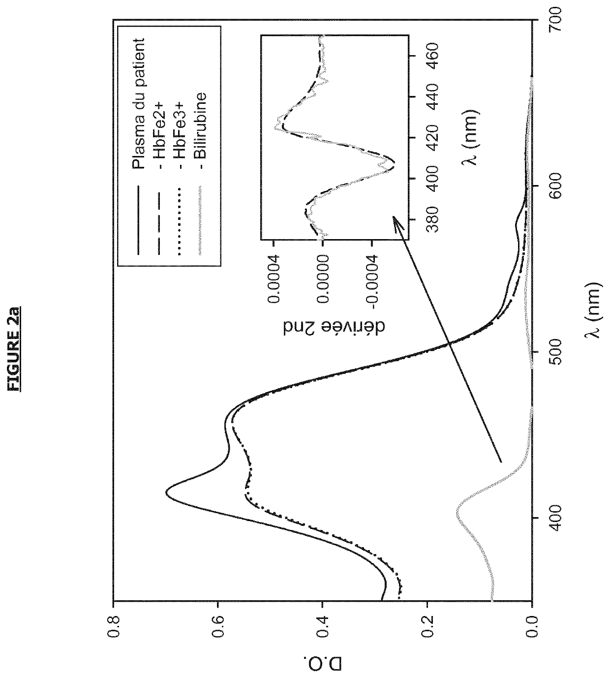
Example 2 (FIGS. 2a, b, c, d): Assay on a Sample of Sickle Cell Patient with a Mechanical Heart Valve Problem

[0305]After subtraction of the different spectral components using the second derivatives for HbFe2+(O2), bilirubin-HSA and the determination of HbFe3+ after addition of KCN, the presence of plasma haem was revealed in purple here in a sickle cell patient with a mechanical heart valve problem (FIG. 2a).

[0306]Materials and methods: plasma diluted ⅙th (optical path 4 mm) in a PBS 50 mM potassium phosphate 50 mM NaCl pH 7.4. Addition of 0.2 mM KCN from a 50 mM stock solution. Or 2.5 μl in 600 μl total of diluted plasma (dilution factor 1.004).

[0307]Results: following the same methodology as in FIG. 1, the biomarker values for haemolysis are: plasma haemoglobin 13 μM (5% Fe3+), total bilirubin 130 μM, plasma haem 15 μM. Likewise, the absence of binding of the haem to haemopexin indicates a virtual depletion of this “scavenger” of the haem.

[0308]In the insert is shown the second d...

example 3 (figs.3a , b)

Example 3 (FIGS. 3a, b): Determination of Total Plasma Bilirubin

[0321]Materials and methods: plasma from a patient with low intravascular haemolysis (hereditary stomatocytosis with dehydration) diluted ⅙th (optical path 1 cm) in a 50 mM PBS 50 mM potassium phosphate NaCl pH 7.4. The low spectral contribution of plasma Hb was subtracted as described in FIG. 1. After dissolution of the lyophilized human HSA “fatty free” in this same buffer then addition of bilirubin (

[0322]Results: The plasma spectrum after subtraction of HbO2 is shown in FIG. 3a. It corresponds to the spectral form of bilirubin (unconjugated and more weakly conjugated) linked to HSA. This spectrum is similar to that obtained from the reference spectra of bilirubin bound to human HSA (commercial products). In fact the second derivatives are almost identical (FIG. 3b).

[0323]Conclusions: The preferable maximum at ...


PUM


Abstract

The aim of the present invention is to propose a novel method for spectral study of a biological fluid from an absorption spectrum of a sample of the biological fluid. The inventors have made an original use of spectroscopy in order to accurately determine the protein content of a biological sample, in particular for oxyhaemoglobin, methaemoglobin, bilirubin and the haem bound to serum albumin. Such a method can be used in a variety of applications concerning haem- or haemoprotein- related diseases, in particular diagnostic methods, treatment follow-up methods, methods for determining biomarkers or screening methods. Such a method is also advantageous for qualifying blood bags.

Description

TECHNICAL FIELD[0001]The invention relates to a method for the spectral study of a biological fluid starting from an absorption spectrum of a sample of the biological fluid. The invention also relates to the use of spectral data obtained by the spectral study method to determine the content of one or more proteins, in particular haemoproteins as well as the haem and its degradation derivatives. The invention relates to a number of applications in the case of diseases related to the haem and to the haemoprotein. The invention also relates to a computer program comprising instructions for the execution of the steps of a method for spectral study of a biological fluid and a recording medium that is readable by a computer and on which the said program is recorded.TECHNOLOGICAL BACKGROUND[0002]Haemolytic diseases including sickle cell anaemia, thalassaemia, membrane pathologies of the red blood cell, enzyme deficits (glucose 6-phosphate dehydrogenase or G6PD), autoimmune haemolytic anaem...

Claims


Application Information

Patent Timeline
IPC IPC(8): G01N21/31G01N33/48
CPCG01N21/31G01N33/48
Inventor KIGER, LAURENTMARDEN, MICHAELBARTOLUCCI, PABLOPIRENNE, FRANCE
Owner INST NAT DE LA SANTE & DE LA RECHERCHE MEDICALE (INSERM)
Why Eureka
Try Eureka