Systems and methods for selectively occluding the superior vena cava for treating heart conditions

a technology of superior vena cava and selective occlusion, which is applied in the field of methods and systems for improving cardiac function, to achieve the effect of increasing the urine flow of patients

Pending Publication Date: 2021-03-18
TUFTS MEDICAL CENTER INC
View PDF0 Cites 3 Cited by
  • Summary
  • Abstract
  • Description
  • Claims
  • Application Information

AI Technical Summary

Benefits of technology

[0040]In another preferred embodiment, the RVAD includes a catheter having a proximal end and a distal region, the distal region having an inflow end and an outflow end, the catheter sized and shaped for placement through a femoral vein of the patient so that the outflow end is disposed in a pulmonary artery of the patient and the inflow end is disposed in an IVC of the patient. The RVAD also includes a pump, e.g., an impeller pump, disposed on the distal region of the catheter, wherein the pump may be selectively actuated to pump blood from the IVC through the inflow end and expel blood into the pulmonary artery via the outflow end, and an RVAD controller operatively coupled to the RVAD to actuate the pump to pump blood from the IVC to the pulmonary artery, thereby unloading the right ventricle. The RVAD controller operatively coupled to the catheter of the system may regulate the activation and deactivation of the flow limiting element to at least partially occlude the SVC simultaneously as the RVAD controller actuates the pump to pump blood from the IVC to the pulmonary artery.
[0041]The system may also be used for modulating a heart of a patient and performing a cardiac procedure. This approach may involve inserting a first catheter comprising a flow limiting element into a superior vena cava (SVC) of the patient such that the flow limiting element is disposed within the SVC and actuating the flow limiting element within the SVC, thereby at least partially occluding the SVC. A cardiac procedure may be performed at the heart of the patient prior to, during, and / or after at least partially occluding the SVC. The flow limiting element may be de-actuated and re-actuated within the SVC prior to or during the cardiac procedure. Actuation of the flow limiting element may stimulate the vagal nerve and increase urine flow of the patient.

Problems solved by technology

Heart failure is a major cause of global mortality.
Heart failure often results in multiple long-term hospital admissions, especially in the later phases of the disease.
Absent heart transplantation, the long-term prognosis for such patients is bleak, and pharmaceutical approaches are palliative only.
Consequently, there are few effective treatments to slow or reverse the progression of this disease.
Heart failure can result from any of multiple initiating events.
While the heart can often initially respond successfully to the increased workload that results from high blood pressure or loss of contractile tissue, over time this stress induces compensatory cardiomyocyte hypertrophy and remodeling of the ventricular wall.
This in turn often leads to overworking of the myocardium, such that the cardiac muscle in the compromised region becomes progressively thinner, enlarged and further overloaded.
Simultaneously, the ejection fraction of the damaged ventricle drops, leading to lower cardiac output and higher average pressures and volumes in the chamber throughout the cardiac cycle, the hallmarks of heart failure.
Not surprisingly, once a patient's heart enters this progressively self-perpetuating downward spiral, the patient's quality of life is severely affected and the risk of morbidity skyrockets.
Depending upon a number of factors, including the patient's prior physical condition, age, sex and lifestyle, the patient may experience one or several hospital admissions, at considerable cost to the patient and social healthcare systems, until the patient dies either of cardiac arrest or any of a number of co-morbidities including stroke, kidney failure, liver failure, or pulmonary hypertension.
Currently, there are no device-based solutions that specifically target a reduction in preload to limit the progression of heart failure.
Pharmaceutical approaches are available as palliatives to reduce the symptoms of heart failure, but there exists no pharmaceutical path to arresting or reversing heart failure.
Moreover, the existing pharmaceutical approaches are systemic in nature and do not address the localized effects of remodeling on the cardiac structure.
Prior to applicants' invention as described herein, there are no effective commercial devices available to treat this disease.
Although the system and methods described in Cedeno appear promising, there are a number of potential drawbacks to such a system that applicants' have discovered during their own research.
Moreover, full inferior vena cava occlusion may increase venous congestion within the renal, hepatic, and mesenteric veins; venous congestion is a major cause of renal failure in congestive heart failure patients.
There are several major limitations to approaches that involve partial or full occlusion of the IVC to modulate cardiac filling pressures and improve cardiac function.
If approached via the jugular or subclavian veins, the apparatus would have to traverse the superior vena cava and right atrium, thereby requiring cardiac penetration, which predisposes to potential risk involving right atrial injury, induction of arrhythmias including supraventricular tachycardia or bradycardia due to heart block.
Second, the IVC approach described by Cedeno and colleagues depends on several highly variable indices (especially in the setting of congestive heart failure): 1) IVC diameter, which is often dilated in patients with heart failure; b) intermittent (full or partial) IVC occlusion may cause harm by increasing renal vein pressure, which reduces glomerular filtration rates and worsens kidney dysfunction; c) dependence on the patient's ability to breathe, which is often severely impaired in HF (A classic breathing pattern in HF is known as Cheynes Stokes respiration, which is defined by intermittent periods of apnea where the IVC may collapse and the balloon will cause complete occlusion resulting in lower systemic blood pressure and higher renal vein pressure); d) if prolonged cardiac unloading is required to see a clinical improvement or beneficial changes in cardiac structure or function, then IVC occlusion will not be effective since sustained IVC occlusion will compromise blood pressure and kidney function.
Fourth, many patients with heart failure have IVC filters due to an increased propensity for deep venous thrombosis, which would preclude broad application of IVC therapy.
Pulmonary hypertension (PH) is also a major cause of morbidity and mortality worldwide.
However, there are drawbacks to pharmacologic treatments such as costs and side effects.
Without proper valve coaptation, the valve will not seal properly and blood may flow back through the valve resulting in regurgitation.
Regurgitation may cause shortness of breath, fatigue and rapid and / or fluttering heartbeat.
Yet another concern with overloading is that it complicates cardiac procedures, such as corrective procedures for regurgitation.
However, if the volume in a heart chamber is too great, deploying the clip may be impossible as the leaflets may be too far apart.
Overloading is also known to occur during and / or after installation of a ventricular assist device (VAD).
For example, during and / or after installation of a left ventricular assist device (LVAD), the right ventricle may become overloaded.
However, this involves installing and / or deploying a second heart pump resulting in added risk of complications and infection.

Method used


Image

  • Systems and methods for selectively occluding the superior vena cava for treating heart conditions
  • Systems and methods for selectively occluding the superior vena cava for treating heart conditions
  • Systems and methods for selectively occluding the superior vena cava for treating heart conditions

Examples


Embodiment Construction

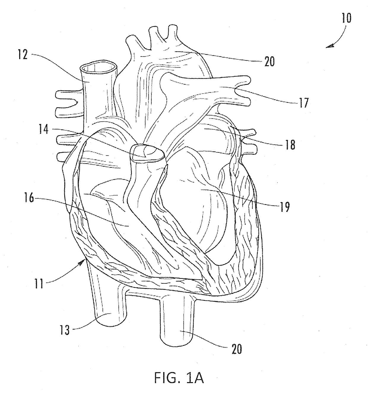
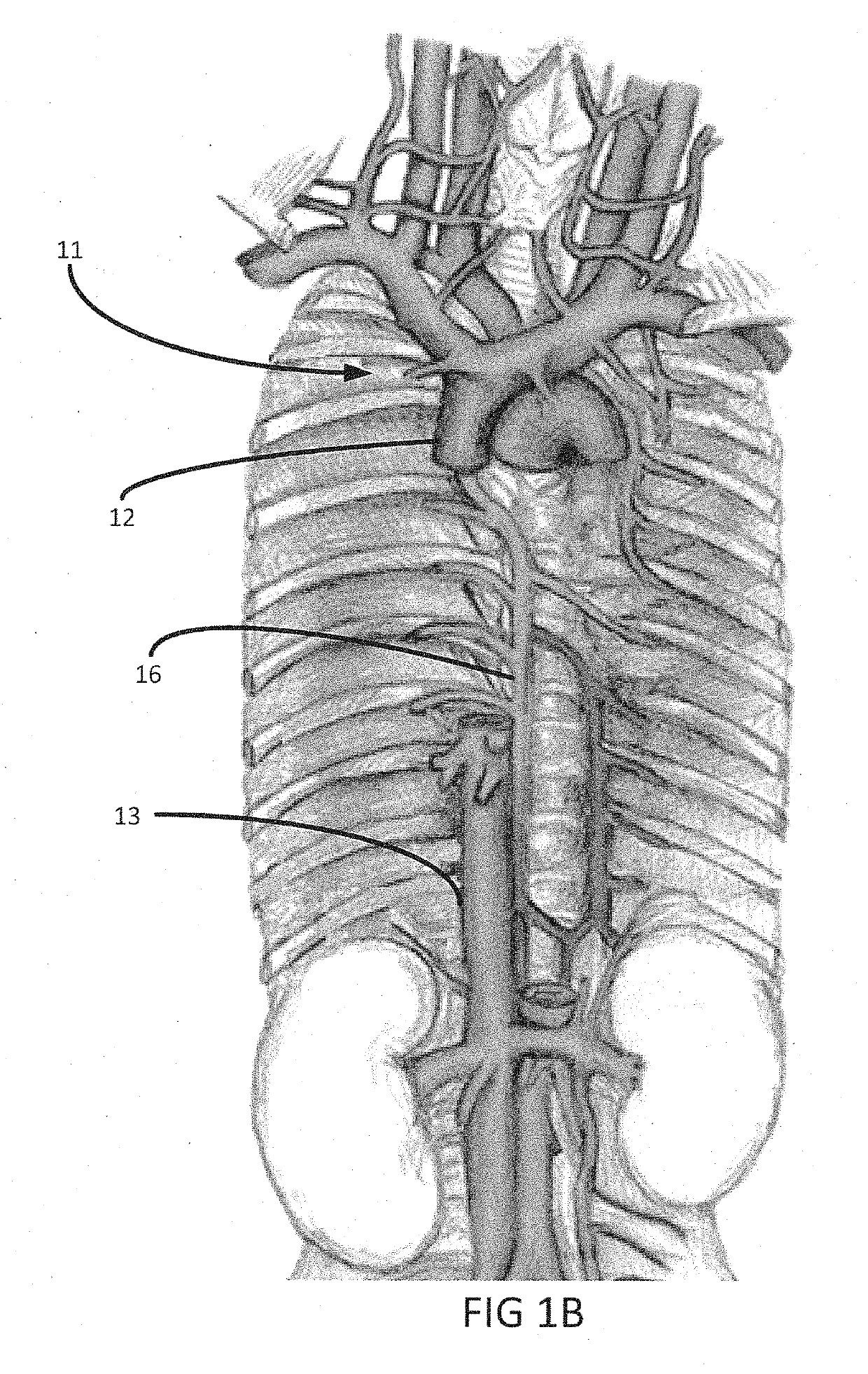
[0107]Referring to FIGS. 1A and 1B, the human anatomy in which the present invention is designed for placement and operation is described as context for the system and methods of the present invention.

[0108]More particularly, referring to FIG. 1A, deoxygenated blood returns to heart 10 through vena cava 11, which comprises superior vena cava 12 and inferior vena cava 13 coupled to right atrium 14 of the heart. Blood moves from right atrium 14 through tricuspid valve 15 to right ventricle 16, where it is pumped via pulmonary artery 17 to the lungs. Oxygenated blood returns from the lungs to left atrium 18 via the pulmonary vein. The oxygenated blood then enters left ventricle 19, which pumps the blood through aorta 20 to the rest of the body.

[0109]As shown in FIG. 1B, superior vena cava 12 is positioned at the top of vena cava 11, while inferior vena cava 13 is located at the bottom of the vena cava. FIG. 1B also shows azygos vein 16 and some of the major veins connecting to the vena...


PUM


Abstract

Systems and methods are provided for treating conditions such as heart failure and/or pulmonary hypertension by at least partially occluding flow through the superior vena cava for an interval spanning multiple cardiac cycles. A catheter with an occlusion device is provided along with a controller that actuates a drive mechanism to provide at least partial occlusion of the patient's superior vena cava, which reduces cardiac filling pressures, and induces a favorable shift in the patient's Frank-Starling curve towards healthy heart functionality and improved cardiac performance. The system may include sensors to determine the degree of occlusion of the superior vena cava. The occlusion system may be used to reduce volume in a heart and facilitate a cardiac procedure. The occlusion system may be used to relieve an overloaded chamber during and/or after deploying a VAD.

Description

CROSS-REFERENCE TO RELATED APPLICATIONS[0001]This application claims priority to U.S. Provisional Application Ser. No. 62 / 939,524, filed on Nov. 22, 2019, the entire contents of which are incorporated herein by reference. This application is also a continuation-in-part of U.S. patent application Ser. No. 16 / 402,194, filed on May 2, 2019, now U.S. Pat. No. 10,842,975, which is a continuation of U.S. patent application Ser. No. 15 / 203,437, filed Jul. 6, 2016, now U.S. Pat. No. 10,279,152, which is a continuation of U.S. patent application Ser. No. 14 / 828,429, filed Aug. 17, 2015, now U.S. Pat. No. 9,393,384, the entire contents each of which are incorporated herein by reference. This application is also a continuation-in-part of U.S. patent application Ser. No. 16 / 168,357, filed on Oct. 23, 2018, now U.S. Pat. No. 10,842,974, which claims priority to U.S. Provisional Application Series No. 62 / 642,569, filed Mar. 13, 2018, and U.S. Provisional Application Ser. No. 62 / 576,529, filed Oct...

Claims


Application Information

Patent Timeline
Patent Type & Authority Applications(United States)
IPC IPC(8): A61M25/10A61B5/00A61B5/0215A61B5/024A61B5/026A61B5/042A61M1/10A61M1/12A61B17/12
CPCA61M25/10184A61B2017/00022A61B5/6858A61B5/746A61B5/0215A61B5/02438A61B5/026A61B5/042A61M1/106A61M1/127A61B17/12036A61B17/1204A61B17/12109A61B17/12136A61M1/1043A61M1/125A61M1/1086A61M25/10188A61B17/12172A61B5/6853A61B5/283A61B2017/00044A61B2017/00115A61B2017/00154A61B2017/00199A61B2017/00221A61B2017/00411A61B2017/00734A61M60/135A61M60/40A61M60/50A61M60/871A61B2017/00243A61B2017/0003A61B2090/065A61B2017/00084A61B17/12013A61B2017/00557A61M60/295A61M60/289A61M60/161A61M60/13A61M60/216A61M60/523A61M60/531A61M60/515A61B2017/00889A61M2025/1052A61M2205/3331A61M2230/005A61M2230/30A61B2017/00398A61M60/268A61B5/29
Inventor KAPUR, NAVIN K.KARAS, RICHARD H.
Owner TUFTS MEDICAL CENTER INC
Why Eureka
Try Eureka