
Method for determining the lipid content of extracellular vesicles

a lipid content and vesicle technology, applied in the field of extracellular vesicles, can solve the problems of insufficient free of co-isolated proteins/protein aggregates in ev preparations to make such approaches, and the availability of standardised quantification approaches for reliable and reproducible standardised quantification of evs is limited

Pending Publication Date: 2021-09-23
RES CENT FOR NATURAL SCI
View PDF0 Cites 1 Cited by
  • Summary
  • Abstract
  • Description
  • Claims
  • Application Information

AI Technical Summary

Benefits of technology

The present invention provides methods for quantifying extracellular vesicles (EVs) and lipids in a sample. The methods involve contacting an EV preparation with sulphuric acid and phospho-vanillin reagent, allowing a color reaction to develop, and measuring the absorbance of the resulting solution. The methods are performed using a reference suspension containing phospholipids and a reference absorbance value. The invention also provides a method for quantifying biological membranes in a sample. The methods are useful for research and diagnostic purposes.

Problems solved by technology

However, the availability of approaches for reliable and reproducible standardised quantification of EVs is limited [4, 5, 6].
Quantifying EVs is not as straightforward as it seems.
One of the main problems of these techniques is that proteins may be co-isolated with EVs as protein aggregates, and EVs share biophysical parameters with them [7].
Therefore EV preparations are usually not sufficiently free of co-isolated proteins / protein aggregates to make such approaches reproducible or accurate (the apparent concentration of EVs in the preparations is therefore overestimated).
However, these methods are error-prone because they cannot distinguish vesicular and non-vesicular structures.
Mihály et al [9] discloses a method for characterization of EVs wherein the protein / lipid (P / L) ratio can be determined using infrared (IR) spectroscopy but the technique is generally not available in most of the biomedical laboratories.
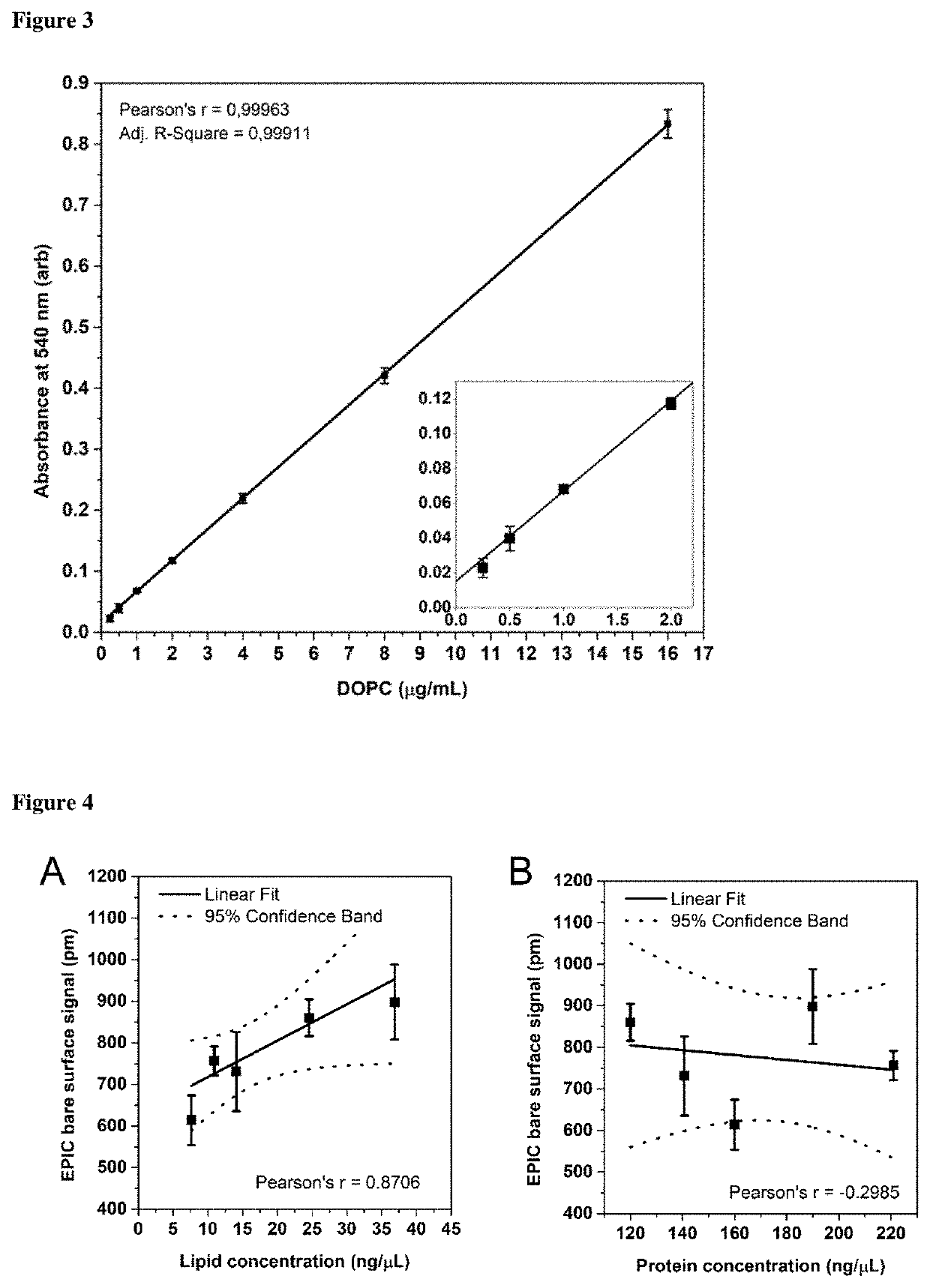
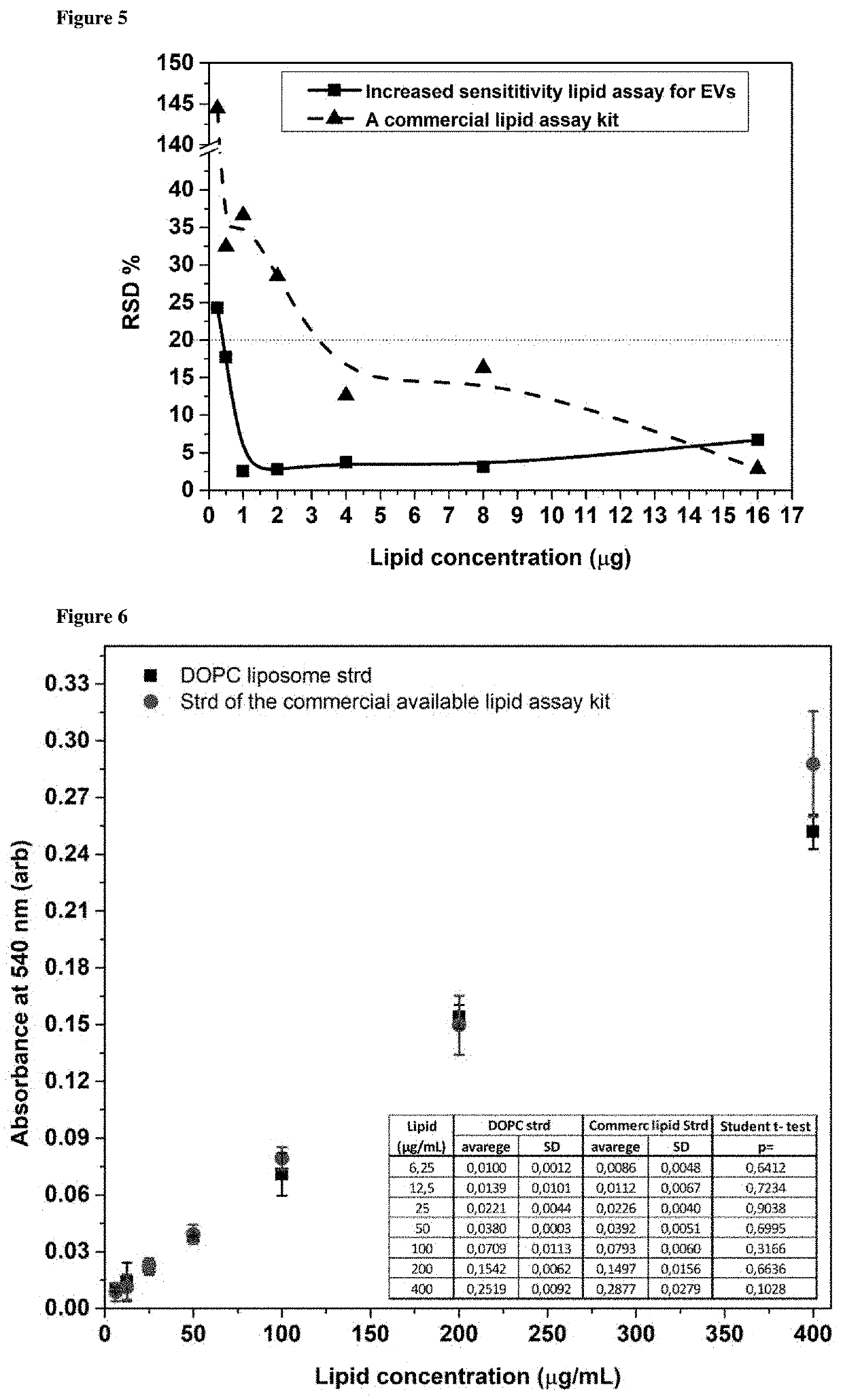
It is also confirmed by a recent review of Théry [6], it states that the broad use of this assay was limited by its relatively low sensitivity which implies that a significant proportion of an EV preparation was required for the lipid test, and it is difficult to keep enough for functional assays.
An alternative approach is mass spectroscopy (MS)-based lipid quantification, however, it still has strong dependence on equipment, requires higher sample amounts, long processing time and the technique may not be readily available to all laboratories [2].

Method used


Image

  • Method for determining the lipid content of extracellular vesicles
  • Method for determining the lipid content of extracellular vesicles
  • Method for determining the lipid content of extracellular vesicles

Examples


example 1

s

[0087]AC16 human cardiomyocyte cell line (SCC109) was purchased from Merck and was cultured according to the instructions of the manufacturer. Before EV isolation, AC16 cells were differentiated according to Davidsons work [11]. Cells were cultured in tissue culture flasks coated with 0.02% gelatine (EMD Millipore) and 5 μg / mL fibronectin (Gibco) up to confluency. Once the cells reached confluency, they were cultured for an additional week in DMEM / F12 medium supplemented with 2% horse serum and 1× Insulin-Transferrin-Selenium (all from Gibco). EVs were isolated from serum free conditioned medium.

[0088]H9c2 (2-1) BDIX rat heart myoblast cell line was ordered from ECACC through Sigma-Merck. Cells were cultured in DMEM medium (Sigma) supplemented with 10% FBS (Gibco), 1% MEM Non-essential amino acid solution (Sigma), antibiotic-antimycotic solution (Gibco); 2 mM L-Glutamine (EMD Millipore) and 3.51 g / L D-Glucose (Sigma). Before EV isolation, H9c2 cells were differentiated according to...

example 2

ation—Isolation from Conditioned Cell Culture Medium

[0091]EVs were isolated based on Osteikoetxea et al. [10] with minor changes. Prior to isolation, cells were washed three times with PBS, and EV production was allowed to take place for 24 hours in serum-free medium or in the presence of 12.5% EV-depleted FBS (Gibco). Three different size-based subpopulations were isolated including large (lEV, here referred to as apoptotic bodies), mid-sized (mEV, here referred to as microvesicles) and small (sEV, here referred to as exosomes) EVs by the combination of gravity driven filtration and differential centrifugation. Briefly, cells were removed by centrifugation at 300 g for 10 min at room temperature (RT), and then the supernatant was filtered by gravity through a 5 μm filter (Millipore, Billerica, Mass.) and submitted to a 2,000 g centrifugation for 30 min at 4° C. to pellet lEVs (Avanti J-XP26 centrifuge, JA 25.15 rotor, Beckman Coulter Inc.). The supernatant was next filtered by grav...

example 3

on of the Lipid Standard—Stock Suspension

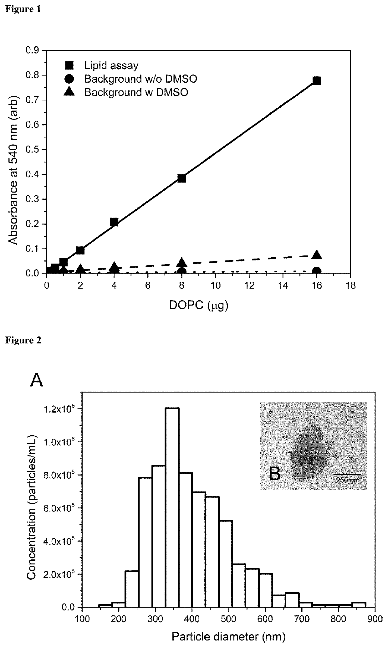
[0092]As a lipid standard, 1,2-Dioleoyl-sn-glycero-3-phosphocholine (DOPC) liposomes were used in 1 mg / mL concentration. DOPC was purchased form Sigma-Aldrich in lyophilised form. 1 mg DOPC was dissolved in 1 mL chloroform (Reanal) in a 2 ml volume test tube. Chloroform was evaporated at 60° C. in a thermoblock (Labnet) under a fume hood. Next, PBS was added to the dried DOPC cake, and the tube was vortexed intensively for 2 min at maximum speed (Fisherbrand). The resulting crude liposome suspension was then sonicated with 35 kHz (Emmi 20, EMAG) at 45° C. for 10 min followed by an additional 2 min intensive vortexing. The obtained liposome standard was stable at 4° C. for at least 3 months, however, immediately before use, intensive vortexing of the standard was essential.


PUM


Abstract

The field of extracellular vesicles (EVs) currently attracts substantial attention in biomedicine due to the proposed role of EVs in various biological processes and their potential of serving as biomarkers for diseases. However, the availability of approaches for reliable and reproducible standardised quantification of EVs is limited. Therefore, current interest in EV research urges reliable tools of standardization and accurate enumeration of EVs, preferably on the basis of lipid quantification. By definition, EVs are surrounded by phospholipid bilayers, therefore, lipids (such as phospholipids and cholesterol) are essential components of all EVs. The method of the invention avoids the overestimation of EV concentration based on the protein measurement, as it focuses on the defining component of EVs, the lipid bilayer. Our method can be used virtually in any standard laboratories where a fume hood, a thermoblock, and a spectrophotometer are available. The application does not require expensive equipment, therefore it can be an easy, reliable and quick method for quantification of EVs and standardisation of EV experiments.

Description

BACKGROUND[0001]The field of extracellular vesicles (EVs) currently attracts substantial attention in biomedicine due to the proposed role of EVs in various biological processes and their potential of serving as biomarkers for disease [1, 2, 3]. However, the availability of approaches for reliable and reproducible standardised quantification of EVs is limited [4, 5, 6]. Therefore, current interest in EV research urges reliable tools of standardization and accurate enumeration of EVs.STATE OF THE ART[0002]Quantifying EVs is not as straightforward as it seems. Commonly used methods for standardisation of EVs rely on measurement of total amount of proteins with colourimetric reactions (e.g. Micro BCA). One of the main problems of these techniques is that proteins may be co-isolated with EVs as protein aggregates, and EVs share biophysical parameters with them [7]. Therefore EV preparations are usually not sufficiently free of co-isolated proteins / protein aggregates to make such approac...

Claims


Application Information

Patent Timeline
IPC IPC(8): G01N33/92G01N21/27G01N21/78
CPCG01N33/92G01N21/278G01N2570/00G01N2405/04G01N2405/08G01N21/78G01N33/5308G01N21/27
Inventor VISNOVITZ, TAMÁSOSTEIKOETXEA VELEZ, XABIERBUZÁS, EDIT IRÉNWERNERNÉ SÓDAR, BARBARAVARGA, ZOLTÁN
Owner RES CENT FOR NATURAL SCI
Learn More