


Quick Research
Generate reliable direction feasibility study reports for your R&D in just a few steps.

Technical Q&A
Discover and master advanced knowledge NOW. Basics, ideas, possibilities, all at once.

Find Solutions
As an expert in R&D theories, this can generate solutions to your technical problems instantly.

Evaluate Feasibility
Analyze your overall solution with one click, know your potential R&D risks in advance.

Monitor Landscape
Get weekly tech updates, stay abreast of the latest tech innovations and key insights.
Wound healing therapeutic hydrogels
- Summary
- Abstract
- Description
- Claims
- Application Information
AI Technical Summary
Benefits of technology
Problems solved by technology
Method used
Image

Examples
example 2
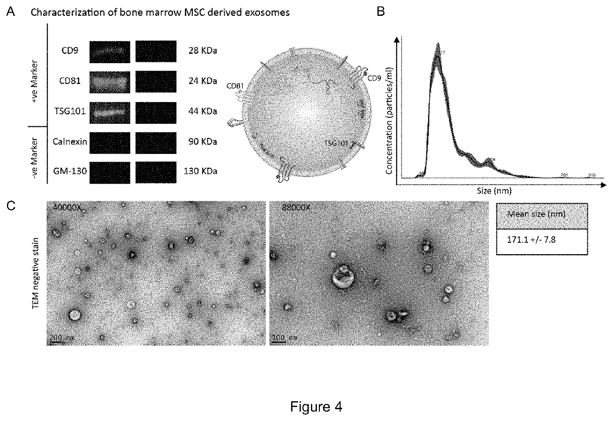
[0084]This example describes isolation of exosomes. At passage 3 (after the first 8-9 population doublings) 70-80% confluent adherent BM-MSCs are washed twice with 1×PBS without Ca2+ / Mg2+. Cells are then incubated for 48 hours at 37° C. / 5%CO2 / 5%O2 in alpha-MEM containing nucleosides, supplemented with 15% exosome-depleted fetal bovine serum (FBS), 1% non-essential amino acids, and 1% penicillin / streptomycin. The conditioned media was then pooled and exosomes were harvested from conditioned media by differential ultracentrifugation. Briefly, conditioned media was centrifuged at 300×g for 5 minutes to remove dead cells and cell debris, 2,000×g for 10 minutes to remove apoptotic bodies, 10,000×g for 30 minutes to remove microvesicles, and then 100,000 g×90 minutes to pellet exosomes. Inclusion of microvesicles or apoptotic bodies alters the size range of our isolated extracellular vesicles, and interferes with isolating a uniform type of vesicle, and subsequently confounds characteriza...
example 3
[0087]This example describes the preparation of hydrogel comprising exosomes. The addition of small molecules prior to gelation hinders network formation (FIG. 6a). In order for the drug to bind the Q gel, we use a stock solution of a small molecule and add it atop the gel. The drug molecules slowly imbibe through the gel and the excess drug is then later removed from the gel.
[0088]Given the larger size of the exosomes, the same approach of adding the exosomes after gelation would not result in its uniform distribution. In an unexpected observation, when a solution of Q protein was mixed with the exosomes, Q successfully formed a gel within the same time period as Q gel by itself (FIG. 6b). Interestingly, the exosomes did not impact the network formation and gelation. The exosomes are uniformly distributed throughout the gel and are interspersed with the Q fibers as observed by transmission electron microscopy (FIGS. 7A and B).
example 4
[0089]This example describes the use of the Exo-Q hydrogel in wound healing. 1×10⁹ exosomes (suspended in phosphate buffered saline containing 0.9 mM CaCl2 and 0.49 mM MgCl2-6H2O) were administered through intradermal injections to the periphery of excisional wounds on a type II diabetic mouse model. This model displays wound healing delays to mimic the chronic nature of human diabetic wounds (Galiano 2004, Wound Repair Regen. 2004 Jul-Aug;12(4):485-92). The results were remarkable when compared to diabetic treated with just normal saline (FIG. 8). Critically important, the single and local exosome dose reduced time to closure of the diabetic wounds within range of wild type non-diabetic mice. Though our results were promising, the administration method was not practical or translatable. For patients to be able to self-administer the exosomes, a suitable delivery vehicle that would not diminish the therapeutic effect of the exosomes was needed.
[0090]Next we tested delivering exosome...
PUM
| Property | Measurement | Unit |
|---|---|---|
| Fraction | aaaaa | aaaaa |
| Percent by mass | aaaaa | aaaaa |
| Molar density | aaaaa | aaaaa |
Abstract
Description
Claims
Application Information
 - R&D Engineer
- R&D Manager
- IP Professional
- Industry Leading Data Capabilities
- Powerful AI technology
- Patent DNA Extraction
Browse by: Latest US Patents, China's latest patents, Technical Efficacy Thesaurus, Application Domain, Technology Topic, Popular Technical Reports.
© 2024 PatSnap. All rights reserved.Legal|Privacy policy|Modern Slavery Act Transparency Statement|Sitemap|About US| Contact US: help@patsnap.com