Separation method and apparatus for microvesicles

Pending Publication Date: 2022-11-03
SAVELIFE BIOTECHNOLOGY CO LTD
View PDF0 Cites 0 Cited by
  • Summary
  • Abstract
  • Description
  • Claims
  • Application Information

AI Technical Summary

Problems solved by technology

However, these methods suffer from the occurrence of adhesion, low efficiency, compromised biological activity, unfavorable downstream experiments, low purity and recovery,

Method used


Image

  • Separation method and apparatus for microvesicles
  • Separation method and apparatus for microvesicles
  • Separation method and apparatus for microvesicles

Examples


example 1

Experimental Method and Materials

[0095]Microfluidic channel and UHF bulk acoustic resonator preparation:

[0096]Microfluidic channels made of polydimethylsiloxane (PDMS) were prepared by soft lithography.

[0097]The bulk acoustic wave resonator devices are prepared by chemical vapor deposition, metal sputtering, and lithography on a silicon wafer. The specific methods are as follows.

[0098]1. The surface of the silicon wafer is thoroughly cleaned using a solution with a 3:1 volume ratio of concentrated sulfuric acid to hydrogen peroxide, which effectively removes organic and inorganic materials from the wafer.

[0099]2. On the cleaned silicon wafer, an aluminum nitride film is formed by surface sputtering, and then a silicon dioxide film is deposited using an ion-enhanced chemical vapor deposition method. Then, using the same method, the aluminum nitride film and the silicon dioxide film are deposited alternately to form a Bragg acoustic reflection structure with alternating layers of alum...

example 2

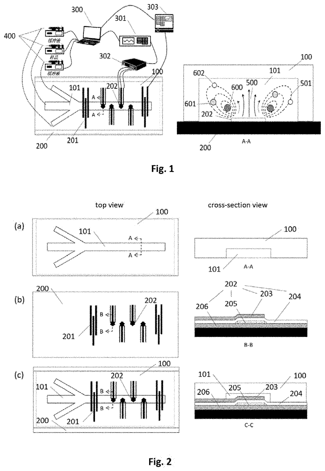
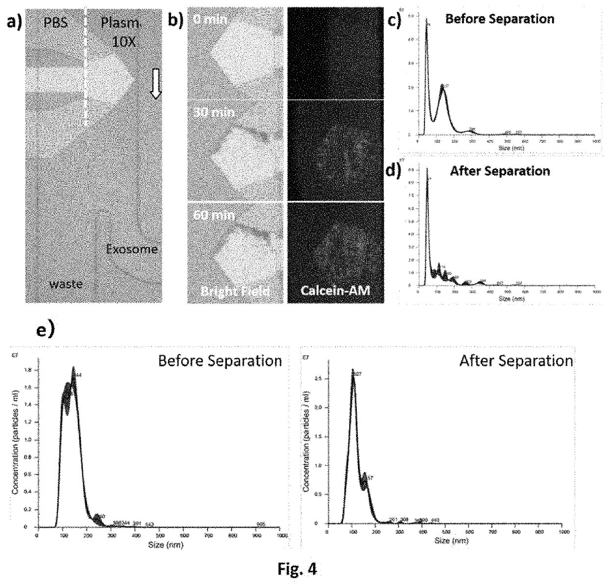
[0109]In this embodiment of the present invention, a microfluidic device is provided which can be used to separate and capture flexible particles in solution. The flexible particles may be artificial or natural. The flexible particles may be biomolecular particles such as nucleic acids. The flexible particles may also be microclusters with a membrane structure, in particular microclusters with a lipid bilayer or a lipid-like bilayer. In one aspect of the present invention, the flexible particles are nature, such as cellular vesicles that are released by cells into the extracellular environment, including exosomes, microvesicles, vesicles, membrane vesicles, prostatic vesicles, microparticles, intraluminal vesicles, intranuclear body-like vesicles, or cytosolic vesicles.

[0110]The method and devices of the present invention can be used to separate and capture flexible particles in solution, for example to separate and obtain microvesicles in blood.

[0111]As shown in FIG. 1, said microf...

example 3

Effect of Flow Channel Height on Acoustic Jets and Vortices Induced by Bulk Acoustic Waves

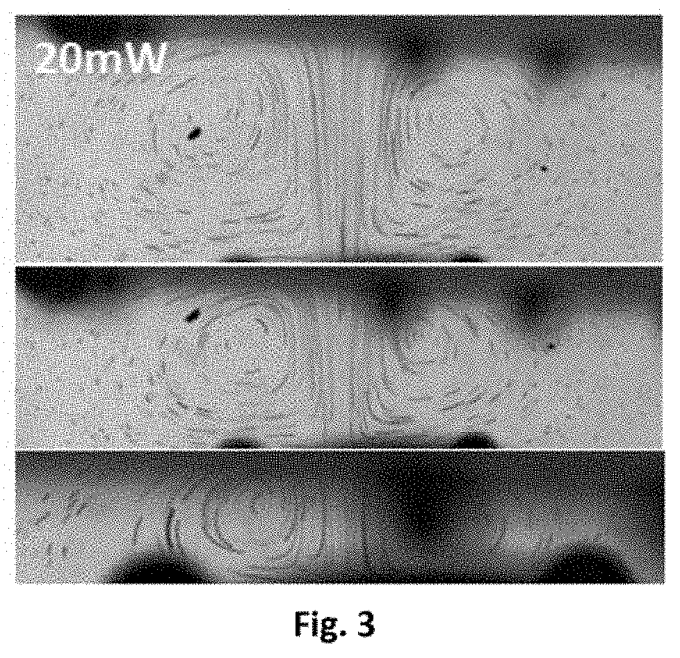
[0142]Observations were made on the acoustic jet and / or vortex currents generated by the UHF bulk acoustic wave resonator used in the present invention in microfluidic channels of different heights. The microfluidic channels of the present invention which are suitable for separating cellular microvesicles (20-800 nm) including exosomes (about 20-250 nm) have a suitable height of no more than 60 μm, e.g. 60 μm, 40 μm, 20 μm.

[0143]In this case, hollow glass beads (with a density close to water) were added to the fluid cavity to characterize the fluid velocity distribution by particle motion trajectories.

[0144]The results are shown in FIG. 3. In the 3 photographs, the microfluidic channel size decreases sequentially from the top, middle to the bottom photograph.

[0145]The photos were taken by a high-speed camera at 5000 fps and combined with 100 frames. Each line segment in the figure represents th...


PUM


Abstract

A microfluidic control system and method for separating flexible particles such as cell vesicles or biomacromolecules such as exosomes in a sample. The system of the present invention comprises one or more ultrahigh frequency acoustic resonators. The ultrahigh frequency acoustic resonators are capable of generating in a fluid channel an acoustic wave of which the frequency is about 0.5-50 GHz and propagated towards a wall opposite the fluid channel. By adjusting the power of the generated acoustic wave and/or the speed at which a conditioning solution flows through an acoustic wave area, flexible particles in a specified range are pushed to and remain at the top part of the flow channel in the acoustic wave area, while flexible particles outside of the specified range go downstream via the acoustic wave area to be collected, thus capturing or releasing the flexible particles in a solution such as cell vesicles or biomacromolecules, particularly exosomes.

Description

[0001]This application claims priority to the following Chinese patent application: Application No. 201910512713.9, filed on Jun. 13, 2019 with a title of “METHOD AND DEVICE FOR SEPARATING MICROVESICLE”, the entire contents of which are incorporated by reference in this application.TECHNICAL FIELD[0002]The present invention relates to the field of cell research methodology and medical devices. Specifically, the invention relates to a microfluidic system for the isolation and analysis of cell microvesicles and a method for using said system to isolate and analyze cell microvesicles.BACKGROUND TECHNOLOGY[0003]The need exists to separate cellular or subcellular particles or biomolecular particles present in body fluids such as blood and tissue fluids, as well as biomolecular particles such as nucleic acids and proteins, which are important for physiological health and research. In the human body, there exist a variety of different cellular vesicles that are released by cells into the e...

Claims


Application Information

Patent Timeline
IPC IPC(8): B01L3/00H03H9/15C12N15/10
CPCB01L3/502761H03H9/15C12N15/1006B01L2400/0436B01L2200/0652B01L2400/082B01L2300/0864C12M23/16C12M47/04C07K1/14C12N15/1003B01L3/5027B01L2200/10B01L2300/0861C12M1/42B01D21/28B01L3/502753B01L2300/0816B01L2400/0439B01D43/00
Inventor DUAN, XUEXINYANG, YANG
Owner SAVELIFE BIOTECHNOLOGY CO LTD
Why Eureka
Try Eureka