Method and equipment for separating microvesicles

A technology of microvesicles and devices for cell research methodology and medical devices

Pending Publication Date: 2020-12-15
SAVELIFE BIOTECHNOLOGY CO LTD
View PDF0 Cites 0 Cited by
  • Summary
  • Abstract
  • Description
  • Claims
  • Application Information

AI Technical Summary

Problems solved by technology

However, there is no method for the separation of nano-sized active particles, especially the controllable precision separation

Method used


Image

  • Method and equipment for separating microvesicles
  • Method and equipment for separating microvesicles
  • Method and equipment for separating microvesicles

Examples


Embodiment 1

[0112] Embodiment 1 Experimental method and material

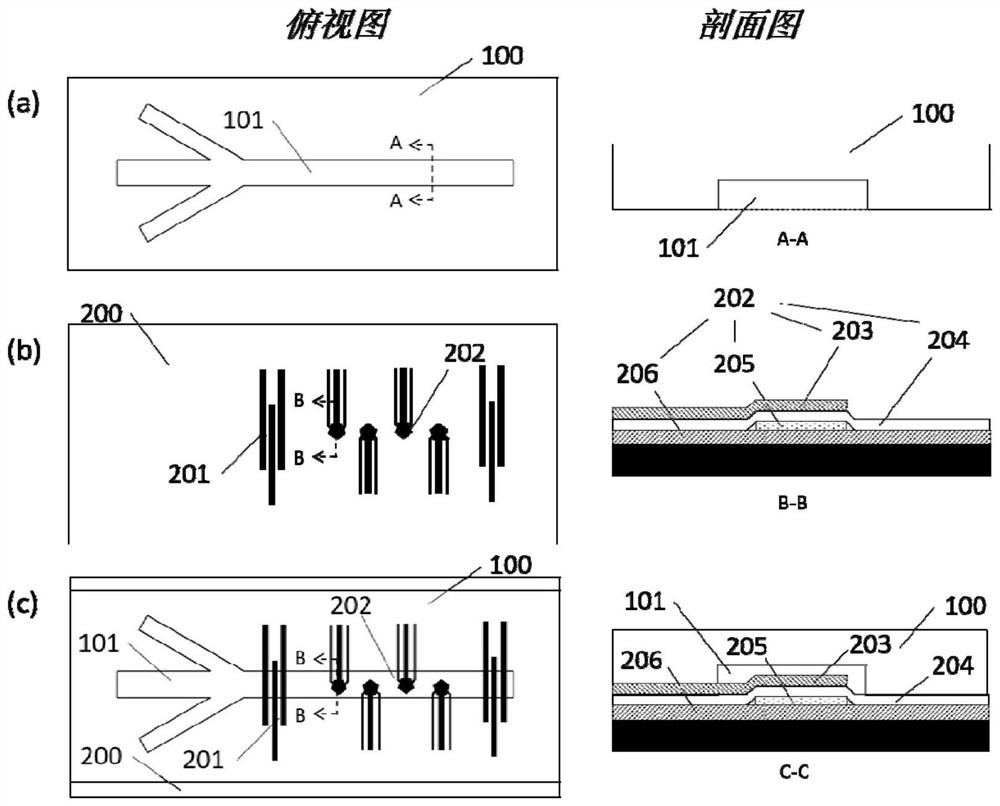
[0113] Fabrication of Microfluidic Channels and UHF Bulk Acoustic Resonators:

[0114] Microfluidic channels made of polydimethylsiloxane (PDMS) were fabricated by soft lithography.

[0115] Bulk acoustic wave resonator devices are prepared by chemical vapor deposition, metal sputtering, and photolithography on silicon-based wafers. The specific method is as follows:

[0116] 1. Use a piranha solution with a volume ratio of concentrated sulfuric acid and hydrogen peroxide of 3:1 to thoroughly clean the surface of the silicon wafer. This method can effectively remove organic and inorganic substances on the silicon wafer.

[0117] 2. On the cleaned silicon wafer, a layer of aluminum nitride film is formed by surface sputtering, and then a layer of silicon dioxide film is deposited by ion-enhanced chemical vapor deposition. Then use the same method to alternately deposit aluminum nitride films and silicon dioxide films to ...

Embodiment 2

[0128] In the specific implementation process of this embodiment, a microfluidic device is provided, which can be used to separate and capture flexible particles in a solution. Flexible particles can be artificial or natural. Flexible particles can be biological macromolecules such as nucleic acids. The flexible particle can also be a micelle with a membrane structure, especially a micelle with a lipid bilayer or a lipid bilayer. In one aspect of the invention, the flexible particle is a naturally occurring particle, such as a cellular microvesicle released by cells into the extracellular environment. Cellular microvesicles include exosomes, vesicles, membrane vesicles, vesicles, air bubbles, prostatic bodies, microparticles, intraluminal vesicles, endosome-like vesicles, or exocytic vesicles, etc.

[0129] The method and device of the present invention can be used to separate and capture flexible particles in solution, for example, to separate and capture target vesicles in...

Embodiment 4

[0166] Example 4 Isolation of exosomes from plasma samples

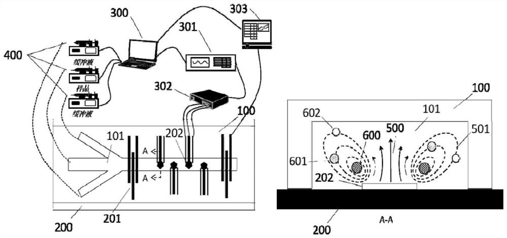
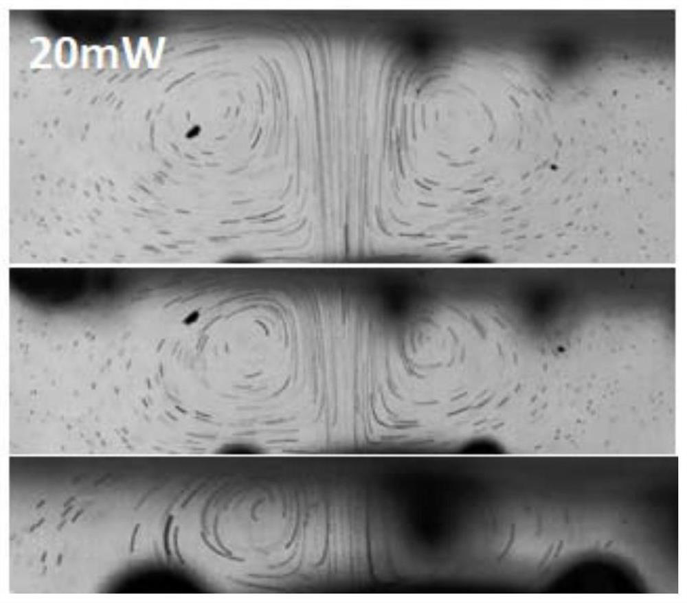
[0167] According to the method described in Examples 1 and 2, prepared and set up as Figure 4 (a) Microchannel and UHF resonator shown. Figure 4 (a) and (b) are top views of the microchannel. The upper part is the inlet of the flow channel, and the arrow on the right indicates the direction of liquid flow. The surface of the ultra-high frequency resonator (that is, the area where the bulk acoustic wave occurs, shown as a five-pointed star in the figure) is located on one side of the channel (the left side in the figure), and the channel inlet of the microchannel includes two solution inlets, the left side The inlet passes through a plasma sample obtained from a volunteer, which is centrifuged at high speed to remove blood cells and some vesicles. Plasma samples were stained by Calcein-AM. The right side is fed with PBS solution, and the dotted line indicates the separation of PBS solution and plasma sample flow...


PUM

PropertyMeasurementUnit
poweraaaaaaaaaa
diameteraaaaaaaaaa
particle diameteraaaaaaaaaa

Abstract

The invention discloses a microfluidic system and method for separating flexible particles such as cell vesicles or biological macromolecules such as exosomes in a sample. The system comprises one ormore ultrahigh-frequency bulk acoustic wave resonators which can generate bulk acoustic waves with the frequency about 0.5-50 GHz in a fluid channel, wherein the bulk acoustic waves are transmitted tothe wall of the opposite side of the fluid channel. By adjusting the power of the generated bulk acoustic waves and / or the speed of a condition solution flowing through a bulk acoustic wave area, theflexible particles in a specified range are pushed to the top of a flow channel and stay in the bulk acoustic wave area, and the flexible particles which do not belong to the specified range enter the downstream through the bulk acoustic wave area to be collected. According to the system and method, the flexible particles such as the cell vesicles or the biological macromolecules in the solution,particularly the exosomes, can be captured or released.

Description

[0001] This application claims the priority of the following Chinese patent application: submitted on June 13, 2019, the application number is 201910512713.9, the title of the invention is "microvesicle separation method and equipment", the entire content of which is incorporated by reference in this application. technical field [0002] The invention relates to the fields of cell research methodology and medical equipment. Specifically, the present invention relates to a microfluidic system for separating and analyzing cell microvesicles and a method for using the system to separate and analyze cell microvesicles. Background technique [0003] Cells or subcellular particles in human body fluids such as blood and tissue fluid, as well as biomacromolecular particles such as nucleic acids and proteins are very important to physiological health and research, so there is a separation of cells or subcellular particles or biomacromolecular particles in body fluids demand. In the ...

Claims


Application Information

Patent Timeline
Patent Type & Authority Applications(China)
IPC IPC(8): C12M1/42C07K1/14C12N15/10B01L3/00
CPCC12M23/16C12M47/04C07K1/14C12N15/1003B01L3/5027B01L2200/10B01L2300/0861B01L2400/0436C12M1/42B01D21/28B01L3/502761B01L3/502753B01L2200/0652B01L2300/0816B01L2400/0439B01D43/00B01L2300/0864B01L2400/082C12N15/1006H03H9/15
Inventor 段学欣杨洋
Owner SAVELIFE BIOTECHNOLOGY CO LTD
Why Eureka
Try Eureka