
Label-Free Hematology and Pathology Analysis Using Deep-Ultraviolet Microscopy

a technology of ultraviolet microscopy and hematology, applied in the field of hematological and histopathological analysis of biological samples, can solve the problems of increasing the cost and labor of these tests, requiring multiple reagents and intensive maintenance, and limiting the monitoring frequency of patients with severe conditions

Pending Publication Date: 2022-11-17
GEORGIA TECH RES CORP
View PDF0 Cites 0 Cited by
  • Summary
  • Abstract
  • Description
  • Claims
  • Application Information

AI Technical Summary

Benefits of technology

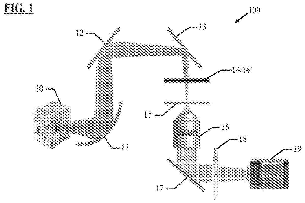
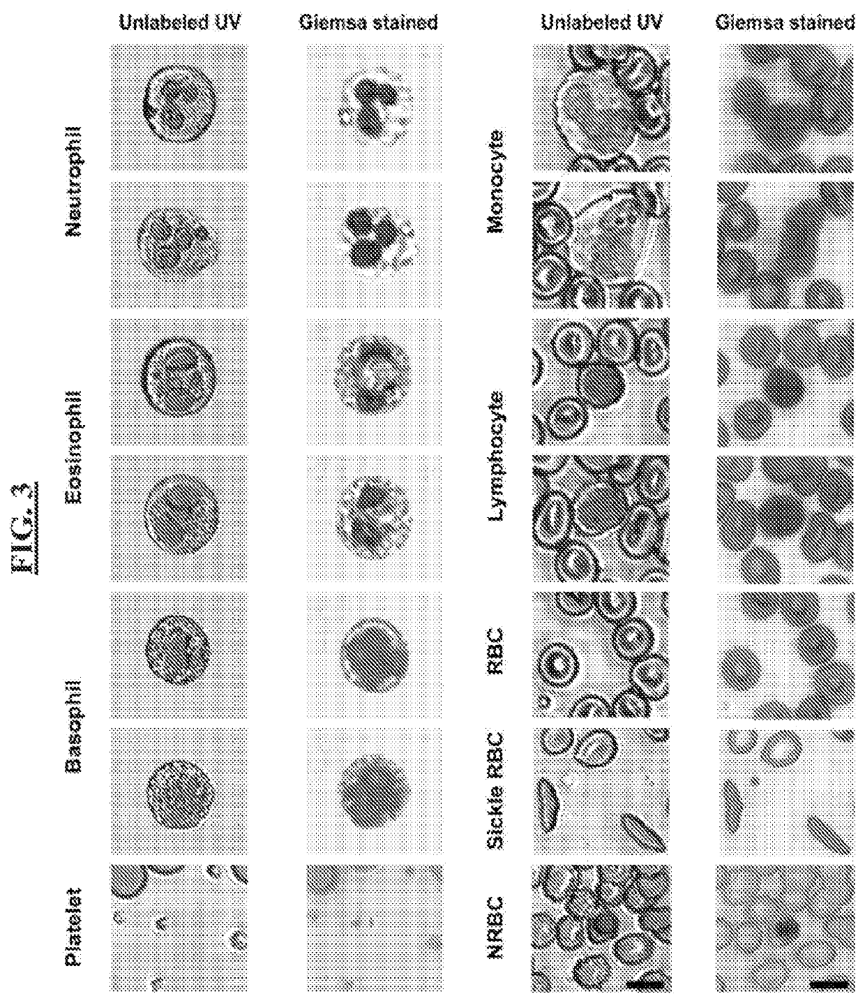
The present invention is a deep-ultraviolet microscopy system that uses a light source to illuminate a biological sample with ultraviolet wavelengths. The system includes an ultraviolet microscope objective for collecting light that has interacted with the sample, an ultraviolet sensitive image capture device for capturing multiple images of the sample, and a short-pass dichroic mirror and parabolic mirror for filtering out non-ultraviolet wavelengths. The system can be used for hematology or histopathology of the sample and can identify and phenotype unique types of cells. The system can also convert the ultraviolet images to a colored image for easier analysis.

Problems solved by technology

Although hematology analyzers are capable of automated and rapid staining and analysis of several samples2, they are costly and require multiple reagents and intensive maintenance.
CBC is one of the most common medical tests3, often performed by trained technicians at healthcare centers, necessitating patients to make several trips for routine tests which further adds to the cost and labor of these tests and limits monitoring frequency for patients with severe conditions.
Accurate evaluation of peripheral blood smears requires well-stained samples and time-consuming microscopic analysis performed by trained personnel and is prone to staining variability.
Alternative techniques such as fluoresce labeling have also been used extensively both for visual assessment4,5 and flow cytometry6-8 of blood cells, however the chemical staining procedures or genetic modifications associated with this method are invasive and suffer from drawbacks such as phototoxicity and photobleaching9.
Although these methods reveal structural and biochemical signatures that can be used for cellular phenotyping and differentiation, the need for careful calibration, complexity of the optical setups, and high equipment costs preclude their use in clinical and point-of-care settings.
Recently, technologies working based on optical imaging of microcirculation in capillaries have enabled point-of-care WBC detection, however such methods are not able to fully differentiate and visualize all WBC subtypes which limits their application to diseases such as neutropenia.
Despite the advances provided to date in the art, there remains no label-free technology suitable for point-of-care settings that can achieve high quality visualization and accurate quantitative differentiation of all blood cell types.

Method used


Image

  • Label-Free Hematology and Pathology Analysis Using Deep-Ultraviolet Microscopy
  • Label-Free Hematology and Pathology Analysis Using Deep-Ultraviolet Microscopy
  • Label-Free Hematology and Pathology Analysis Using Deep-Ultraviolet Microscopy

Examples


Embodiment Construction

[0028]The following disclosure discusses the present invention with reference to the examples shown in the accompanying drawings, though does not limit the invention to those examples.

[0029]The use of any and all examples, or exemplary language (e.g., “such as”) provided herein is intended merely to better illuminate the invention and does not pose a limitation on the scope of the invention unless otherwise claimed. No language in the specification should be construed as indicating any non-claimed element as essential or otherwise critical to the practice of the invention, unless made otherwise clear in context.

[0030]As used herein, the singular forms “a,”“an,” and “the” include plural referents unless the context clearly dictates otherwise. Unless indicated otherwise by context, the term “or” is to be understood as an inclusive “or.” Terms such as “first”, “second”, “third”, etc. when used to describe multiple devices or elements, are so used only to convey the relative actions, po...


PUM

PropertyMeasurementUnit
wavelengthaaaaaaaaaa
wavelengthsaaaaaaaaaa

Abstract

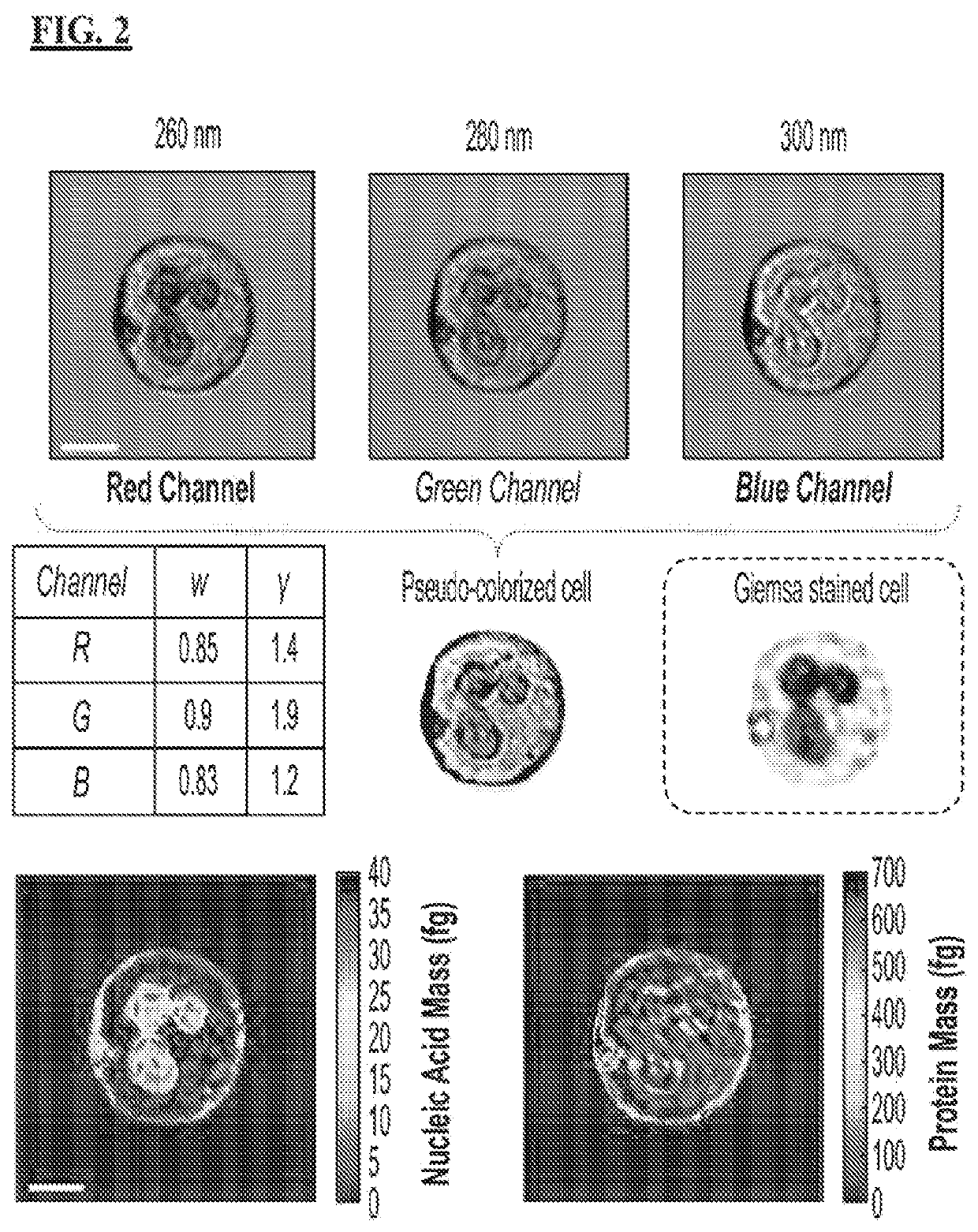
A deep-ultraviolet microscopy system includes a light source for outputting a light beam for illuminating a biological sample, the light beam being inclusive of ultraviolet wavelengths; a reception space for reception of a biological sample for illumination by the light beam; an ultraviolet microscope objective for collecting and relaying light that interacts with the biological sample to an image capture device; and an ultraviolet sensitive image capture device for capturing images of the biological sample, with the microscopy system configured to capture multiple images of the biological sample at one or more ultraviolet wavelengths. A method of processing ultraviolet images of biological samples includes receiving a plurality of multi-spectral ultraviolet images of a biological sample; normalizing and scaling the images; and assigning each image to a channel in the RGB color-space based on wavelength.

Description

ACKNOWLEDGEMENT OF GOVERNMENT SUPPORT[0001]The present inventions were made with government support under Award No. 1752011, awarded by the National Science Foundation. The government has certain rights in the inventions set forth herein.FIELD OF THE INVENTION[0002]The present invention relates to an apparatus and method for hematological and histopathological analysis of biological samples. In particular, the present invention is directed to systems and methods for label-free hematology and histopathology assessments of live and fixed cells and tissues using deep-ultraviolet microscopy.BACKGROUND OF THE INVENTION[0003]Hematological analysis of blood cells has been the standard method for routine clinical diagnosis and monitoring of many blood diseases such as sickle cell anemia, neutropenia, and thrombocytopenia. Current hematological practices rely on assessing alterations in morphology, population, and molecular or cytogenetic properties of blood cells to diagnose diseases. In th...

Claims


Application Information

Patent Timeline
Patent Type & Authority Applications(United States)
IPC IPC(8): G06V20/69G01N21/33G01N33/49H04N5/225G06T3/40G06V10/70H04N9/64G02B21/16G02B21/36
CPCG06V20/698G01N21/33G01N33/49G06V20/693H04N5/2256G06V20/695G06T3/40G06V10/70H04N9/64G02B21/16G02B21/365G01N2201/0621G16H30/40G16H50/20G02B27/0025G01N2201/0627G01N2015/1006G01N2015/1472G01N2015/1488G06T7/0012G06V10/143G06T2207/10056G06T2207/30024H04N23/84H04N23/56
Inventor ROBLES, FRANCISCO E.OJAGHI, ASHKAN
Owner GEORGIA TECH RES CORP
Learn More