Method and apparatus for breathing during anesthesia

a technology for breathing and anesthesia, applied in mechanical equipment, valves, respirators, etc., can solve the problems of general anesthesia, decrease in respiratory muscle strength, and increase in arterial blood carbon dioxide tension

Inactive Publication Date: 2000-09-26
DOWNS JOHN B
View PDF3 Cites 39 Cited by
  • Summary
  • Abstract
  • Description
  • Claims
  • Application Information

AI Technical Summary

Problems solved by technology

Most general anesthetic agents cause a decrease in central drive for respiration, which unimpeded may cause a decrease in oxygenation and increase in arterial blood carbon dioxide tension (PaCO.sub.2).
In addition, general anesthetic agents decrease respiratory muscle strength.
This especially is true of paralytic agents, such as curare, which may remove any ability of the patient to breathe.
In addition, general anesthesia has been shown to decrease compliance of the lung and thoracic cage.
Anesthetic agents are known to have several effects, which may impede the efficiency of oxygenation.
Release of HPVC will cause an increase in perfusion to poorly ventilated lung units, causing PaO.sub.2 to decrease.
General anesthesia is likely to result in inadequate alveolar ventilation and hypercarbia (increase PaCO.sub.2) and inadequate arterial oxygenation (arterial hypoxemia), unless active intervention is applied.
Conventional positive pressure ventilation produced with a mechanical ventilator is associated with several undesirable side-effects.
As a result, any surgical field except for the head, neck and extremities will be subject to undesired movement for a considerable portion of the respiratory cycle.
Decrease in V.sub.A / Q will cause decline in efficiency of oxygenation of the arterial blood.
During standard positive pressure ventilation with an anesthesia ventilator, no spontaneous respiratory activity may occur, due to lack of sufficient flow of respiratory gases from the anesthesia circuit.
Furthermore, standard positive pressure ventilation from an anesthesia ventilator often results in excessive removal of CO.sub.2, increase in arterial blood pH and the well known adverse effects of respiratory alkalosis.
Unfortunately, standard mode positive pressure ventilation during general anesthesia with existing equipment makes such monitoring relatively inaccurate and difficult.
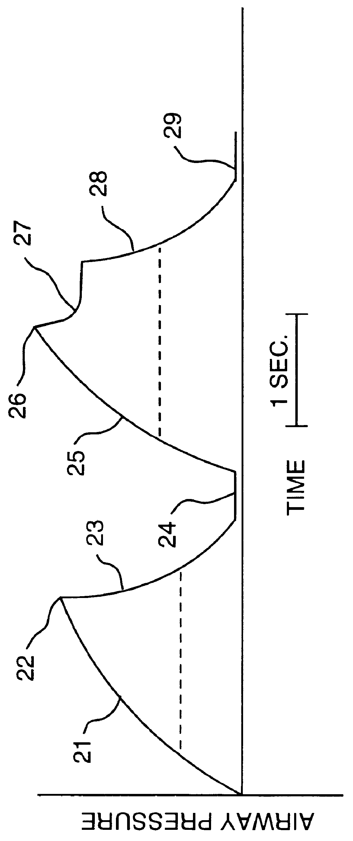
Most anesthesia ventilators deliver inspirable gas at a flow rate such that airway pressure is increased excessively, secondary to resistance of the tracheal tube and the patients' large airways.
Thus, assessment of small airways resistance is extremely difficult.
However, application of such an inspiratory hold will result in a marked increase in mean airway pressure and intrapleural pressure and decrease in venous return and cardiac output.
In addition, such an inspiratory hold significantly decreases the time of stability of the surgical field.
However, studies have repeatedly shown that positive pressure ventilation applied with a standard anesthesia ventilator tends to cause an increase in alveolar dead space and inaccuracy of monitoring of alveolar ventilation.
During the expiratory phase of the respiratory cycle, currently existing means of mechanical ventilation do not allow an active flow of gas from the reservoir bellows.
Thus, any inspiratory effort by the patient during the expiratory phase of the ventilator cycle will result in a decrease in airway pressure.
This decrease in airway pressure will cause undesirable decrease in intrapleural pressure, which may cause significant deterioration of cardiovascular function, secondary to afterload of the left ventricle of the heart and will increase work of breathing.
For this reason, spontaneous ventilation is not allowed when a mechanical ventilator is employed during general anesthesia.
In addition, anesthesia circuitry does not permit the application of a continuous positive airway pressure (CPAP) during spontaneous respiration.
Such an increase in V.sub.A / Q will increase physiologic dead space, with its attendant undesirable effects.
Some systems employ sufficiently high gas flow to prevent significant rebreathing of anesthetic gases, so that CO.sub.2 absorption is unnecessary.
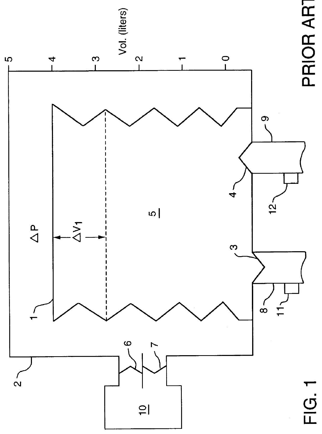
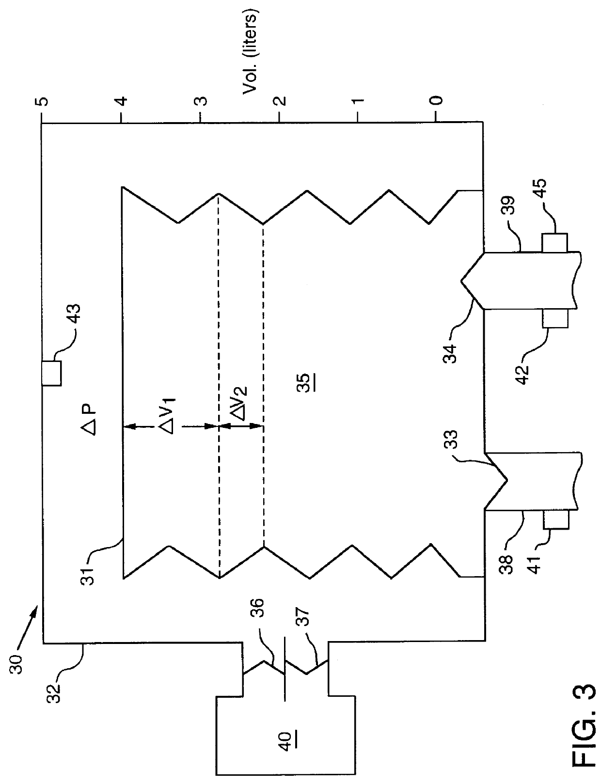
Exhalation from the patient results in gas entering the bellows, causing it to rise within the container.
These include regulation of flow into the rigid chamber, time allowed for inspiration, and mechanical limitation of the excursion of the bellows.
Thus, exchange of oxygen between alveolar space and pulmonary capillary blood is unimpeded.
Further, because of relative absence of alveolar dead space, end-tidal CO.sub.2 monitoring renders analysis of arterial blood for determination of PaCO.sub.2 unnecessary, with rare exception.

Method used


Image

  • Method and apparatus for breathing during anesthesia
  • Method and apparatus for breathing during anesthesia
  • Method and apparatus for breathing during anesthesia

Examples


Embodiment Construction

Operation of the AAV apparatus of the present invention was studied as follows. Nonsedated ASA physical status I and II patients scheduled for general anesthesia, intra-abdominal operations and insertion of an intra-arterial catheter for blood pressure monitoring, signed an Institutional Review Board approved consent. Patients with unstable cardiovascular function or severe obstructive lung disease were excluded from the study. Chest leads were attached to monitor EEG and heart rate was determined electronically. A probe was positioned around a finger tip and connected to a pulse oximeter for determination of oxygen saturation (SpO.sub.2).

Anesthesia and neuromuscular blockade were induced with propofol (1 to 2 mg / kg IV) or thiopental (2 to 5 mg / kg IV) and succinylcholine (1.5 mg / kg) and the patients were intubated orotracheally. Anesthesia and neuromuscular blockade were maintained with isoflurane, nitrous oxide and oxygen and vecuronium. An intravenous narcotic was administered whe...


PUM


Abstract

A method and apparatus are provided for ventilation of patients during general anesthesia. Breathing gas is supplied to the patient during anesthesia at a controlled volume above the functional residual capacity of the patient's lungs. The patient is allowed to spontaneously respire when the volume of breathing gas is above the functional residual capacity. The pressure of the breathing gas is periodically reduced to facilitate expulsion of carbon dioxide-containing gas from the patient. The system promotes alveolar ventilation, carbon dioxide excretion, oxygenation and respiratory monitoring in patients who receive general anesthesia.

Description

1. Field of the InventionThe present invention relates to ventilation of patients during anesthesia, and more particularly relates to a method and apparatus for maintaining and monitoring alveolar ventilation, carbon dioxide excretion and oxygenation in patients who receive general anesthesia.2. Background InformationGeneral anesthesia induces a state of respiratory insufficiency. Most general anesthetic agents cause a decrease in central drive for respiration, which unimpeded may cause a decrease in oxygenation and increase in arterial blood carbon dioxide tension (PaCO.sub.2). In addition, general anesthetic agents decrease respiratory muscle strength. This especially is true of paralytic agents, such as curare, which may remove any ability of the patient to breathe. In addition, general anesthesia has been shown to decrease compliance of the lung and thoracic cage. A decrease in the compliance of these structures requires an increase in muscle strength to produce adequate ventila...

Claims


Application Information

Patent Timeline
Patent Type & Authority Patents(United States)
IPC IPC(8): A61M16/00
CPCA61M16/00A61M16/0081A61M2016/0039A61M2016/0042A61M2230/432A61M16/024
Inventor DOWNS, JOHN B.
Owner DOWNS JOHN B
Why Eureka
Try Eureka