Non-invasive monitoring of hemodynamic parameters using impedance cardiography

a technology of impedance cardiography and hemodynamic parameters, which is applied in the field of non-invasive monitoring of hemodynamic parameters using impedance cardiography, can solve the problems of reducing affecting etc., and achieves the effect of enhancing the accuracy of ejection time detection and reducing the cost of operation

Inactive Publication Date: 2000-12-12
RHEO GRAPHIC PTE
View PDF24 Cites 141 Cited by
  • Summary
  • Abstract
  • Description
  • Claims
  • Application Information

AI Technical Summary

Benefits of technology

The invention discloses a method of measuring hemodynamic parameters using a novel combination of bioimpedance cardiography and electrocardiography which allows medical practitioners to obtain an accurate, substantially continuous assessment of a patient's cardiac performance. The bioimpedance and electrocardiogram signals are measured over a common time interval of interest, preferably more than ten (10) heartbeats.
Concurrently, a method of the invention may utilize electrocardiography to enhance the accuracy of the ejection time detection from the measured bioimpedance signal. The electrocardiogram can be obtained in any standard position, well known in electrocardiography. To eliminate the overall quantity of electrodes at the patient's skin, this invention may use the same set of electrodes for bioimpedance and ECG measurements (see FIG. 6B).
The invention may also involve the step of continuously adjusting the calculation of cardiac parameters according to changes in the red blood cell count and according to varying bodily compositions of different patients, and can thus be used under a variety of different circumstances without a loss of accuracy.
In addition, the invention comprises a method of improved bioimpedance signal processing. It may employ a computer system to analyze the both the bioimpedance signal and the electrocardiogram in a variety of ways to provide an accurate report of cardiac parameters. The computer system may be used to make corrections in the gain-phase-frequency characteristics caused by the transducers used to measure the bioimpedance and ECG. The computer system communicates a sinusoidal test curve to the transducer receiver and may then measure and record the gain-phase-frequency distortions created by the receiver. The "real" bioimpedance signals subsequently received by the computer system through that transducer may pass through filters which remove the transducer's characteristic distortions as well as breath and movement artifacts. Accuracy of gain-frequency response and phase-frequency response may be corrected to within 5%.

Problems solved by technology

The invasive methods require that a medical practitioner insert a measuring device into the patient's body, such as a catheter in the throat, and present numerous disadvantages to both patient and physician.
The patient must often endure substantial pain and discomfort and the physician must perform a relatively complicated procedure and occasionally expose himself or herself to the risk of contact with infectious blood.
Many of the electrode configurations currently in use fail to adequately take into account the paths followed by the lines of electrical potential through the thorax and thus create a distortion in the cardiac measurement.
These band electrodes typically wrap around a patient like a belt and further limit access to the patient, an especially undesirable condition curing reanimation procedures.
The movements associated with respiration also make band electrodes very inconvenient when placed on the neck and chest.
Perhaps the most significant problem with the presently existing bioimpedance methods in the imprecise mathematical derivation of cardiac parameters from bioimpedance measurements.
The prior art does not teach a method for determining ventricular ejection time with sufficient accuracy.
Furthermore, the prior art fails to account for the fact that VET is not a single event.
The measurements of left VET and right VET for most patients are generally very close, but even slight differences between them can create errors in bioimpedance readings under the methods currently in use.
It is not accurate for patients under physical training or other physical stress, or for critically ill patients, such as those typically in intensive care units.
Without further refinement, however, the Kubicek equation frequently given inaccurate measurements.
In addition, the prior art does not disclose a method for adjusting R in accordance with the fluctuation of a patient's hematocrit (red blood cell count).
Many of the methods for bioimpedance cardiography require that the patient hold his or her breath during each measurement because respiration causes interference in the bioimpedance signal.
Such methods are inconvenient for some patients and completely useless for other patients who are unconscious or otherwise unable to hold their breath.

Method used


Image

  • Non-invasive monitoring of hemodynamic parameters using impedance cardiography
  • Non-invasive monitoring of hemodynamic parameters using impedance cardiography
  • Non-invasive monitoring of hemodynamic parameters using impedance cardiography

Examples


Embodiment Construction

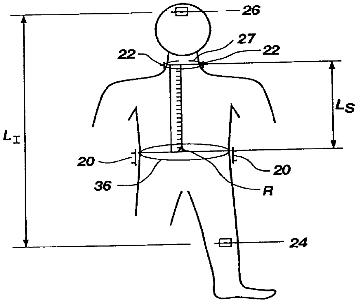
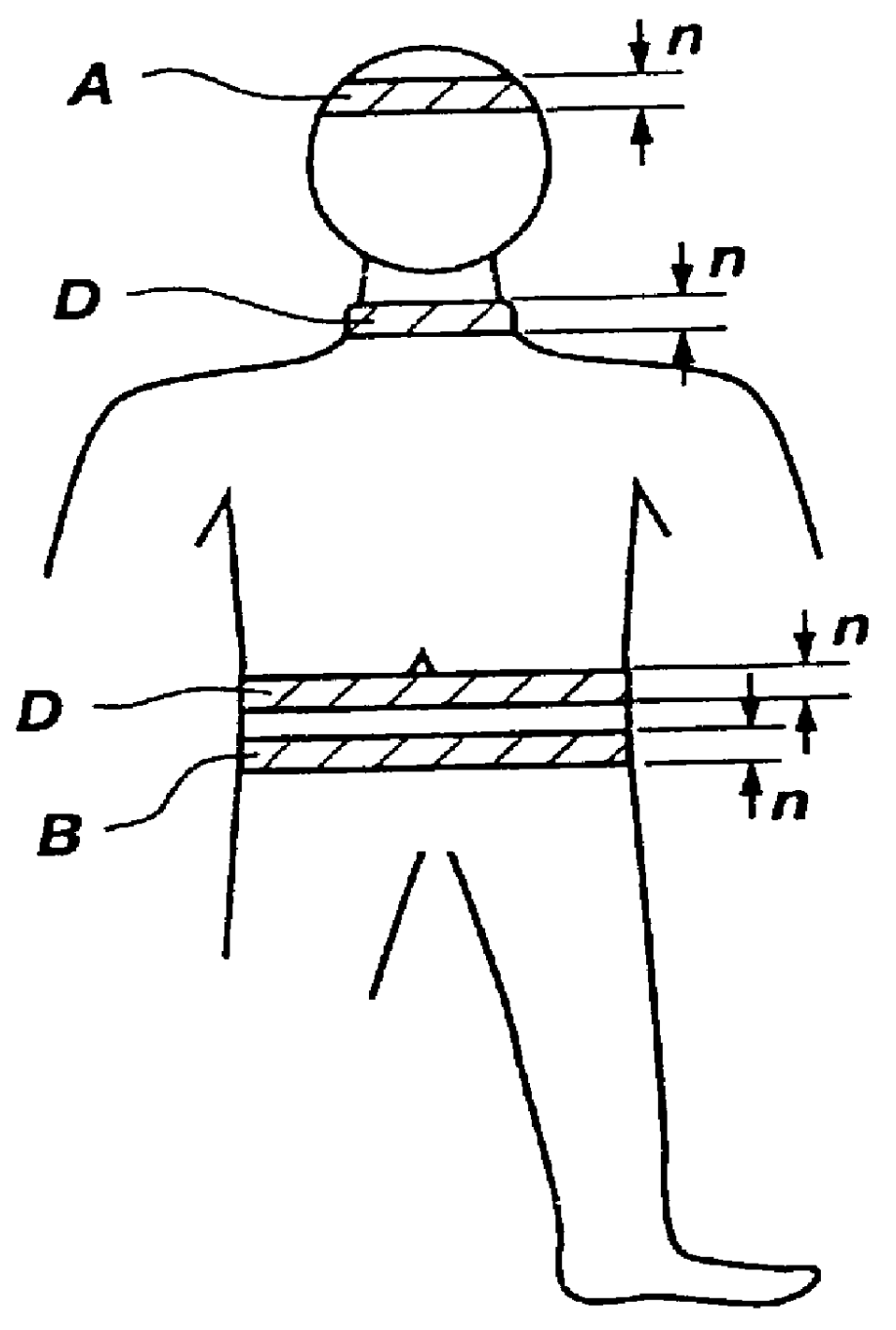
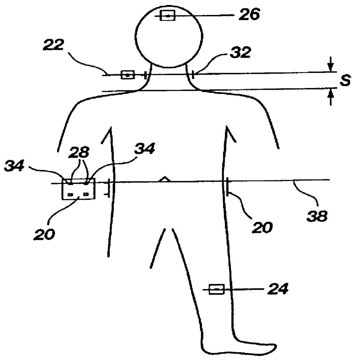
The first step in the present invention involves taking bioimpedance measurements over segments of tissue on a patient's body. Electrodes must be placed at appropriate points on the surface of the skin to generate a high frequency, low amplitude electric current and to detect changes in the generated current after it passes through the segments of tissue (see FIGS. 2A, 2B, 3 and 4). The electrodes are "spot electrodes" rather than "band electrodes" in order to maximize the free area on the patient's body. The spot electrodes are preferably of the disposable, one-use type. The patient thus has increased freedom of movement and medical practitioners have more access to the patient's skin for other medical procedures, such as the introduction of catheters and the administration of anesthesia.

The bioimpedance electrode system employs a total of six electrodes: a pair of detecting (measuring) electrodes 20 at the xiphoid process level, a pair of detecting (measuring) electrodes 22 positi...


PUM


Abstract

PCT No. PCT / SG97 / 00013 Sec. 371 Date Oct. 8, 1998 Sec. 102(e) Date Oct. 8, 1998 PCT Filed Apr. 7, 1997 PCT Pub. No. WO97 / 37591 PCT Pub. Date Oct. 16, 1997A method and apparatus for determination of heart rate, heart stroke volume, heart stroke volume, and cardiac output from thoracic bioimpedance signals and electrocardiograms. A unique bioimpedance electrode arrangement is employed, and the bioimpedance signals are corrected for gain-phase-frequency distortion through the use of sinusoidal test signals through the measuring or detection electrodes to identify distortions and correct for same during actual measurements. Time-derivative bioimpedance signals are employed, the power spectrum calculated, and a novel autoconvolution procedure used to emphasize the heart rate harmonic. Breath waves and other signals not indicative of the patient's cardiocycles are removed. Left ventricular ejection time is derived from the bioimpedance signals, and an improved version of Kubicek's equation is employed to derive heart stroke volume and thus cardiac output.

Description

The present invention relates generally to cardiac monitoring and specifically to the determination of heart rate (HR), heart stroke volume (SV), and cardiac output (CO) according to detection and complex analyses of thoracic bioimpedance and electrocardiograph (ECG) signals, which permit precise detection of the start of left ventricular ejection.Heart rate is the number of times the heart beats per minute. Heart stroke volume is the volume of blood pumped during each heart stroke. Cardiac output is the volume of blood pumped in one minute and is generally considered to be the most significant gauge of cardiac fitness. Physicians must frequently rely upon such cardiac parameters to diagnose heart disease, to assess a patient's overall health, to determine the most appropriate method of treatment, and to quickly discover sudden lapses in cardiac performance.The currently existing methods for measuring cardiac output and other cardiac parameters may be divided into two categories: in...

Claims


Application Information

Patent Timeline
Patent Type & Authority Patents(United States)
IPC IPC(8): A61B5/08A61B5/053A61B5/0245A61B5/02A61B5/026A61B5/0295A61B5/296
CPCA61B5/0535A61B5/7239A61B5/0809A61B5/7257A61B5/0295
Inventor SCHOOKIN, SERGEI I.ZUBENKO, VIATCHESLAV G.BELIAEV, KONSTANTIN R.MOROZOV, ALEKSANDR A.YONG, WEN H.
Owner RHEO GRAPHIC PTE
Why Eureka
Try Eureka