

Rapid quantitative measurement of soluble fibrin in opaque body fluids
a technology of opaque body fluid and quantitative measurement, which is applied in the direction of instruments, peptides/protein ingredients, peptides, etc., can solve the problems of difficult to achieve the effect of rapid quantitative measurement of soluble fibrin in opaque body fluid, and difficult to achieve the effect of rapid quantitative measuremen
- Summary
- Abstract
- Description
- Claims
- Application Information
AI Technical Summary
Benefits of technology
Problems solved by technology
Method used
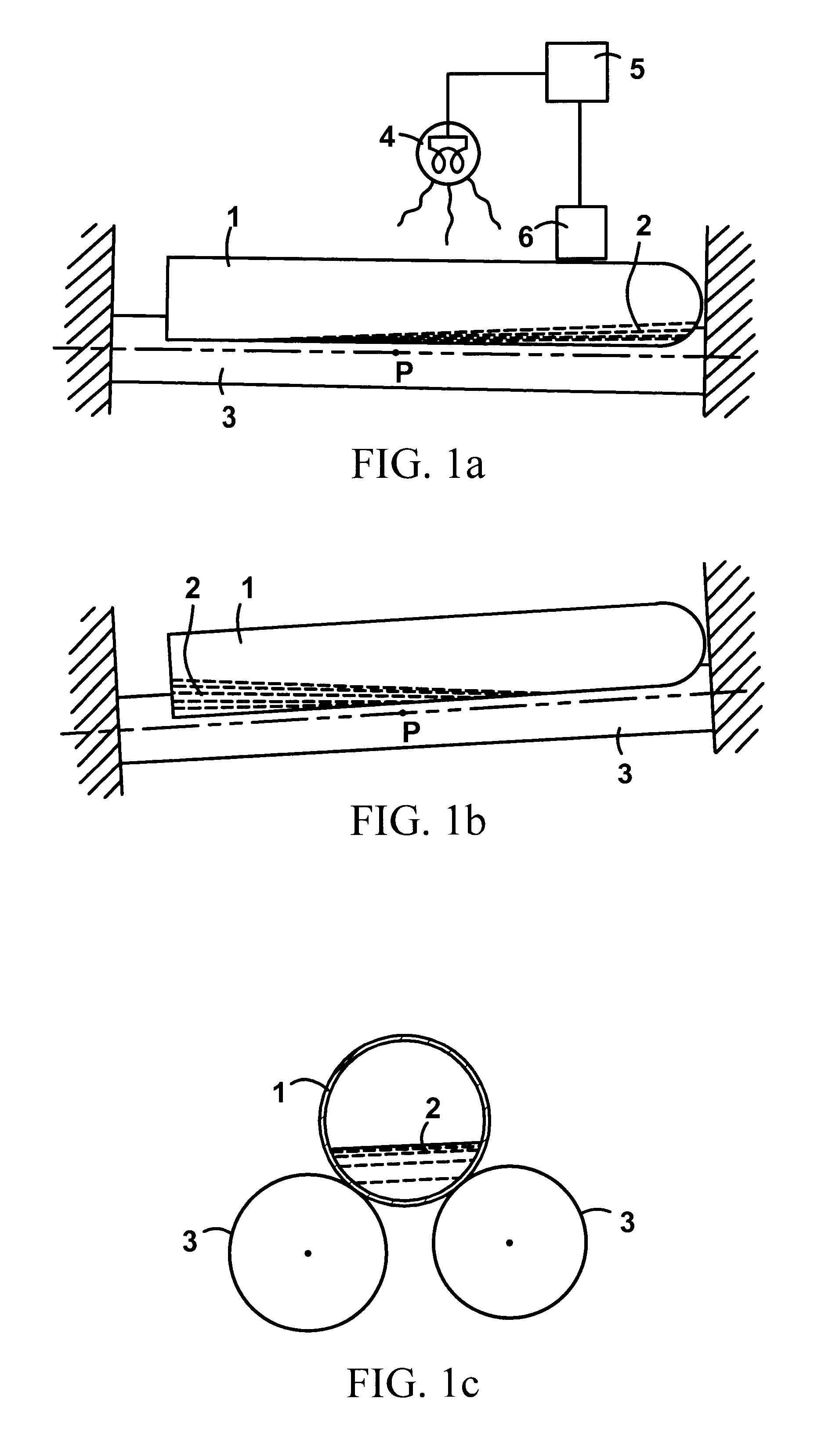
Image

Examples
example ii
SF Measurement in Whole Blood
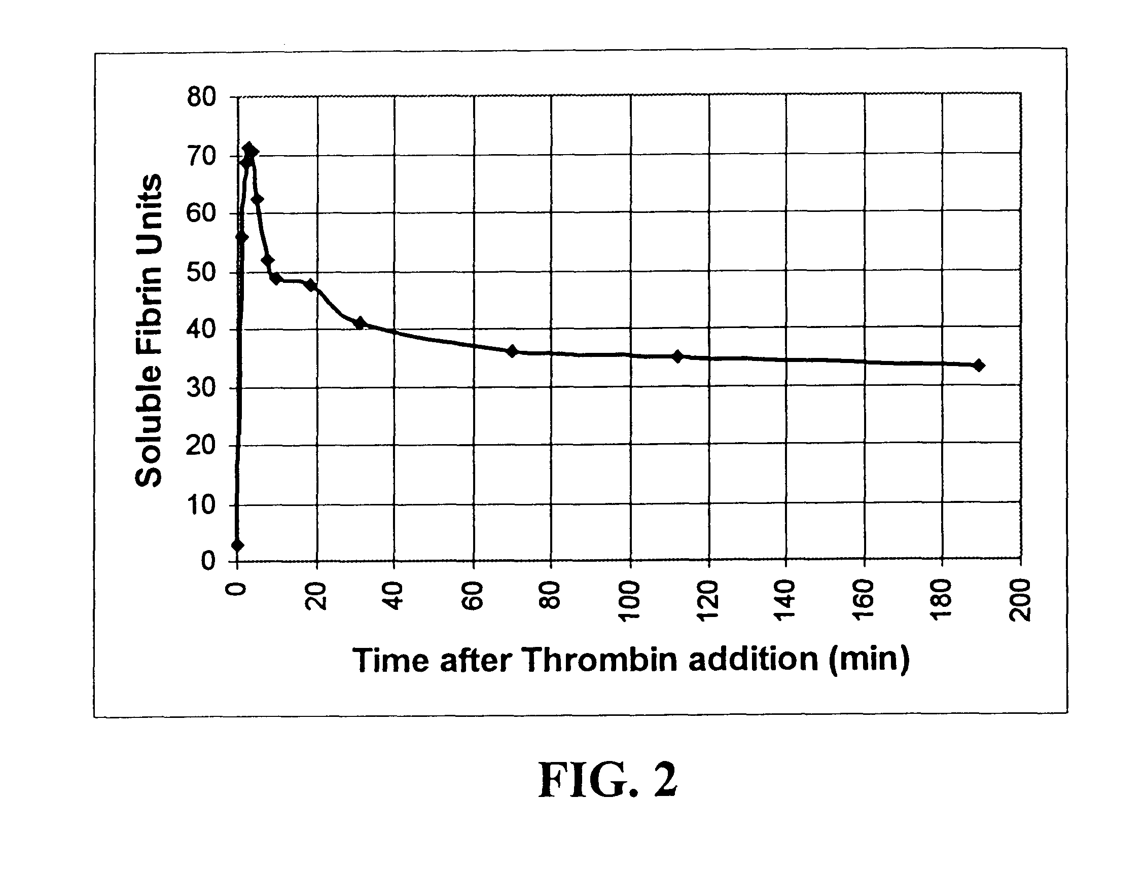
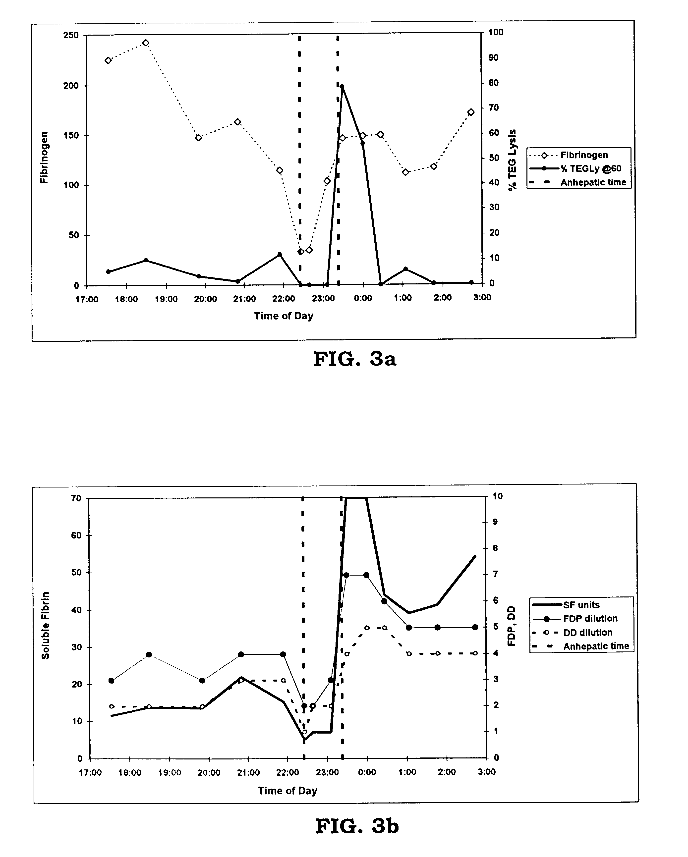
In a second test of the system, a series of 150 .mu.l whole blood samples were, at thirty minute intervals, removed from a patient undergoing liver transplantation. These samples were suspended in about 450 .mu.l saline, and were placed in a specimen container together with about 20 .mu.l of the precipitating reagent, protamine sulfate. The whole blood sample was taken from a patient at the start of the operation, then every 30 minutes with samples spaced more closely during the critical reperfusion phase of the transplantation process. The mixture of protamine sulfate and whole blood was placed in a hemostatic analysis apparatus as described in the '188 patent at a temperature of about 37.degree. C. to allow the mixture to react until precipitates formed and adhered to the inner tube surface. The time that it took from the start of the process to the first-end point, and the second end-point were then measured, and the measurements were correlated to th...
PUM
| Property | Measurement | Unit |
|---|---|---|
| temperature | aaaaa | aaaaa |
| pH | aaaaa | aaaaa |
| transparent | aaaaa | aaaaa |
Abstract
Description
Claims
Application Information