
Medical probe device and method

a medical probe and probe technology, applied in the field of medical probe devices and methods, can solve the problems of frequent urination, difficult precise placement of treatment probes, obstruction of urinary tract, etc., and achieve the effect of minimizing trauma and achieving greater medical benefits

Inactive Publication Date: 2005-02-08
MEDTRONIC VIDAMED
View PDF61 Cites 307 Cited by
  • Summary
  • Abstract
  • Description
  • Claims
  • Application Information

AI Technical Summary

Benefits of technology

"The invention is a device and method for penetrating tissue to perform medical actions, such as tissue destruction and fluid delivery, with precision and ease. The device includes a catheter with a control end and a probe end, and a stylet guide housing with a port for introducing a flexible stylet. The stylet can be an electrical conductor enclosed in a non-conductive layer, which can be moved longitudinally through the catheter to the target tissue. The device also includes a temperature sensor for better positioning and control. The method involves introducing the catheter and stylet through a curved path, and the non-conductive layer can have a leading tip and a flexible portion extending from it. The non-conductive layer can also have a movement means for longitudinally moving the stylet. The device and method provide greater medical effectiveness with reduced trauma and improved precision."

Problems solved by technology

Often, precise placement of a treatment probe is difficult because of the location of a target tissue in the body or the proximity of the target tissue to easily damaged, critical body organs, nerves, or other components.
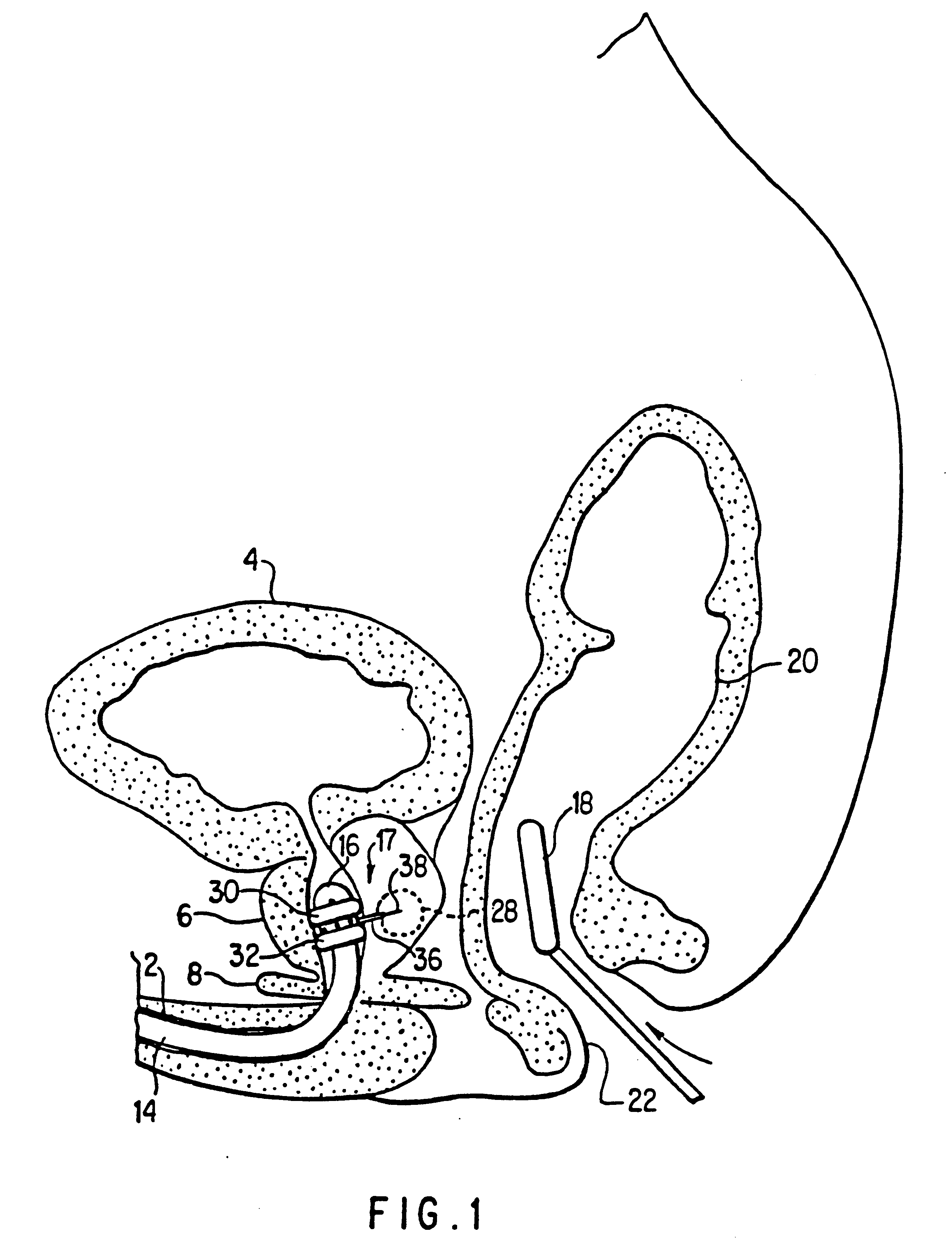
Hyperplastic enlargement of the prostate gland often leads to compression of the urethra, resulting in obstruction of the urinary tract and the subsequent development of symptoms including frequent urination, decrease in urinary flow, nocturia, pain, discomfort, and dribbling.
Currently there is no proven effective nonsurgical method of treatment of BPH.
In addition, the surgical procedures available are not totally satisfactory.
Those suffering from BPH are often elderly men, many with additional health problems which increase the risk of surgical procedures.
Surgical procedures for the removal of prostatic tissue are associated with a number of hazards including anesthesia associated morbidity, hemorrhage, coagulopathies, pulmonary emboli and electrolyte imbalances.
These procedures performed currently can also lead to cardiac complications, bladder perforation, incontinence, infection, urethral or bladder neck stricture, retention of prostatic chips, retrograde ejaculation, and infertility.
Due to the extensive invasive nature of the current treatment options for obstructive uropathy, the majority of patients delay definitive treatment of their condition.
This circumstance can lead to serious damage to structures secondary to the obstructive lesion in the prostate (bladder hypertrophy, hydronephrosis, dilation of the kidney pelves, etc.) which is not without significant consequences.
In addition, a significant number of patients with symptoms sufficiently severe to warrant surgical intervention are poor operative risks and are poor candidates for prostatectomy.
In addition, younger men suffering from BPH who do not desire to risk complications such as infertility are often forced to avoid surgical intervention.
Thus the need, importance and value of improved surgical and non-surgical methods for treating BPH is unquestionable.
Healthy cells of the duct wall and healthy tissue between the nodules and duct wall are also indiscriminately destroyed in the process and can cause unnecessary loss of some prostate function.
Furthermore, the added cooling function of some microwave devices complicates the apparatus and requires that the device be sufficiently large to accommodate this cooling system.
Application of liquids to specific tissues for medical purposes is limited by the ability to obtain delivery without traumatizing intervening tissue and to effect a delivery limited to the specific target tissue.

Method used


Image

  • Medical probe device and method
  • Medical probe device and method
  • Medical probe device and method

Examples


Embodiment Construction

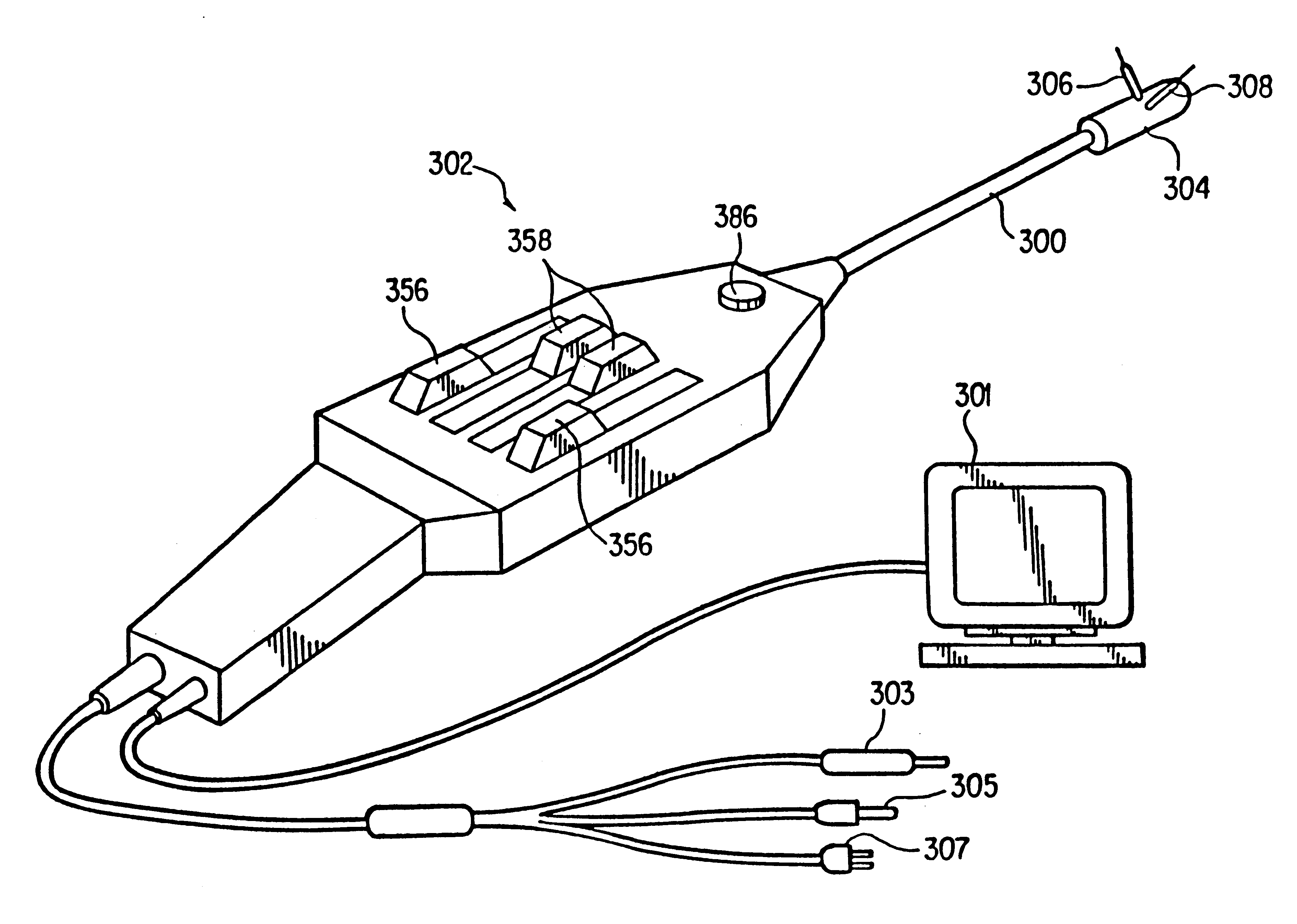
The device of this invention provides a precise controlled positioning of a treatment stylet in a tissue targeted for treatment, destruction or sampling from a catheter positioned in the vicinity of the target tissue.

The term “stylet” as used hereinafter is defined to include both solid and hollow probes which are adapted to be passed from a catheter port through normal tissue to a target tissue. The stylet is shaped to facilitate easy passage through tissue. It can be a solid wire, thin rod, or other solid shape or it can be a thin hollow tube or other shape having a longitudinal lumen for introducing fluids to or removing materials from a site. The stylet can also be a thin hollow tube or other hollow shape, the hollow lumen thereof containing a reinforcing or functional rod or tube such as a laser fiberoptic cable. The stylet preferably has a sharpened end to reduce resistance and trauma when it is pushed through tissue to a target site.

The stylet can be designed to provide a var...


PUM


Abstract

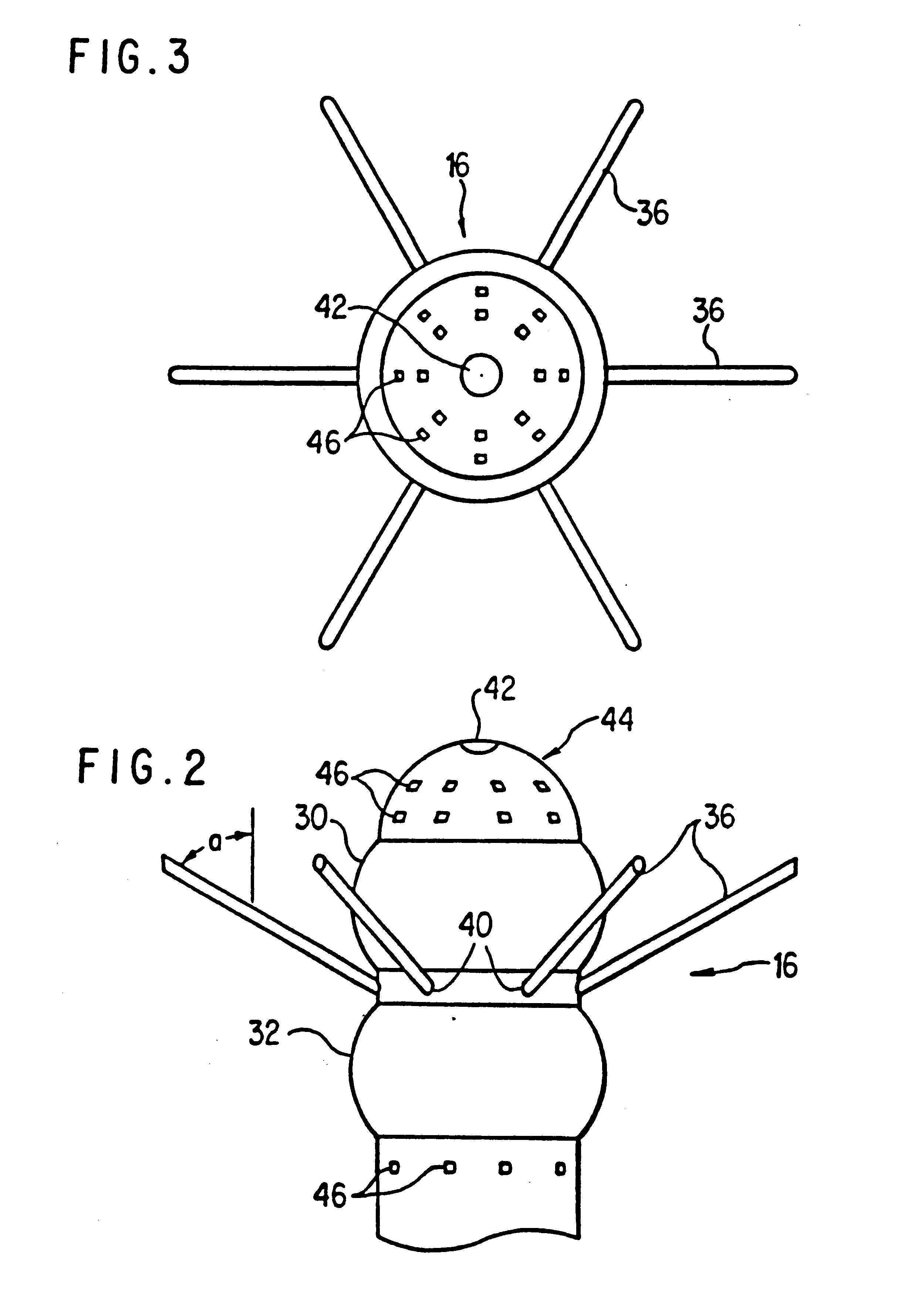
A medical probe device comprises a catheter having a stylet guide housing with one or more stylet ports in a side wall thereof and a stylet guide for directing a flexible stylet outward through the stylet port and through intervening tissue at a preselected, adjustable angle to a target tissue. The total catheter assembly includes a stylet guide lumen communicating with the stylet port and a stylet positioned in said stylet guide lumen for longitudinal movement from the port through intervening tissue to a target tissue. The stylet can be an electrical conductor enclosed within a non-conductive layer, the electrical conductor being a radiofrequency electrode. Preferably, the non-conductive layer is a sleeve which is axially moveable on the electrical conductor to expose a selected portion of the electrical conductor surface in the target tissue. The stylet can also be a microwave antenna. The stylet can also be a hollow tube for delivering treatment fluid to the target tissue. It can also include a fiber optic cable for laser treatment. The catheter can include one or more inflatable balloons located adjacent to the stylet port for anchoring the catheter or dilation. Ultrasound transponders and temperature sensors can be attached to the probe end and / or stylet. The stylet guide can define a stylet path from an axial orientation in the catheter through a curved portion to a lateral orientation at the stylet port.

Description

FIELD OF THE INVENTIONThis invention is directed to a unique device and method for penetrating body tissues for medical purposes such as tissue destruction and fluid substance delivery, for example. The device penetrates tissue to the precise target selected in order to deliver energy to the tissue and / or deliver substances. It limits this activity to the precise preselected site, thereby minimizing trauma to normal surrounding tissue and achieving a greater medical benefit. This device is a catheter-like device for positioning a treatment assembly in the area or organ selected for medical treatment with one or more stylets in the catheter, mounted for extension from a stylet port in the side of the catheter through surrounding tissue to the tissue targeted for medical activity.BACKGROUND OF THE INVENTIONTreatment of cellular tissues usually requires direct contact of target tissue with a medical instrument, usually by surgical procedures exposing both the target and intervening tis...

Claims


Application Information

Patent Timeline
Patent Type & Authority Patents(United States)
IPC IPC(8): A61B18/18A61B18/00A61B18/14A61B10/00A61N1/06A61N1/40A61N5/04A61N5/02A61B17/22A61B17/28A61B17/24A61B18/08A61B18/04A61B18/20A61B17/34A61B18/24A61B18/22A61B19/00A61F7/00A61M1/00A61M25/00A61B17/00A61M3/02A61M3/00A61B5/01A61B10/02A61B10/06A61M25/092A61M25/14A61N5/06
CPCA61B18/00A61B18/1477A61B18/1485A61B18/18A61B18/1815A61N1/06A61N1/40A61N1/403A61N5/02A61N5/045A61B10/02A61B10/0241A61B18/082A61B18/1206A61B18/14A61B18/1442A61B18/148A61B18/24A61B2017/00084A61B2017/00092A61B2017/00106A61B2017/00274A61B2017/00292A61B2017/003A61B2017/00867A61B2017/22072A61B2017/22077A61B2017/22082A61B2017/248A61B2017/2939A61B2017/3488A61B2018/00005A61B2018/00011A61B2018/00083A61B2018/00095A61B2018/00196A61B2018/0022A61B2018/00547A61B2018/00577A61B2018/00678A61B2018/00702A61B2018/00726A61B2018/00744A61B2018/0075A61B2018/00761A61B2018/00791A61B2018/00797A61B2018/00821A61B2018/00869A61B2018/00886A61B2018/0091A61B2018/00916A61B2018/00946A61B2018/00982A61B2018/1253A61B2018/126A61B2018/1273A61B2018/128A61B2018/1412A61B2018/1425A61B2018/1861A61B2018/2238A61B2019/4873A61B2019/5217A61B2019/5278A61B2019/5425A61F2007/0054A61M3/0279A61M25/0068A61M25/0069A61M25/008A61M25/0082A61M2025/0086A61M2025/0087A61M2025/0089A61M2025/009A61M2025/0096A61M2025/018A61N5/04A61B2090/3782A61B2090/3614A61B2090/0814A61B2090/3925
Inventor EDWARDS, STUART D.LAX, RONALD G.LUNDQUIST, INGEMAR H.SHARKEY, HUGH R.
Owner MEDTRONIC VIDAMED
Learn More